INTRODUCTION
It is well known that Kakiemon-style porcelains have very elegant and bright colored overglazes having its original color tone and its characteristic pattern-distribution drawn on their porcelain basic body of white-color. The Kakiemon-style porcelains had been exported from the
later 17th century and were estimated to be very famous for interior decorations and dinner-sets in Europe, as Chinese porcelains. However, the Kakiemon-style porcelains were one kind of HIZEN colored porcelains produced at Arita and Ohkawachi (Imari) areas, which sited at a Nabeshima domain of HIZEN-Han in the early Edo period (at present, Arita and Ohkawachi are in Saga prefecture of Kyushu
Local structures and electronic band states of α−Fe
2O
3polycrystalline
particles in the glazes of the HIZEN celadons produced in the Edo period
of Japan, by means of X-ray absorption spectra (II)
(Estruturas locais e estados de banda de partículas policristalinas nos
esmaltes de celadons Hizen produzidos no período Edo do Japão, por meio
de espectros de absorção de raios X (II))
M. Hidaka1, K. Ohashi2, R. P. Wijesundera1,6, L. S. R. Kumara1, S. Sugihara3, N. Momoshima3, S. Kubuki4, N. E. Sung5
1Department of Physics, Graduate School of Science, Kyushu University, Fukuoka 812-8581, Japan 2The Kyushu Ceramic Museum, Arita, Saga, 844-8585, Japan
3Radioisotope Center, Kyushu University, Fukuoka 812-8581, Japan
4Department of Chemical and Biological Engineering, Ube National College of Technology Yamaguchi, 755-8555, Japan
5Department, Pohang Accelerator Laboratory, Pohang University of Science and Technology, Pohang 790-784, Korea 6Department of Physics, University of Kelaniya, Kelaniya, Sri Lanka
Abstract
HIZEN celadon glazes produced in 1630’s to 1790’s (Edo period, Japan) have been investigated by means of X-ray absorption spectra (XAS) near a Fe-K edge by using synchrotron radiation and a Mössbauer spectrum. The XAS suggest that the local structure around Fe2O3 ine powders is slightly different between the Izumiyama ceramics of mainly the Quartz-SiO2 and Ohkawachi ceramics of mainly the feldspar of (K,Na)Si3O8 (Sanidine), and that the glazes of the HIZEN celadons include the Fe2O3 ine powders in
the glassy state, though the X-ray diffraction patterns of the glassy celadon glazes do not show any peaks of the Fe2O3 structure.
The Mössbauer spectrum suggests that the celadon glaze of Seiji (m) includes only Fe3+ ions, but not Fe2+ ions. This indicates
the existence of Fe2O3 in the celadon glaze. It is interpreted that the colored brightness of the HIZEN celadons is induced by the
structural properties of the used raw celadon ceramics and the other transition-metal ions of Cr, Cu, Zn in the celadon glazes, but not by the chemical reaction from Fe2O3 to FeO under the deoxidizing thermal treatment at higher temperature in a kiln.
Keywords: HIZEN celadon glaze, local structure, electronic band states.
Resumo
Esmaltes de celadon Hizen produzidos dos anos 1630 a 1790 (período Edo, Japão) foram investigados por meio de espectros de absorção de raios X (XAS) próximos da linha Fe-K usando radiação síncrotron e espectro Mossbaues. Os resultados de XAS sugerem que a estrutura local em pós inos de Fe2O3 é levemente diferente entre as cerâmicas Izumiyama principalmente o quartzo e cerâmicas Ohkawachi principalmente do feldspato (K,Na)Si3O8 (Sanidine), e que os esmaltes dos celadons Hizen incluem inos
pós de Fe2O3 no estado vítreo, embora os difratogramas de raios X dos esmaltes celadon não mostrem picos da estrutura do
Fe2O3. O espectro Mossbauer sugere que os esmaltes celadon de Seiji (m) incluem somente íons Fe3+, mas não Fe2+. Isto indica a existência de Fe2O3 no esmalte celadon. É feita a interpretação que o brilho nas cores dos celadons Hizen é induzido pelas propriedades estruturais das cerâmicas básicas de celadon e os outros metais de transição Cr, Cu, Zn nos esmaltes celadon, mas não pela reação química entre Fe2O3 para FeO sob tratamento térmico desoxidante em forno a altas temperaturas.
island, Japan). The HIZEN colored porcelains are generally classiied by four kinds of porcelains: Shoki-Iroe (Kokutani-style), Kakiemon-style, Kinran-style, and Nabeshima ware [1-9]. The Nabeshima wares were as gifts only to the Tokugawa Shogun (Edo period). The raw porcelain ceramics of high-quality ferromagnetic used as the basic body were discovered by Korean potters at a small mountain, called Izumiyama (Arita), in 1610’s. The basic porcelain body was consisted of ferromagnetic porcelain ceramics, called Hakujikou.
In the Arita and Ohkawachi areas, the porcelain ceramics including slightly larger content of Fe2O3 than that of Hakujikou were collected and used as a raw material of Hizen celadons, where the celadon is called with “Seiji” in Japanese. In 1610~1640’s the Shoki-Imari porcelains were produced with a Korea porcelain technique. From 1640~50’s, the colored underglazes and overglazes were made by the porcelain technique due to Keitokuchin-kiln (China), while the celadons were due to Ryusen-kiln (China) [10, 11]. The producing technique of the HIZEN celadons at Arita was developed in 1650~1660’s and increased its completion in 1670’s. After 1700’s the technique was gradually declined [12]. In 1630~1650’s Mitunomata celadons of elegant and bright were also produced at Mitunomata of Hasami, which is about 10 km away from Arita [13, 14].
It is known that the iron oxides in the celadon glaze show blue-green color under the deoxidizing thermal treatment at high-temperature. However, the celadon glaze changes gradually its color from yellow-brown to black color as increasing the iron oxides, even if under the deoxidizing thermal treatment. J. S. Larid (1918) [15] and R. R. Hunghan (1950) [16] reported that the blue-green color of the celadon glazes results from chemical reaction from Fe2O3 to FeO in the celadon glazes under the deoxidizing thermal treatment. At the present, many Japanese celadon potters thought that ine air-bubbles in the celadon glaze contribute to a diffused relection of blue-green light. Recently, we have studied the correlation between the red-colored brightness and the structural and electronic properties for the overglaze of the HIZEN colored porcelains, which were produced at Arita areas from the 17th to 18th century, by means of X-ray diffraction (XRD) and X-ray absorption spectrum (XAS),
using synchrotron radiation [17-19]. In this paper, we study the geographical and historical correlations among the glaze-colors of the HIZEN celadons produced at Aita and Imari areas in the Edo period.
MATERIALS
In order to study the structural deformation localized around Fe ions and the partially electronic band states of Fe ions in the glazes of the HIZEN celadons, we carried out measurements of X-ray absorption spectra (XAS) at the K-edge of Fe ions. Before the present celadon broken pieces, we measured the XAS of the raw ceramics for the Izumiyama celadon ceramics, the Ohkawachi celadon ceramics, and the Izumiyama basic ceramics (Hakujikou), of which the ratios of the oxide components are listed in Table I of Ref. [20]. Three kinds of the raw ceramics contained Fe2O3 of about 1.52, 4.21, and 0.56 wt% for the Izumiyama ceramics, the Ohkawachi ceramics, and Hakujikou, respectively. All of their X-ray diffraction patterns showed very small peaks of the Fe2O3 structure. The component ratio in the raw ceramics at Izumiyama (Arita) and Ohkawachi (Imari) were not so large difference, as shown in Table I [20]. However, we found from X-ray diffraction that the Izumiyama ceramics are mainly the quartz (SiO2), while the Ohkawachi ones are mainly the feldspar of (K,Na)Si3O8 (sanidine). This suggests that both materials are grown in the different mechanism from the earth crust, though Izumiyama and Ohkawachi areas are about 10 km away each other. As described later, their different structural properties are partially related to the characteristic colored brightness of the celadon glazes in the HIZEN celadons produced at Arita and Ohkawachi areas, as shown in photo 1 in Ref. [20].
EXPERIMENTAL
In the present investigations the XAS near the Fe–K edge were measured for the raw ceramics, the HIZEN celadons, and the modern Nabeshima celadons (Choshun celadon) in photo 1 in Ref. [20], by using synchrotron radiation at the Pohang Light Source (2.5 GeV). A double crystal monochromator of Si (111) gave a relative energy
Table I - Oxide compositions (wt.%) of the celadon glazes of the HIZEN celadons, Seiji (c), (e), (j), (h), and (ℓ), in addition to the red-colored overglaze of the Kakiemon-style porcelain.
[Tabela I - Composições de óxidos (peso%) dos esmaltes celadon dos celadons Hizen, Seiji (c), (e), (j), (h), e (ℓ), em complemento aos esmaltes de cor vermelha da porcelana estilo Kakiemon.]
kiln remain SiO2 Al2O3 CaO K2O Na2O MgO Fe2O3 MnO TiO2 PbO
c Hyakuken 62.8 14.9 11.8 5.39 0.78 0.86 2.73 0.32 0.18 ─
e Nagayoshidani 64.5 15.8 10.8 4.06 0.57 1.39 2.85 0.28 0.26 ─
h Shimoshirakawa 67.3 15.2 9.42 3.91 0.35 1.44 2.02 0.26 ─ ─
j Nabeshima kiln 69.3 16.2 5.79 5.61 1.32 ─ 1.59 0.07 ─ ─
ℓ Higuchi 66.6 15.4 8.13 5.74 2.02 ─ 1.89 0.07 ─ ─
resolution ΔE to be less than about 0.2 eV at each respective monochromatic incident X-ray beam for the XAS near the Fe-K edge. The incident X-ray photons (Io) were detected with an ionic chamber set in front of the specimen holder, while the X-ray luorescence photons (IF) emitted from the front surface of the specimens were simultaneously detected with an X-ray luorescence detector. The surface was always set with about 45.0° to the incident X-ray beam, of which the size was about 3 mm in horizontal and 1 mm in vertical on the specimen surface to the electron orbital of the accelerator. The ratio μ(I) (=IF / Io) gives the XAS near the Fe-K edge. We normalized the measured raw data with software programs of Artemis and Athena, which was developed by C. H. Booth and F. Bridges (2005) to analysis of the observed XANES and EXAFS spectra [21].
RESULTS AND DISCUSSION
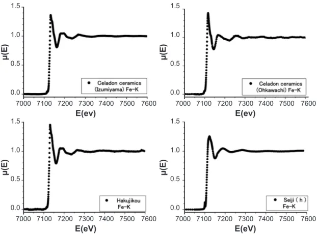
Fig. 1 shows the XAS around Fe-K edge of the raw ceramics; the Izumiyama celadon ceramics, the Ohkawachi celadon ceramics, and Hakujikou, in addition to the HIZEN celadon of Seiji (h). Seiji (h) was taken at the kiln remain of Shimoshirakawa closed to Izumiyama in 1655-1660’s [20]. The X-ray diffraction patterns suggested from the very weak intensity of the observed peaks that the Izumiyama and Ohkawachi celadon ceramics, and Hakujikou include
the α-Fe2O3 ine powders of small amount, as Table I in Ref. [20]. Thus, the XAS of the raw ceramics in Fig.1 results from the α-Fe2O3 particles. On the other hand, the celadon glaze of Seiji (h) shows the XAS to be similar to the raw ceramics, though the celadon glaze is in the glassy state. Fig. 2 shows the EXAFS spectra in the energy region of 7150to 7500 eV, which are expanded the XAS in Fig. 1. The spectra suggest that the local structure around Fe ions relects on the structural difference between the Izumiyama ceramics of mainly the quartz and the Ohkawachi one of mainly the sanidine of (K,Na)Si3O8 [20]. The similarity of the EXAFS spectrum between the Izumniyama celadon ceramics and Seiji (h) also suggests that the glaze material of Seiji (h) was based on the Izumniyama celadon ceramics, though an X-ray absorption threshold (Eo) depends on the structural circumstance around Fe ions in the ceramics.
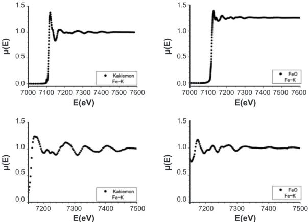
For comparison we measured the XAS around K-edge of the red-colored overglaze of the Kakiemon-style porcelain, which was produced at Arita in 1710’s-1740’s, and the marketed powders of FeO. The red-colored overglaze includes much amount of α-Fe2O3 (19.1 wt.%) and lead oxide PbO (24.2 wt.%). The results are shown in Fig. 3. The left and right spectra in the lower igures are expanded in the EXAFS region of the XAS in the upper igures for the Kakiemon-style overglaze and FeO, respectively. We found the clear difference of the XAS between the α-Fe2O3
1.5 1.5
1.5 1.5
1.0 1.0
1.0 1.0
0.5 0.5
0.5 0.5
0.0 0.0
0.0 0.0
7000 7000
7000 7000
E(ev) E(ev)
E(eV) E(eV)
7200 7200
7200 7200
7400 7400
7400 7400
7100 7100
7100 7100
7300 7300
7300 7300
7500 7500
7500 7500
7600 7600
7600 7600
Figure 1: X-ray absorption spectra around Fe-K edge of the Izumiyama celadon ceramics, the Ohkawachi celadon ceramics, the Izumiyama basic ceramics (Hakujikou), and the HIZEN celadon of Seiji (h).
[Figura 1: Espectros de absorção de raios X em torno da borda Fe-K das cerâmicas celadon Izumiyama, cerâmicas celadon Okhawachi, cerâmicas básicas (Kahujikou) Izumiyama e celadon Hizen de Seiji (h).]
µ(E)
µ(E)
µ(E)
structure and the FeO one. As described before, the XAS of the raw ceramics also result from Fe ions of the α-Fe2O3 structure. Thus, we know from the different oscillated amplitude of the EXAFS spectrum that the XAS also gives structural information of the local circumstance around the Fe2O3 ine powders in a material. That is, it is considered that the Fe2O3 ine particles make a large cluster of long-range order in the red-colored overglaze and a ine cluster of short-range order or independently co-exist in the raw ceramics. The XAS in Figs. 1 and 2 suggest that Seiji (h) includes the Fe2O3 ine particles in its celadon glaze, though the celadon glaze is in the glassy state. The melting temperature of Fe2O3 and FeO are 1570 °C and 1370 °C, respectively. In 1630’s to 1790’s the maximum temperature in the used kilns was about 1200-1250 °C.
In the present investigations the used specimens were 12 broken pieces of the monochromatic HIZEN celadons (photo 1(a) to (ℓ)) produced in 1630’s to 1730’s at Arita areas (Arita, Yamauchi) and Imari, and 2 ones of the modern Nabeshima celadons (Photo 1(m) and (n)) produced by Choshun kiln sited at Ohkawachi (Imari), as photo 1 in our other paper [20]; (a) Kamanotsuji (Yamauchi, 1630-1640’s), (b) Kamanotsuji (Yamauchi, 1630-1640’s), (c) Hyakuken (Yamauchi, 1630-1640’s), (d) Hyakuken (Yamauchi,
1630-1640’s), (e) Chokichidani (Arita, 1655-1660’s), (f) Chokichidani (Arita, 1655-1660’s), (g) Maruo (Arita, 1650-1660’s), (h) Shimoshirakawa (Arita, 1655-1660’s), (i) Nabeshima-domain kiln (Imari, 1690-1720’s), (j) Nabeshima-damain kiln (Imari, 1690-1720’s), (k) Higuchi (Arita, 1760-1790’s), (ℓ) Higuchi (Arita, 1760-1790’s), (m) Choshun kiln (Imari, 2007), (n) Choshun kiln (Imari, 2007). All of the present kiln remains are famous and historic in the Edo period. It was deduced that the Nabeshima-domain kiln, called Odougu-yama, was transferred from Nangawara (Arita) to Ohkawachi (Imari) in 1673-1681’s. Ohkawachi is about 10 km away from Arita, while Yamauchi is about 5 km away from Izumiyama at Arita. The broken pieces of the Nabeshima celadons in Photo 1(i) and (j) were produced at Ohkawachi Odougu-yama. The Nabeshima wares including the celadons show excellently elegance and brightness. Table I lists the ratios of the component oxides in the celadon glazes of the HIZEN celadons, called Seiji (c), (e), (j), and (ℓ), in Photo (c), (e), (j), and (ℓ) of our other paper [20], in addition to those in the red-colored overglaze of the Kakiemon-style porcelain. In that time, Isubai (natural wood ash) including much CaCO3 was traditionally added into the raw ceramics of the HIZEN celadon glazes. CaCO3 is chemically changed to CaO at about 825 °C, and CaO has a catalytic function to
1.2 1.2
1.2
1.0 1.0
1.0
0.8 0.8
0.8
E(eV) E(eV)
E(eV)
7200 7200
7200
7400 7400
7400
7300 7300
7300
7500 7500
7500
Figure 2: EXAFS spectra around Fe-K edge of the Izumiyama celadon ceramics, the Ohkawachi celadon ceramics, and the HIZEN celadon of Seiji (h) in Figure 1.
[Figura 2: Espectros EXAF em torno da borda Fe-K das cerâmicas celadon Izumiyama, cerâmicas celadon Okhawachi, cerâmicas básicas (Kahujikou) Izumiyama e celadon Hizen de Seiji (h) na Figura 1.]
µ(E) µ(E)
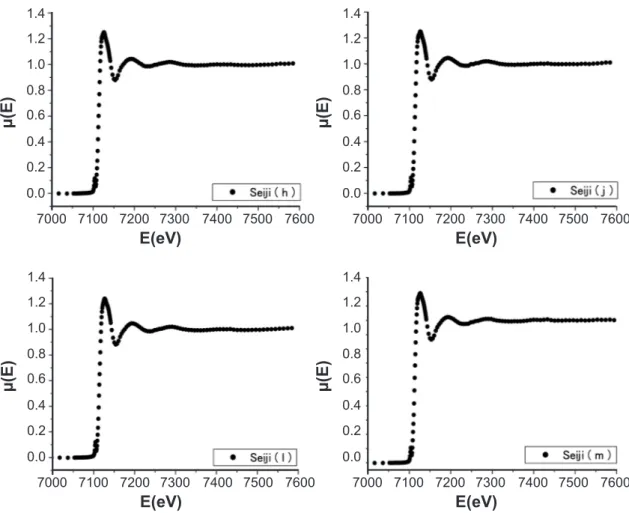
reduce the glassy temperature of the raw celadon ceramics. Fig. 4 shows the Hizen celadon glazes, called Seiji (a), (c), (e), and (f), as in Photo 1(a), (c), (e), and (f) in Ref. [20], respectively. In this paper, all of the igures related to the XAS data were drawn by using only the data points at speciic intervals from a lot of observed data points for igure easiness to see. The XAS of the present celadon glazes are very similar each other. Eo slightly shifted from 7111 eV (a free Fe ion) to about 7114 eV. Eo depends on the crystallographic structure. Since the X-ray diffraction patterns in Figs. 3 and 4 of Ref. [20] show the halo-like pattern, it is considered that the XAS data in Fig. 4 relect the short-range order of the ionic arrangements in the celadon glazes. The XAS spectra do not result from Fe metallic particles in the glazes, because of the largely different spectrum of the Fe particles. However, at a moment, it can not be determined whether all of the component oxides are melted or resolved to make the glassy glaze. As will be described later, we should reine the observed EXAFS spectrum of the present celadon glazes to determine whether the glazes include the ine particles of Fe2O3 or FeO. Fig. 5 shows the XAS of the HIZEN celadon glazes, called Seiji (h), (j), (ℓ), and (m), as in Photo 1(h), (j), (ℓ), and (m) in Ref. [20], respectively. The spectra in Fig. 5 are very similar to those in Fig. 4. Thus, it is found that the HIZEN celadons and the modern Nabeshima celadon (Choshun kiln) have the similar local structure and the electronic band states to the Fe ions in the glazes, where the
local structure around X-ray absorbing atoms and the band states of the absorbing atoms are obtained from the EXAFS spectrum and the XANES one, respectively. It is usually known that the XANES spectrum is in the region of about –100 to + 50~80 eV, while the EXAFS one is in the higher region of about 50~80 to 700 eV above its X-ray absorption thresholds Eo, when Eo= 0.0 eV.
In order to study the local structure around Fe ions in the celadon glaze, we carried out tthe analysis of the EXAFS spectra of the HIZEN celadons and the modern Nabeshima celadon (Choshun kiln), more quantitatively. We obtained an observed Fourier transformation spectrum │F(R)│ to the observed oscillating EXAFS spectrum Хobs(E), after representing an energy of X-ray photons with a wave-number k{= 8π2m
e(E-Eo)/h2}, where me and h are an effective mass of electron and Planck’s constant, respectively. The details of its analysis were already reported in our recent paper [17]. After obtaining the observed oscillating EXAFS spectrum Хobs(E), we calculated a theoretical oscillating EXAFS spectrum Хcal(E) by using the optical interference theory between the X-ray photoelectron waves emitted from the X-ray absorbing Fe ions and its back-scattering waves induced by the surrounding ions. In the present investigations, we used software programs of Artemis and Athena to analysis the XAS data and reine the Хobs(K) with the theoretical Хcal(K) [21]. We carried out to do a best it between the Хobs(K) and the Хcal(K) by mean of a least squares Figure 3: X-ray absorption spectra around the Fe-K edge for the red-colored overglaze of the Kakiemon
colored porcelain (Kakiemon) and the marketed powders of FeO.
[Figura 3: Espectros de absorção de raios X em torno da borda Fe-K para os esmaltes de cor vermelha das porcelanas coloridas (Kakiemon) e de pó comercial de FeO.]
1.5
1.5
1.5
1.5 1.0
1.0
1.0
1.0 0.5
0.5
0.5
0.5 0.0
0.0
0.0
0.0
7000 7000
E(eV)
E(eV)
E(eV)
E(eV)
7200 7200
7200 7200
7400 7400
7400 7400
7100 7300 7100 7300
7300 7300
7500 7500
7500 7500
7600 7600
µ(E) µ(E)
1.4 1.4 1.2
1.2
1.2
1.2 0.8
0.8
0.8
0.8 0.6
0.6
0.6
0.6 0.4
0.4
0.4
0.4 0.2
0.2
0.2
0.2 0.0
0.0
0.0
0.0 1.0
1.0
1.0
1.0 7000
7000
7000
7000
E(eV)
E(eV)
E(eV)
E(eV)
µ(E)
µ(E)
µ(E)
µ(E)
7200
7200
7200
7200 7400
7400
7400
7400 7100
7100
7100
7100 7300
7300
7300
7300 7500
7500
7500
7500 7600
7600
7600
7600
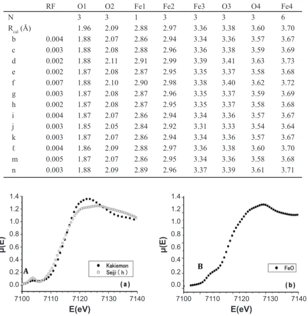
method with several reined parameters, which consist of numbers (Nj) of ions sited on the shell having the same radial distance Rj, Debye-Waller factors (σj), characteristic temperature (QDj), a passive electron reduction factor (S2o). The reinements were always monitored by a convergence factor RF; RF = {Σ|Хobs(K) – kХcal(K)|2}/Σ|Хobs(K)|2, where k is a scale factor. The details were reported in our paper [17]. Fig. 6 shows the reinements of the EXAFS spectra for the Fe ions of the α-Fe2O3 structure of Seiji (c), (e) in Fig. 4 and Seiji (j) and (ℓ) in Fig. 5. The abscissa refers to a radial distance (R(Å)) from a X-ray absorbing Fe ion, as a coordinate origin, to its surrounding shells including the cations or anions of the α-Fe2O3 structure. The peaks of the observed │F(R)│ approximately represent the shells being away from the X-ray absorbing Fe ions by Rj (Ǻ). Each shell includes the equivalent surrounding ions.
In Fig. 6 the solid lines and the solid circles represent the theoretical and observed Fourier transformation spectrum │F(R)│, respectively. The RF-values were about 0.003, 0.02, 0.03, and 0.04 for Seiji (c), (e), (j), and (ℓ), respectively. The theoretical calculation was based on an ideal α-Fe2O3 structure, which has a hexagonal symmetry with its lattice constants of a=5.035 Ǻ and b=13.72 Ǻ. The present spectral analyses were successfully done.Thus, we found from Fig. 5 that the celadon glazes of Seiji (c), (e), (j), and (ℓ) have the α-Fe2O3 structure. In the present investigations, we carried out reinements of the EXAFS data for all of the HIZEN celadons and the modern Nabeshima celadons in Photo 1
in Ref. [20]. The results are shown in Table II, except Seiji (a). In Table II, Rcal is a radial distance of the ideal α-Fe2O3 structure, while the Oj and Fej (j=integer) represent the surrounding oxygen and Fe ion in the shell sited at the radial distance Rj away from the origin of the X-ray absorbing Fe ion, respectively. When studying the local structure by the reinement of the EXAFS spectrum, it is important to regard a phase between the photoelectron waves emitted from the X-ray absorbing Fe ions and the backscattering waves produced by the surrounding shells. Thus, we also reined the phase parameter in the present investigations. The calculated radial distances Rcalj are due to the ideal α−Fe2O3 structure. Thus, a slight difference between Rj and Rcalj results from the effect of the phase parameters. It is found in Table II that the Fe components of the HIZEN celadons and the modern Nabeshima celadon (Choshun kiln) should be the α-Fe2O3 structure. However, we could not succeed the reinements of the EXAFS data for the present celadons with the FeO structure.
It is known that the pre-edge XANES spectrum gives information of unoccupied electronic band states in a top region of the valence band just below EF, while the main XANES spectrum gives empty electronic band states in a bottom region of the conduction band just above EF. Both XANES spectra sensitively depend on the crystallographic structure. In order to study the structural and electronic properties of Fe ions in the red-colored overglaze of the Kakiemon-style porcelain and the HIZEN celadons, we Figura 4: X-ray absorption spectra around Fe-K edge of α−Fe2O3 in the of HIZEN celadon glazes, Seiji (a), (c), (e), (f).
[Figura 4: Espectros de absorção de raios X em torno da borda Fe-K de a-Fe2O3 nos esmaltes celadon Hizen, Seiji (a), (c), (e), (f).]
Figure 5: X-ray absorption spectra around Fe-K edge of α-Fe2O3 in the HIZEN celadon glazes, Seiji (h), (j), (ℓ), in
addition to the modern Nabeshima one (Seiji (m)).
[Figura 5: Espectros de absorção de raios X em torno da borda Fe-K de a-Fe2O3 nos esmaltes celadon Hizen, Seiji (h), (j), (ℓ), alem de um Nabeshima moderno (Seiji (m)).]
1.4
1.4
1.4
1.4 1.2
1.2
1.2
1.2 0.8
0.8
0.8
0.8 0.6
0.6
0.6
0.6 0.4
0.4
0.4
0.4 0.2
0.2
0.2
0.2 0.0
0.0
0.0
0.0 1.0
1.0
1.0
1.0 7000
7000
7000
7000
E(eV)
E(eV)
E(eV)
E(eV)
µ(E)
µ(E)
µ(E)
µ(E)
7200
7200
7200
7200 7400
7400
7400
7400 7100
7100
7100
7100 7300
7300
7300
7300 7500
7500
7500
7500 7600
7600
7600
7600
also carried out measurements of XAS around Fe-K edge for marketed FeO powders by using the present XAS spectrometer. Fig. 7 shows a comparison of the pre-edge and main XANES spectra between Fe2O3 of the Kakiemon-style porcelain and the HIZEN celadons and FeO in the marketed powders. The sample of the marketed FeO powders was a pellet of 10 mm in diameter and 4 mm in height. As described before, Eo of the Kakiemon-style porcelain and Seiji (h) are about 7114.0 and 7113.5 eV in Fig. 7a, respectively. Eo of FeO is about 7118.8 eV in Fig. 7b. The pre-edge XANES peaks are observed at about 7103.6 eV on Fe2O3 (A) and 7113.4eV on FeO (B). The XANES peak in the pre-edge region of the Fe-K absorption edge is induced by an electronic transition between a K-electron band and an unoccupied hybridized valence band of Fe ions in the Fe2O3 structure and the FeO one. The electronic hybridization results from an electronic coniguration interaction between the unoccupied electronic bands of cations, Fe ions, and the full occupied bands of anions, O ions, in the crystalline solid of Fe2O3 and FeO structure [22]. In Fig.7 the relativistic energy difference ΔE (=Eo−Ep) between Eo and the pre-edge peak (Ep) are about 10.4 eV and 9.9 eV for the Kakiemon-style porcelain and Seiji (h), respectively, while ΔE of FeO is about 5.4 eV. Thus, the pre-edge XANES peaks suggest
that the valence electrons of Fe ions in the celadon glaze is 3+, but not 2+ [22]. This is not consistent with the color emission mechanism of the celadon glaze reported [15, 16]. They suspected that the celadon glaze was colored by the chemical reaction from Fe2O3 to FeO under the deoxidizing thermal treatment at higher temperature in a kiln.
Figure 6: Theoretical |F(R)|(solid line-it) of the EXAFS spectrum, taken by the least squares reinement to the observed |F(R)| (solid circles) for the Fe ions in the α−Fe2O3 structure of Seiji (c), (e) in Figure 4 and Seiji (j) and (ℓ) in Figure 5.
[Figura 6: Ajuste teórico |F(R)|(linha sólida-ajuste) do espectro EXAFS, obtido por reinamento por mínimos quadrados do espectro |F(R)| observado (círculos sólidos) dos íons Fe na estrutura α-Fe2O3 de Seiji (c), (e) na Figura 4 e Seiji (j) e (ℓ) na Figura 5.]
ray or conversion electrons is in the Mössbauer spectrum of iron systems. The Mössbauer spectrum is affected by temperature and three other factors of isomer shift, quadrupole splitting, and magnetic splitting. In the present investigations, we regard only the effects of the isomer shift and the quadrupole splitting because of the measurement at room temperature and no applied magnetic ield.
The isomer shift (δ) results from the difference in the electron densities at the nuclear sites in the emitting and absorbing atoms. This difference in density changes the Mössbauer transition-energy and so the Mössbauer spectrum is shifted. The isomer shift is sensitive to the oxidation state and can therefore be used to study valence electrons of Fe ions in the celadon glaze. If the nuclei do not have a charge distribution of spherically symmetric, the nucleus will possess an electric nuclear quadrupole moment. This moment interacts with an asymmetric electronic charge distribution splits the degeneracy of the excited state into two levels, which are separated by the quadrupole splitting (Δ). The Mössbauer spectrum is a doublet. The quadrupole splitting can be broken down into two contributions of a valence contribution from the atom itself and a lattice contribution from neighboring atoms. Fe3+ (3d5) ions
have no contribution to the electric ield gradient from the 3d electron orbitals. This means that Fe3+ ions have the relatively low quadrupole splitting. On the hand, Fe2+ (3d6) ions have the large electric ield gradient contribution from the 6th 3d-electron, and give the large quadrupole splittings of about 1.5-3 mm/s (with a maximum of > 4 mm/s). Thus, the isomer shift and the quadrupole splitting is a useful tool to determine whether Fe ion has the valence electrons of 3+ (Fe2O3) or 2+ (FeO). If Fe2O3 and FeO particles coexist in the glaze, we can observe a mixed Mössbauer spectrum induced by Fe3+ and Fe2+ ions.
When measuring the Mössbauer spectrum, the incident gamma rays penetrate a powder specimen in front of its detector. Thus, to make ine powders, we must remove mechanically the celadon glazes of the Hizen celadons from its basic body and crush the broken pieces. However, all of the broken pieces of the Hizen celadons in Photo 1 in Ref. [20] were rented from at the public museum are very important cultural assets in Japan. We were strongly requested to use nondestructive methods for studying the structural and electronic properties of the Hizen celadons. Thus, we measured only the powdered celadon glaze of the modern Nabeshima celadons, Seiji (m), in Photo 1-m 0.25
0.25
0.25
0.25 0.20
0.20
0.20
0.20 0.15
0.15
0.15
0.15 0.10
0.10
0.10
0.10 0.05
0.05
0.05
0.05 0.00
0.00
0.00
0.00 0.5
0.5
0.5
0.5 1.0
1.0
1.0
1.0 2.5
2.5
2.5
2.5 1.5
1.5
1.5
1.5 3.0
3.0
3.0
3.0 4.0
4.0
4.0
4.0 2.0
2.0
2.0
2.0
R(Å)
R(Å)
R(Å)
R(Å)
3.5
3.5
3.5
3.5
F(R)
F(R)
F(R)
Table II - Reined parameters Rj of the surrounding ions (oxygen ions Oj and Fe ions Fej) around the X-ray absorbing Fe ion to the ideal radial distance Rcalj(Ǻ) of the α-Fe2O3 structure in the glazes of the HIZEN celadons; Seiji (b) to (ℓ) and the modern Nabeshima celadons of Choshun kiln; Seiji (m) and (n).
[Tabela II - Parâmetros reinados Rj dos íons (íons oxigênio Oj e íon Fe Fej) em torno do íon Fe absorvedor de raios X para a distância radial ideal Rcalj(Ǻ) da estrutura do α-Fe2O3 nos esmaltes de celadons Hizen; Seiji (b) a (ℓ) e os celadons modernos Nabeshima de forno Choshun; Seiji (m) and (n).]
RF O1 O2 Fe1 Fe2 Fe3 O3 O4 Fe4
N 3 3 1 3 3 3 3 6
Rcal (Å) 1.96 2.09 2.88 2.97 3.36 3.38 3.60 3.70
b 0.004 1.88 2.07 2.86 2.94 3.34 3.36 3.57 3.67
c 0.003 1.88 2.08 2.88 2.96 3.36 3.38 3.59 3.69
d 0.002 1.88 2.11 2.91 2.99 3.39 3.41 3.63 3.73
e 0.002 1.87 2.08 2.87 2.95 3.35 3.37 3.58 3.68
f 0.007 1.88 2.10 2.90 2.98 3.38 3.40 3.62 3.72
g 0.003 1.87 2.08 2.87 2.96 3.35 3.37 3.59 3.69
h 0.002 1.87 2.08 2.87 2.95 3.35 3.37 3.58 3.68
i 0.004 1.87 2.07 2.86 2.94 3.34 3.36 3.57 3.67
j 0.003 1.85 2.05 2.84 2.92 3.31 3.33 3.54 3.64
k 0.003 1.87 2.07 2.86 2.94 3.34 3.36 3.57 3.67
ℓ 0.004 1.86 2.09 2.88 2.97 3.36 3.38 3.60 3.70
m 0.005 1.87 2.07 2.86 2.95 3.34 3.36 3.58 3.68
n 0.003 1.88 2.09 2.89 2.96 3.37 3.39 3.61 3.71
Figure 7: X-ray absorption spectra around Fe-K edge for the red-colored overglaze of the Kakiemon-style porcelain, the HIZEN celadon Seiji (h), and the marketed FeO powders.
[Figura 7: Espectros de absorção de raios X em torno da borda Fe-K para os esmaltes de cor vermelha das porcelanas de estilo Kakiemon, celadon Seiji Hizen (h), e de pó comercial de FeO.]
1.4 1.4
0.6 0.6
1.0 1.0
0.2 0.2
1.2 1.2
0.4 0.4
0.8 0.8
0.0 0.0
7100 7110 7100 7110
E(eV) E(eV)
µ(E) µ(E)
7120 7130 7140 7120 7130 7140
in Ref. [20]. The Choshun kiln succeeds to produce the Nabeshima celadons traditionally at Ohkawachi (Imari), until now. The Nabeshima-domain kilns, called Odougu-yama, were set up at Ohkawachi, after transferred from
Nangawara (Arita) to Okawachi (Imari) in 1673-1681’s. The observed Mössbauer spectrum taken at room temperature is shown in Fig. 8. The abscissa refers to a shift velocity of the incident 57Co gamma ray radiation, while the ordinate to a penetrating ratio of the incident gamma ray to the powder specimen. After reining the Mössbauer spectrum, we obtained that the isomer shift δ
CONCLUSIONS
In order to study the historical and technical correlation of the Hizen celadon glazes including the modern Nabeshima celadon (Choshun kiln), we carried out the measurements of the X-ray absorption spectra (XAS) near the Fe-K edge by using the synchrotron radiation, and the Mössbauer spectrum. Although the oxide components in the raw ceramics at Izumiyama (Arita) and Ohkawachi (Imari) were not so large difference [20], we found from X-ray diffraction that the Izumiyama ceramics are mainly quartz, while the Ohkawachi one are mainly the feldspar of (K,Na) Si3O8 (sanidine). The XAS near the Fe-K edge results also showed that there is the slight difference of the EXAFS spectra between the Izumiyama and Ohkawachi ceramics. As regarding the Fe-XAS of the red-colored overglaze of the Kakiemon-style porcelain and the marketed powders of FeO, this suggested that the α-Fe2O3 particles are structurally affected by the surrounding other oxides of the raw ceramics. All of the present celadon glazes show the similar Fe-K XAS to that of the Izumiyama ceramics. The spectrum similarity suggested that the celadon glazes include the Fe2O3 ine powders, since the melting temperature of Fe2O3 is about 1570 °C, while FeO is about 1370 °C. In 1630’s to 1790’s, the used kiln temperature was about 1200-1250 °C. After reining the EXAFS spectra, it was found that the celadon glazes include the Fe2O3 ine powders, but not FeO, even in the glassy state. The pre-edge XANES peaks also suggested that the valence electrons of Fe ions in the celadon glaze is 3+, but not 2+ [22]. The Mössbauer spectrum suggested that the celadon glaze of Seiji (m) includes only Fe3+ ions, but not Fe2+ ions. This indicates the existence of Fe
2O3 in the celadon glaze. Thus, the present investigations are not
consistent with that reported [15, 16]. They suspected that the celadon glaze was colored by the chemical reaction from Fe2O3 to FeO under the deoxidizing thermal treatment at higher temperature in a kiln. We considered that the colored brightness of the Hizen celadons depends on the structural properties of the used raw celadon ceramics and the other transition-metal ions of Cr, Cu, Zn in the celadon glazes [20]. However, in the present investigations, we could not determine what kinds of the oxides of Cr, Cu, Zn exist in the celadon glaze, because of their very small amounts and the renting condition of the nondestructive methods for studying the structural and electronic properties of the Hizen celadons from the public museum.
REFERENCES
[1] The Kyushu Ceramic Museum, Polychrome porcelain in Hizen-its early type and change of style (1991).
[2] T. Tanigawa, Y. Kawabata, Koimari, Nippon Toji Zenshu 8, Choukouronsha (1992).
[3] H. Nishida, Y. Ohhashi, Old Imari ware, The Sun special issue 63, Autum ’88, Heibonsha (1994).
[4] I. Ogi, Imari, Ribun Shuppan (2000).
[5] C. Shimizu, La Porcelaine japonaise, Massin (2002). [6] The Society of Kyshu Early Modern Ceramic Studies, Hizen Porcelains in Japan (2002).
[7] K. Ohashi, M. Arakawa, Early Imari, the origins of Underglaze Cobalt-blue and Overglaze Polychrome Enamels, Japan Broadcasting Corporation (NHK) (2004). [8] Y. Imura, Kakiemon, Rokusho 5, Maria Shobou (2005). [9] The Kyushu Ceramic Museum, Nabeshima, Porcelain for the Shogunate (2006).
[10] A. Imai, Celadon 4, Chinese wares, Heibonsha (2002). [11] Wang Qing-zheng, Fan Dong-quing, Zhou Li-li, The Discovery of Ru Kiln, a Famous Song-ware Kiln of China, The Woods Piblishing Co. (1991).
[12] K. Ohashi, Arita and Imari porcelains, Tankosha (2002).
[13] K. Takeuchi, T. Nobayashi, S. Morokuma, H. Tsuzuki, Development of old-fashined porcelain with traditional raw material II - Characteristic feature on the body and glaze of Hasami celadon, Annual report of Ceramic Reserch
Center of Nagasaki, 48 (2001).
[14] Y. Nakano, Mitumata Seiji Kiln, Hasami Cultural properties, report 10, Hasami-cho Boad of Education (1998). [15] J. S. Larid, The composition of Chinese celadon pottery, J. Am. Ceram. Soc. 1 (1918) 675.
[16] R. R. Hunghan, Early Chinese ceramics glazes, Ceram. Age 56 (1950) 40.
[17] M. Hidaka, K. Ohashi, S. Kajihara, R. P. Wijesundera, L. S. R. Kumara, Jae-Young Choi, Nark Eon Sung, Structural properties of the red-color overglaze for the HIZEN porcelains produced in the early Edo period of Japan, Ceram. Int. 35 (2009) 875.
[18] M. Hidaka, H. Horiuchi, K. Ohashi, R. P. Wijesundera, L. S. R. Kumara, Jae-Young Choi, Yong Jun Park, Structural properties of the red-color overglazes on the Kakiemon-style -6
100.2
100.0
99.6 99.8
99.4
99.2
99.0
98.8
6
-2 2
-4 0
Velocity (mms-1)
T (%)
4
Figure 8: Mössbauer spectrum of the Seiji (m) celadon glaze.
porcelains produced in the later 17th century by means of X-ray diffraction, Cerâmica 55 (2009) 120.
[19] M. Hidaka, H. Horiuchi, K. Ohashi, R. P. Wijesundera, L. S. R. Kumara, Nark Eon Sung, Local structures and electronic band states of α−Fe2O3 polycrystalline particles included in the red-color overglazes and the transparent glazes of the Kakiemon-style porcelains by means of X-ray absorption spectra, Cerâmica 55, 335 (2009) 223.
[20] M. Hidaka, K. Ohashi, R. P. Wijesundera, L. S. R. Kumara, M. Watanabe, K. Koga, Jae-Young Choi, Nark Eon Sung, Young Jun Park, Correlation between glaze-colors and structural properties of the HIZEN celadons produced in the Edo period of Japan, by means of X-ray diffraction (І),
Cerâmica 56, 340 (2010) 106.
[21] C. H. Booth, F. Bridges, Physical Scripta T115 (2005) 202.
[22] J. Zaanen, G. A. Sawatzky, J. W. Allen, Band Gaps and Electronic Structure of Transition-Metal Compounds, Phys. Rev. Lett. 55 (1985) 418.
[23] E. De Grave, A. Van Alboom, Evaluation of ferrous and ferric Mössbauer fractions, Phys. Chem. Minerals 18 (1991) 337.
[24] M. Darby Dyar, Mössbauer spectroscopy (2009), http://serc.carleton.edu/research_education/geochemsheets/ techniques/mossbauer.html.