J Bras Pneumol. 2013;39(1):98-101
well as to determine the position of the hyoid bone, the configuration of the mandible, the posterior pharyngeal airspace, the dimensions of the tongue, the length and thickness of the uvula, etc. Anatomical changes in these sites can predispose patients to OSA.(3)
Magnetic resonance imaging (MRI) and CT have been highlighted for their ability to perform multiplanar imaging. Because it allows a better anatomical resolution and it does not use ionizing radiation, MRI has been used as the major imaging method for this type of investigation.(4) This article aims to describe the major MRI and CT Obstructive sleep apnea (OSA) is characterized
by recurrent upper airway obstruction occurring at the level of the pharynx during sleep.(1) It is a chronic progressive disease associated with high rates of cardiovascular morbidity.(2)
The diagnosis of OSA is confirmed by polysomnography. However, imaging studies are highly relevant supporting methods in the evaluation of patients with OSA.
Cephalometric analysis, performed using X-rays, is an important method in the diagnosis of craniofacial deformities. With this method, it is possible to measure the skull base, as
Radiological findings in patients with obstructive sleep apnea*
Achados radiológicos em pacientes portadores de apneia obstrutiva do sono
Carlos Fernando de Mello Junior, Hélio Antonio Guimarães Filho, Camila Albuquerque de Brito Gomes, Camila Caroline de Amorim Paiva
Abstract
Obstructive sleep apnea (OSA) is characterized by recurrent upper airway obstruction occurring at the level of the pharynx during sleep. Although cephalometric analysis is an important method in the diagnosis of craniofacial deformities, CT and magnetic resonance imaging have been highlighted as the major imaging methods to investigate the possible causes of OSA, which, in most cases, is multifactorial. Magnetic resonance and CT both allow an excellent evaluation of the various anatomical planes of the site of obstruction, which enables better clinical assessment and surgical approach. This pictorial essay aims to describe the aspects that must be evaluated in the diagnostic imaging of patients presenting with the major predisposing factors for OSA.
Keywords: Sleep apnea syndromes; Magnetic resonance imaging; Tomography, X-ray computed.
Resumo
A apneia obstrutiva do sono (AOS) é caracterizada por obstruções recorrentes das vias aéreas superiores durante o sono que ocorrem no nível da faringe. Apesar de a análise cefalométrica ser um importante método no diagnóstico das deformidades craniofaciais, a TC e a ressonância magnética vêm se destacando como os principais métodos de imagem para a investigação das eventuais causas da AOS que, na maioria das vezes, é multifatorial. Esses métodos permitem uma excelente avaliação nos diversos planos anatômicos do eventual sítio da obstrução, o que permite uma melhor avaliação clínica e abordagem cirúrgica. O presente ensaio pictórico tem como objetivo descrever os aspectos que devem ser avaliados no diagnóstico por imagem dos principais fatores predisponentes para a AOS.
Descritores: Síndromes da apneia do sono; Imagem por ressonância magnética; Tomografia computadorizada por raios X.
* Study carried out at the Universidade Federal da Paraíba – UFPB, Federal University of Paraíba – João Pessoa, Brazil. Correspondence to: Carlos Fernando de Mello Junior. Rua Waldemar Chianca, 365, apto. 1001, Bessa, CEP 58047-255, João Pessoa, PB, Brasil.
Tel. 55 83 3049-4444. E-mail: [email protected] Financial support: None.
Submitted: 26 March 2012. Accepted, after review: 10 October 2012.
Radiological findings in patients with obstructive sleep apnea
J Bras Pneumol. 2013;39(1):98-101 99
the pattern that is observed in normal subjects is laterolateral (Figure 2).(2) Increased soft tissue (fat, muscle, or lymphoid tissue) volume in the oropharyngeal region can make the oropharynx exhibit a pathological pattern anteroposteriorly (Figure 3).
findings in the diagnostic evaluation of patients with OSA.
In most cases, OSA is multifactorial,(5) being the result of upper airway collapse or narrowing occurring during sleep. In general, the greatest narrowing of the pharyngeal air passage is seen at the level of the lower soft palate. In the protocols to be used, axial and sagittal images of the oropharyngeal and hypopharyngeal air passages should always be included, and imaging studies should be performed as soon as possible.
Midsagittal slices are of fundamental importance, because they make it possible to characterize the airway contour, the maxillomandibular relationship (to evaluate patients for retrognathia and micrognathia), the volume of the soft palate, the palate shapes, the position of the hyoid bone, and the position and volume of the dorsum of the tongue. Figure 1 shows a midsagittal slice of a normal MRI scan. Axial slices should be acquired at the levels of the nasopharynx, hypopharynx, palates, dorsum of the tongue, and vocal cords.
One of the most important changes that must be investigated on imaging studies in patients with OSA is the pattern of the airway passage on the axial CT or MRI slices. Physiologically,
Figure 1 - Midsagittal slice of a magnetic resonance image from a normal subject. Note the maxillomandibular relationship, the symmetry of the palate, and the diameter of the air passage.
Figure 2 - Axial CT slice showing the physiological pattern of the air passage, the laterolateral axis being the longest.
100 Mello Jr CF, Guimarães Filho HA, Gomes CAB, Paiva CCA
J Bras Pneumol. 2013;39(1):98-101
The conditions that can predispose to OSA include the following:
• Vocal cord paralysis
• Micrognathia and retrognathia (Figure 4) • Soft palate abnormalities
• Increased soft palate size (Figure 5) • Deviation of the nasal septum
• Tonsil or adenoid hypertrophy, or both • Tumors or cysts in the pharyngeal region
(Figure 6)
• Macroglossia (Figure 7)
• Hard palate deformities: high-arched palate (Figure 7)
• Obesity(2) (because an excess of soft tissue in the pharynx hampers the pharyngeal air passage from remaining open)
• Glossoptosis (tongue ptosis has been highlighted as a cause of OSA in children)(6-8)
• A low hyoid bone, ectopic thyroid, and craniofacial abnormalities(8)
• Acromegaly
Although polysomnography is the method used to confirm the diagnosis of OSA,(2) CT and especially MRI have established themselves as important supporting methods in the clinical diagnosis, preoperative evaluation, and post-treatment follow-up of patients who do not respond well to initial therapy. Both CT and MRI can provide an excellent evaluation of the various anatomical planes of the site of obstruction, which enables better clinical assessment as well as better planning for a possible surgical approach.
Figure 4 - Sagittal slice of a magnetic resonance image showing retrognathia (arrow), the dorsum of the tongue in contact with the soft palate (arrowheads), and narrowing of the nasopharyngeal air passage.
Figure 5 - Sagittal slice of a magnetic resonance image showing a pathological pattern of the soft palate. The patient had a high-arched palate (arrow) and increased soft palate size (arrow heads), with consequent narrowing of the air passage.
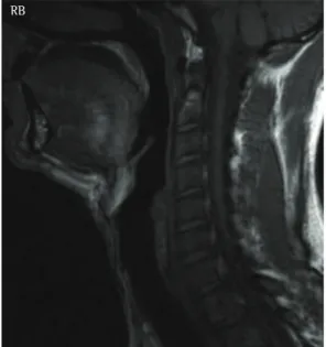
Figure 6 - Sagittal reconstruction of a CT scan showing a massive polyp protruding toward the nasopharynx and oropharynx, causing significant narrowing of the lumen of the air passage (arrow).
Radiological findings in patients with obstructive sleep apnea
J Bras Pneumol. 2013;39(1):98-101 101
5. Suto Y, Matsuda E, Inoue Y, Suzuki T, Ohta Y. Sleep apnea syndrome: comparison of MR imaging of the oropharynx with physiologic indexes. Radiology. 1996;201(2): 393-8. PMid:8888230.
6. Donelly LF. Obstructive sleep apnea in pediatric patients: evaluation with cine MR sleep studies. Radiology. 2005;236(3): 768-78. PMid:16014437. http:// dx.doi.org/10.1148/radiol.2363040306
7. Donnelly LF, Strife JL, Myer CM 3rd. Glossoptosis (posterior displacement of the tongue) during sleep: a frequent cause of sleep apnea in pediatric patients referred for dynamic sleep fluoroscopy. AJR Am J Roentgenol. 2000;175(6):1557-60. PMid:11090374. 8. Donnelly LF, Surdulescu V, Chini BA, Casper KA, Poe
SA, Amin RS. Upper airway motion depicted at cine MR imaging performed during sleep: comparison between young patients with and those without obstructive sleep apnea. Radiology. 2003;227(1):239-45. PMid:12616001. http://dx.doi.org/10.1148/radiol.2271020198
References
1. Suto Y, Matsuo T, Kato T, Hori I, Inoue Y, Ogawa S, et al. Evaluation of the pharyngeal airway in patients with sleep apnea: value of ultrafast MR imaging. AJR Am J Roentgenol. 1993;160(2):311-4. PMid:8424340. 2. Mancini MC, Aloe F, Tavares S. Apnéia do sono em
obesos. Arq Bras Endocrinol Metab. 2000;44(1):81-90. http://dx.doi.org/10.1590/S0004-27302000000100013 3. Salles C, Campos PS, Andrade NA, Daltro C. Obstructive
sleep apnea and hypopnea syndrome: cephalometric analysis. Rev Bras Otorrinolaringol. 2005;71(3):369-72. http://dx.doi.org/10.1590/S0034-72992005000300018 4. Abbott MB, Donnelly LF, Dardzinski BJ, Poe SA, Chini BA,
Amin RS. Obstructive sleep apnea: MR imaging volume segmentation analysis. Radiology. 2004;232(3): 889-95. PMid:15333801. http://dx.doi.org/10.1148/ radiol.2323031581
About the authors
Carlos Fernando de Mello Junior
Adjunct Professor. Department of Radiology, Universidade Federal da Paraíba – UFPB, Federal University of Paraíba – School of Medicine, João Pessoa, Brazil.
Hélio Antonio Guimarães Filho
Physician. Ecoclínica, João Pessoa, Brazil.
Camila Albuquerque de Brito Gomes
Medical Student. Nova Esperança School of Medicine, João Pessoa, Brazil.
Camila Caroline de Amorim Paiva