© 2012 Dental Press Journal of Orthodontics 2 Dental Press J Orthod. 2012 Sept-Oct;17(5):2-3
Matheus Melo Pithon1
How to cite this article: Pithon MM. Orthodontics highlights. Dental Press J Or-thod. 2012 Sept-Oct;17(5):2-3.
Submitted: August 20, 2012 - Revised and accepted: September 05, de 2012 1 Professor, Southwest Bahia University (UESB, Brazil). MSc and PhD in
Orthodontics, Federal University of Rio de Janeiro (UFRJ, Brazil). Diplomate by the Brazilian Board of Orthodontics and Dentofacial Orthopedics.
Contact address: Matheus Melo Pithon E-mail: [email protected]
PARAMETERS USED FOR REFERRING PATIENTS FOR ORTHOGNATHIC SURGERY
During the course of decades, orthodontics was based on linear and angular static measurements to define the orthodontic treatment plan. Nowadays, a great deal of attention has been paid to facial analysis. In line with this trend, an interesting article1 was published, in which
the proposal was to verify whether it was necessary or not to refer Class II, division 1 patients for orthognathic surgery, based on observation of the face in lateral view. The conclusions drawn from this study were that the dis-placement of the soft tissues of the pogonium and point B in the posterior direction and the diminished facial profile angle were decisive factors for orthodontists to indicate these patients for orthognathic surgery. These results suggested that in the presence of cases with limitations it is always important to take a profile photo-graph of the patient and show it to the patient, so that he/ she can evaluate his/her face and together with the or-thodontist, define whether or not it would be convenient to undergo surgical treatment.
SELF-ETCHING AGENTS
PRODUCE FEWER CHANGES IN ENAMEL COLOR AFTER DEBONDING
When bonding orthodontic accessories, a con-stant concern is whether this bonding will or will not change the color of the enamel on the conclusion of orthodontic treatment. There are various materials and techniques used for tooth enamel conditioning before bonding orthodontic accessories. In a recent study2 Egyptian researchers performed an
evalu-ation of whether or not the different methods and materials would alter enamel color after debonding, in addition to verifying the degree of penetration of bonding materials into enamel. The results found showed that self-etching agents presented lower
lev-els of penetration and coloring of enamel after de-bonding, whereas etching with 37% phosphoric acid for 60 seconds presented the highest level of coloring and infiltration, and showed differences when com-pared with the same phosphoric acid for a period of 15 seconds. These findings add further advantages to the use of self-etching agents, which are outstanding for their practicality and the possibility of perform-ing bondperform-ing in a humid environment.
PATIENTS ARE THE PRINCIPAL RESPONSIBLE FOR THE APPEARANCE OF WHITE STAINS
In spite of the many advancements achieved by or-thodontics over the last few years, decalcifications and white stain lesions continue to be frequent problems in orthodontic dental offices. The growing trend to re-place banding of teeth by bonding associated with the use of fluoride-releasing bonding materials has cer-tainly reduced the appearance of these lesions, how-ever, there is still a great deal to be done. While giving some thought to this problem, researchers at the Uni-versities of Jordan and Virginia in the USA3 decided to
verify, by means of questions put to patients and their parents, orthodontists and general clinicians, about who would be the responsible for the appearance of white stains, what would be the methods to prevent them, and who should treat them. All the groups eval-uated were unanimous that white stain lesions depre-ciate the orthodontic patient’s general appearance,
Orthodontics Highlights
Figure 1 -Intraoral photographs of two patients after orthodontic treat-ment: A) teeth with no white stain lesions, B) teeth with white stain le-sions (Source: Maxfield et al.3, 2012).
© 2012 Dental Press Journal of Orthodontics 3 Dental Press J Orthod. 2012 Sept-Oct;17(5):2-3
Pithon MM
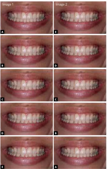
a persistent question arises: How acceptable will this treatment method be to the patient? With the inten-tion of elucidating this doubt, Brazilian researchers,4
using photographs with changes made by means of an image edition software, created images in which the mandibular incisor had been extracted. The results found showed that when there are other options avail-able, the option to extract mandibular incisors should be discarded, due to the esthetic impairment caused and perceived by the evaluated groups.
TOOTH EXTRACTIONS CHANGE THE LIP VERMILION
Recognized worldwide for her esthetic attributes, Angelina Jolie is outstanding because of her delicate facial lines and full lips. As she is a Hollywood actress her esthetic attributes encourage women all over the world to copy her. In this context, an increasing num-ber of women appear in medical consulting services ev-ery day seeking to have lips like those of Angelina Jolie. You may, however, ask, what does orthodontics have to do with lips? It was in an endeavor to explain this re-lationship that Japanese authors developed a study5
evaluating the influence of orthodontic treatment with extraction, in cases of bimaxillary protrusion, on lips vermilion. The results showed evidences of the influ-ence of tooth extractions on lip vermilion, which could thereby favor improvements in facial esthetics.
Figure 2 -Modified images assessment. Image 1: A) With four incisors; B) without any alteration as regards the width of the three remaining incisors.; C) with increase in the three mandibular incisors with the same proportion; D) with a mesiodistal increase in the central incisor and no alteration in the lateral incisors; E) image with a mesiodistal increase in the lateral incisors and the central without any alteration. Image 2: E) image with a mesiodistal increase in the lateral incisors and the central without any alteration; D) with a mesiodistal increase in the central incisor and no alteration in the lateral incisors; C) with increase in the three mandibular incisors with the same proportion; B) without any alteration as regards the width of the three remaining incisors; A) with four incisors (Source: Pithon et al.4, 2012).
1. Hodge TM, Boyd PT, Munyombwe T, Littlewood SJ. Orthodontists’ perceptions of the
need for orthognathic surgery in patients with Class II Division 1 malocclusion based on extraoral examinations. Am J Orthod Dentofacial Orthop. 2012 Jul;142(1):52-9.
2. Zaher AR, Abdalla EM, Abdel Motie MA, Rehman NA, Kassem H, Athanasiou AE.
Enamel colour changes after debonding using various bonding systems. J Orthod. 2012 Jun;39(2):82-8.
3. Maxfield BJ, Hamdan AM, Tüfekçi E, Shroff B, Best AM, Lindauer SJ. Development
of white spot lesions during orthodontic treatment: perceptions of patients, parents, orthodontists, and general dentists. Am J Orthod Dentofacial Orthop. 2012 Mar;141(3):337-44.
4. Pithon MM, Santos AM, Couto FS, da Silva Coqueiro R, de Freitas LM, de Souza RA,
dos Santos RL. Perception of the esthetic impact of mandibular incisor extraction treatment on laypersons, dental professionals, and dental students. Angle Orthod. 2012 Jul;82(4):732-8. Epub 2011 Dec 12.
5. Trisnawaty N, Ioi H, Kitahara T, Suzuki A, Takahashi I. Effects of extraction of four
premolars on vermilion height and lip area in patients with bimaxillary protrusion. Eur J Orthod. 2012 May 9. [Epub ahead of print].
REFERENCES
and that the patient is the principal responsible for the appearance and prevention of these lesions. These results bring us some relief, but does not exonerate us from our responsibility towards these patients.
EXTRACTION OF MANDIBULAR INCISOR AFFECTS ESTHETICS WHEN SMILING
One of the options widely disseminated in the literature for the treatment of anterior mandibular crowding is the extraction of a mandibular incisor. In spite of being an extensively practiced procedure,
A
B
C
D
E
E
D
C
B
A