Fisioter. Mov., Curitiba, v. , n. , p. - , Jan./Mar. Licenciado sob uma Licença Creative Commons DO): http://dx.doi.org/ . / - . . .AO
[T]
Thermographic and anthropometric assessment of
electrical stimulation on localized body fat
Avaliação termográi ca e antropométrica da
eletrolipólise na adiposidade localizada
Graciele Guimarães Pitelli Aroca, Larissa Granato Viana, Rafaela Ferreira de Araújo Costa, Dalilia Schmildt, Ligia de Sousa*
Universidade Federal de Alfenas UN)FAL , Alfenas, MG, Brazil
Abstract
Introduction: Adiposity is de ined as the accumulation of energy reserves within the adipose tissue at
spe-ci ic body sites. Low-frequency electrical stimulation elispe-cits lipolysis. When applied by insertion of needles into the dermis-hypodermis junction, it leads to a modi ication of the interstitial space, favoring metabolic changes and lipolysis. Objective: To investigate the effects of electrical stimulation on body fat localized to
the abdomen and lanks. Methods: Randomized, controlled clinical trial consisted of two groups of women
with body fat localized to the abdomen and lanks. The intervention group )G was made up of women ± , years who received ten sessions of electrical stimulation, whereas the control group CG was made up of women ± , years who did not receive electrical stimulation. Perimetric, adipometric and thermographic data were collected before and after the intervention. Data were analyzed using the Shapiro-Wilk test, t test, one-way ANOVA. The signi icance level was set at p < , . Results: There were statistically
signi icant differences between the intervention and control groups in the assessment immediately follow-ing intervention )G: . ± . ; CG: . ± . ; p = . , minutes following intervention )G: . ± . ; CG: . ± . ; p < . and at the endpoint )G: . ± . ; CG: . ± . ; p= . for the thermographic data. For the anthropometric variables, there were no statistically signi icant differences before and after treatment. Conclusion: Electrical stimulation evokes a signi icant increase in the
tempera-ture of the subcutaneous tissue.
Keywords: Abdominal Fat. Electric Stimulation Therapy. Lipolysis.
30
Resumo
Introdução: Adiposidade é o acúmulo de reservas energéticas no tecido adiposo, em regiões especíϔicas. A estimu-lação elétrica de baixa frequência como geradora da lipólise, quando aplicada com agulhas inseridas na junção derme-hipoderme, gera modiϔicação do meio intersticial, favorecendo as trocas metabólicas e a lipólise. Objetivo: Investigar o efeito da eletrolipólise na adiposidade localizada em região abdominal e de ϔlancos. Métodos: Ensaio clínico controlado e randomizado constituído de dois grupos: grupo intervenção GI, constituído de 9 mulheres (± 24,77 anos) com adiposidade localizada nas regiões abdominal e de ϔlancos, que realizaram dez sessões de eletro-lipólise; e o grupo controle GC, constituído de 7 mulheres (± 21,8 anos) que não realizaram as sessões de eletrolipó-lise. Prévia e posteriormente à intervenção foram coletados a perimetria, a adipometria e a análise termográϔica. Para análise dos dados foram utilizados os testes Shapiro-Wilk, teste T, teste One-Way ANOVA; considerou-se nível de signiϔicância de p < 0,05. Resultados: Para os dados termográϔicos, observou-se diferença estatística entre os grupos intervenção e controle na reavaliação imediata (GI: 33,08 ± 1,00; GC: 30,83 ± 1,5; p = 0,002), reavaliação após 15 minutos de aplicação da técnica (GI: 33,05 ± 0,48; GC: 30,40±1,24; p < 0,0001) e na reavaliação ϔinal (GI:32,22 ± 14,20; GC: 30,53 ± 1,34; p=0,005). Para as variáveis antropométricas, não houve resultado signiϔicativo antes e após o tratamento. Conclusão: A eletrolipólise promove incremento signiϔicativo na temperatura do tecido subcutâneo.
Palavras-chave: Gordura Abdominal. Terapia por Estimulação Elétrica. Lipólise.
Introduction
Adiposity is de ined as the accumulation of energy reserves within the adipose tissue. )t is caused by ca-loric intake above the needed energy requirements for body metabolism. )t refers to the excess of adipose tissue in the subcutaneous layer, known as adipose panicle, which stores % of the body fat. The adipose tissue is a differentiated connective tissue. )t is formed by adi-pocytes, which have a thermal and illing function, and help in the maintenance of bodily structures and energy reserves , , .
Adipose cells are remarkably diverse, depending on their site of concentration. Some cells are more ef i-cient in the absorption of excess calories into the blood-stream, while others release their stored energy to other tissues. This explains the higher resistance of some fat deposits to reduction , . Adiposity gets stored in different regions of the body depending on sex, age, and metabolic, genetic, environmental and nutritional factors. Localized adiposity is not directly associated with obesity and can be present in nonobese individuals with fat accumulation in speci ic regions of the body . Obesity is classi ied into android and gynecoid ac-cording to fat distribution and location. )n gynecoid obe-sity, there is a greater amount of fat cells in the gluteal and trochanteric region, but mainly in the abdominal region, as seen in the women who participated in this study .
)n the clinical setting, there are speci ic treatments that promote lipolysis at the application site . One of these resources is electrical stimulation, which uses nee-dles as electricity conductors. Electrical stimulation has already been presented in the literature as a technique that can help reduce localized adiposity and edemas, im-prove metabolic response, stimulate nerves and vessels, and has anti-in lammatory, vasodilating, and, above all, hydrolytic properties , . The physiological effect of the application of the electrical current is explained by the activation of the sympathetic nervous system, which causes the release of catecholamines epinephrine and norepinephrine . Catecholamines activate adrenergic receptors, leading to the release of adenylate cyclase and stimulating intracellular conversion of ATP into cyclic AMP, which brings about lipolysis degradation of triglycerides into glycerol and fatty acids .
31
Methods
Study Population and Sample
This pilot, single-blind, randomized, controlled clinical trial was submitted to, and approved by, the Ethics Committee of the Federal University of Alfenas UN)FAL , opinion number . Prior to data collection, all volunteers were informed of the objectives and methods of the study, and agreed to participate in the trial. They all signed an informed consent form. The data were collected between April and June .
)nclusion criteria were: being a female undergradu-ate student at the Federal University of Alfenas UN)FAL, MG , aged between and years, Caucasian, and sedentary for at least the previous months; having adi-posity localized to the abdomen and lanks evidenced by a protrusion of the abdomen and lanks even during abdominal contraction while standing in the upright position; having normal vital signs blood pressure = / mm(g, heart rate up to beats per minute, and respiratory rate up to breaths per minute , and with body mass index classi ied as normal weight be-tween . and kg/m² or overweight bebe-tween and kg/m² .
Exclusion criteria were having already undertaken or being currently on dermatological-functional treat-ment to reduce localized adiposity; having cardiac or respiratory diseases, diabetes mellitus, hypertension or cancer; being unable to give a sensitive feedback or showing changes in skin sensitivity; and missing three consecutive days of treatment.
The participants were instructed to maintain their eating and exercise habits until completion of data collection.
Data collection procedures: Assessment and Treatment
Data were collected using an assessment form de-veloped by the researchers, which contained the fol-lowing items: age, body mass index, smoking habits, alcohol consumption, body shape, and presence of grade geloid ibrous edema in the abdominal area. Prior to data collection, we measured the vital signs heart rate, blood pressure and respiratory rate of the participants. )n case of changes, the subject would be excluded from the trial.
The low-frequency current stimulates the ibrils of the connective tissue, improving skin tone .
Azevedo et al. have measured localized abdomi-nal adiposity using bioimpedance, perimetry and mea-surement of the abdominal fold before and after electri-cal stimulation associated or not with aerobic exercise training. They have found that electrical stimulation is effective in reducing body fat and waist circumference. Mello-Carpes et al. , in turn, have assessed the ef ica-cy of percutaneous electrical stimulation in reducing the fat layer in the abdomen and lanks, by measuring the fat layer with the aid of ultrasound imaging and perimetric measurements. Their study has shown that percutane-ous electrical stimulation was effective in reducing fat localized to the abdomen and lanks. We searched the literature in order to ind studies on the physiological effects of electrical stimulation, but found no studies analyzing these effects by using infrared thermography. Thus, this study aims to investigate electrical stimula-tion using this technique.
)nfrared thermography is a simple, safe and non-invasive method that allows the understanding of the physiology of the human body thermoregulation in different situations , . )t displays an image based on the amount of heat infrared radiation emitted by the body . Skin thermography is widely used in physical therapy to assess various treatment resources
, , .
Despite the lack of statistical data, there is a great demand for aesthetic and dermatological-functional treatments in clinical practice , . Notwithstanding this demand, only a few treatments have been studied. There is a gap in the literature, especially with regard to the main stimulation parameters of electrical stimula-tion, which does not allow the performance of a proce-dure with adequate scienti ic support. There are only a small number of studies on this issue. )t is therefore important to carry out studies to investigate the stimula-tion parameters of electrical stimulastimula-tion, using objective measures such as skin thermography, in order to ensure its effective use in clinical practice.
32
were performed by the same examiner main investiga-tor , and met all the prede ined data collection criteria.
Intervention procedures
Volunteers were randomly divided into two groups. The )ntervention Group )G attended two sessions of electrical stimulation per week, for ive weeks totaling sessions . Electrical stimulation was applied with the participant in the supine position, with the application sites free from clothing. First, the application site surface was sterilized with % ethanol. Then sterile needles . x mm were inserted into the subcutaneous tissue using four channels: two channels on the right and two on the left below the umbilicus. )n total, needles were used per procedure. Needles corresponding to the same channels were positioned cm apart from each other. Only sterile needles were used in the treatment sessions. After insertion of the needles, we used an electric stimulator (TM® , "normal mode" TENS, a -minute application time, (z frequency, microseconds pulse width and the participant's sensitivity threshold intensity . The entire treatment protocol was carried out by the same researcher assistant investigator .
The control group CG consisted of volunteers who were monitored during the same period of time and met all the inclusion criteria, but received no treatment.
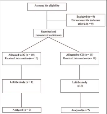
Figure shows the lowchart of the study design and allocation of participants.
Figure 1 - Study design flowchart. Two researchers were involved in data collection:
a main investigator assessments and an assistant in-vestigator intervention .
Perimetry
Perimetry was performed according to the stan-dards recommended by GU)RRO and GU)RRO , to identify the location of fat distribution in the body, as well as changes in body shape using a measuring tape. The tape was placed on the skin surface, but not too tight itting, on the umbilicus, two centimeters below the umbilicus, and two centimeters above the umbilicus. All measurements were performed by the same examiner
principal investigator .
Adipometry
Suprailiac and abdominal skinfold thickness were measured by lifting and folding the skin and fat with one's thumb and fore inger, detaching it from the muscle tissue. All measurements were performed three times, in the right hemisphere, with the subjects in the stand-ing position. The result reported was the average of the three measurements .
Perimetry and adipometry were carried out be-fore the irst treatment session baseline assessment and after the last session end point assessment . All measurements were performed by the same examiner
principal investigator .
Infrared Thermography
Thermal analysis was performed using an infrared camera Flir Systems® with digital image processing, thermal sensitivity of . °C and spectral range of - micrometers. We used the support software Velocity . , with automatic calibration. The images were captured in a room with °C temperature controlled by a ther-mometer , no sunlight and illuminated using cold light
luorescent bulbs. Participants were assessed in the standing position with the site of assessment free from clothing . The camera was positioned . meters away from the site of assessment, with the reference being the umbilicus.
33
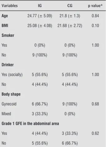
Table 1 - Clinical variables of the study population
Variables IG CG p value*
Age 24.77 (± 5.09) 21.8 (± 1.3) 0.84
BMI 25.08 (± 4.08) 21.68 (± 2.72) 0.10
Smoker
Yes 0 (0%) 0 (0%) 1.00
No 9 (100%) 9 (100%)
Drinker
Yes (socially) 5 (55.6%) 5 (55.6%) 1.00
No 4 (44.4%) 4 (44.4%)
Body shape
Gynecoid 6 (66.7%) 9 (100%) 0.68
Mixed 3 (33.3%) 0 (0%)
Grade 1 GFE in the abdominal area
Yes 4 (44.4%) 3 (33.3%) 0.62
No 5 (55.6%) 6 (66.7%)
Note: P < 0.05; Data presented as frequency and percentage. *t test; GFE: geloid fi brous edema; BMI: Body Mass Index; IG: interven-tion group; CG: control group.
Table 2 - Pre- and post-intervention anthropometric data for both groups, regarding adipometry and perim-etry of the abdominal area
IG (mean and SD)
CG
(mean and SD) p value*
Abdominal adipometry
Baseline 35.45 (± 6.95) 25.58 (± 8.95) 0.68
End Point 34.36 (± 7.72) 26.49 (± 7.21)
Suprailiac adipometry
Baseline 40.40 (± 3.85) 28.50 (± 5.63) 0.94
End Point 40.44 (± 8.93) 29.89 (±
10.74)
Waist circumference perimetry
Baseline 83.83 (± 12.94) 73.40 (± 5.94) 0.51
End Point 84.38 (± 11.94) 73.75 (± 4.19)
Data processing and statistical analysis
For the analysis of the photos, the abdominal region was divided into four quadrants two upper and two lower using the umbilicus as the center point. The aver-age value obtained from the four quadrants was used for data analysis.
We irst performed a descriptive analysis to charac-terize the study participants. The sample was found to be normally distributed using the Shapiro-Wilk normal-ity test, and then parametric tests were performed. We used the t test to compare pre- and post-intervention perimetry, adipometry and thermography between-group data. Comparative intrabetween-group analysis of the four thermography assessments was performed using paired t test.
All data were analyzed using the statistical program SPSS Statistical Package for Social Sciences version
. . The signi icance level was set at % p < . .
Results
The initial sample consisted of volunteers who were divided into two groups: an intervention group )G = and a control group CG = . (owever, as one participant from the )G and three from the CG decided to leave the study at some point, the inal sample was composed of participants )G = and CG = .
The mean age of the sample was . ± . years )G = . ± . ; CG = . ± . . The following clinical characteristics were analyzed: smoking habit, alcohol consumption, previous pregnancies, body shape, presence of abdominal geloid ibrous edema GFE , and BM). The values are depicted in Table . The t test showed that there were no statistical differ-ences between groups; both groups were considered similar with respect to the characteristics analyzed. The anthropometric data related to pre- and post-intervention suprailiac and abdominal adipometry, and waist circumference, abdomen and lanks pe-rimetry are shown in Table . There was no statisti-cal difference between the )G and CG regarding the decrease in abdominal measurements.
34
Discussion
Electrical stimulation elicits physiological changes in the adipocyte, evokes changes in the polarity of the cell membrane, activates microcirculation, and improves skin tone and lipolysis . The Joule effect elicits an increase in the temperature and contributes to an in-crease in local blood low and vasodilation, contributing to an increased metabolism, the burning of calories and an improved cell trophism . The literature indicates that the temperature at sites with quantitatively higher amounts of fat tissue is usually lower than in other sites, so the use of certain resources is needed to stimulate blood low and increase the metabolism in the area . According to Guedes and Guedes , changes in body fat distribution are an important morphological indicator of endocrine and metabolic complications. )ndividuals with higher concentrations of fat in the ab-dominal area have a higher incidence of diabetes mel-litus, hypertension and dyslipidemia. We opted therefore to apply electrical stimulation to the abdominal area.
)t is believed that the increase in cutaneous tem-perature after the use of functional and dermatological physical therapy resources is one of the main mecha-nisms to improve body shape and to reduce measures of adiposity. This is because local heating facilitates the spontaneous output of triglycerides — as fatty acid and glycerol — from the cytoplasm of fat cells into the blood-stream .
A clinical trial whose objective was to analyze the behavior of fat cells in women undergoing electrical stimulation has found an increase in plasma levels of fatty acid and glycerol in the region close to the stimu-lated cells. This proves the release of such molecules and, consequently, the reduction in the volume of adipocytes in the body region that received the intervention , . Blood tests were not performed in the aforemen-tioned study. They would have probably improved the results found.
The effectiveness of electrical stimulation using TENS has been con irmed by Araújo et al. and Assumpção et al. , . These authors claim that the stimulus gener-ated by TENS devices was able to reproduce the same physiological effects as when classic electrical stimula-tion devices were used. This justi ies the use of TENS in this study.
With regard to the application technique, Mello et al. assert that it is applied by insertion of small-diameter needles into the adipose panicle or through the application of electrodes to the skin surface. Parient Perimetry on the umbilicus
Baseline 89.66 (± 12.65) 78.80 (± 6.68) 0.32
End Point 89.57 ± 12.87 78.00 (± 5.47)
Flanks perimetry
Baseline 92.38 (± 12.09) 81.50 (± 4.6) 0.47
End Point 90.50 (± 14.16) 81.30 (± 4.03)
Note: p < 0.05. *T test; IG: intervention group; CG: control group; Baseline: baseline assessment; End Point: end point assessment.
Thermographic indings are shown in Table . We found statistical differences between groups in all as-sessments after baseline, regarding the increase in temperature. There were statistical differences in the )G but not in the CG.
Table 3 - Pre- and post-intervention thermographic assess-ment data for both groups
IG
(mean and SD) CG
(mean and SD) P value*
Baseline 31.85 ± 0.74a, b 30.48 ± 1.62 0.47
Imm.Ass 33.08 ± 1.00a, c 30.83 ± 1.5 0.002
15mAss 33.05 ± 0.48b, d 30.40 ± 1.24 < 0.0001
End Point 32.22 ±
14.20c,d 30.53 ± 1.34 0.005
Note: P < 0.05. *T test; IG: intervention group; CG: control group; Base-line: Baseline assessment; Imm.Ass: Assessment immediately follow-ing intervention; 15mAss: Assessment 15 min followfollow-ing intervention; End Point: End point assessment. Intragroup analysis (columns): paired t test - intervention group:a = statistical difference between Baseline
and Imm.Ass (p < 0.001). b = statistical difference between Baseline
and 15mAss (p < 0.001). c = statistical difference between Imm.Ass
and End Point (p < 0.001). d = statistical difference between 15mAss
and End Point (p < 0.001).
Regarding the thermographic data, there were in-tragroup statistical differences between baseline and the assessment immediately following intervention p < . ; between baseline and minutes assessment p < . ; between immediate assessment and end point assessment p < . ; and between the minutes assessment and the end point assessment p < . .
35 in this study (z , and min application time. The authors reported satisfaction with the new body shape acquired after using electrical stimulation .
)n a study with volunteers who received six ses-sions of electrical stimulation, Paula et al. have also found a signi icant reduction in waist circumference
about % .
According to Assumpção et al. the adoption of a hypocaloric diet combined with aerobic physical activ-ity can contribute to better treatment outcomes. )n our study there was no such association because the par-ticipants maintained their eating habits and sedentary lifestyles, which has probably limited the results.
Most of the previous studies with electrical stimula-tion , have used treatment sessions and found positive results. We also used only sessions and found positive results only in the thermographic assessment but not in anthropometric assessment. These data sug-gest that further treatment sessions could be conducted in order to enhance the effects of electrical stimulation and achieve better results with the use of this technique.
The different results found in the studies reported above may be associated with the use of different appli-cation techniques and stimulation parameters. Of note, the literature on this subject is still scarce. There are only a few randomized controlled trials with good method-ological design. Thus, further studies on this topic are needed. The small size of the sample, the fact that some subjects left the study at some point, and the lack of studies and of standardization of current parameters are recognized as limitations of this study.
We recommend that further studies investigate the use of other current modulation parameters, as well as the complementation with other techniques, such as electrical bioimpedance, ultrasound imaging, among others, to assess the metabolic effects of the intervention.
Conclusion
We found that electrical stimulation evokes a signi i-cant increase in the temperature of the subcutaneous tissue. This results in an increased metabolism, and thus facilitates lipolysis. )n the anthropometric assess-ment, however, there was no statistically signi icant dif-ferences before and after treatment. This is probably due to the small sample size. Thus, further studies are required to scienti ically prove the effects of electrical stimulation on abdominal fat and improve the provision of evidence-based care.
, however, claims that thicker needles show higher effectiveness than small-diameter needles. The author suggests the use of . to . mm diameter stainless steel needles with a length variation of to centime-ters. (e also suggests that the needles are separated by more than centimeters for improved coverage of the area to be treated. Assumpção et al. , on the other hand, asserts that the distance between needles should not exceed centimeters in order to prevent the dis-sipation of current.
)n this study electrical stimulation was performed with TENS, using . to mm needles centime-ters away from one another, and at a frequency of (z, in accordance with the aforementioned studies. Scorza et al. have used the same current, frequency and distance between needles and found a reduction of abdominal fat in the area to which electrical stimula-tion was applied. This inding was not repeated in the anthropometric data from this study.
Azevedo et al. have used electrical stimulation at the acupuncture points E Liangmen , E Taiyi , E Tianshu and E Daju , together with treatment sessions sessions per week for weeks . Participants were divided into two groups: Group one G consisted of subjects who only received electrical stimulation, while Group two G comprised subjects who re-ceived electrical stimulation and performed aerobic training. The eletrical stimulation device was set at a frequency of (z, and the intensity was set according to the tolerance of the individual patient. There were favorable perimetry and adipometry results for both groups, and the most signi icant results were found for the G . The study results have been optimized with the combined use of electrical stimulation and aerobic ex-ercise, because the latter enhances tissue lipolysis and increases blood low, thus increasing cellular activity and the excretion of cellular metabolites . )n this study, however, no signi icant perimetry and adipometry results were found. )t differs from the study by Azevendo et al. in respect to the parameters used frequency of (z, pulse width of microseconds and min ap-plication time , and because we did not use acupuncture points nor aerobic exercising.
36
. Brioschi ML, Yeng LT, Pastor EM(, Colman D, Silva FMRM, Teixeira MJ. Documentação da síndrome dolo-rosa miofascial por imagem infravermelha. Acta Fisiatr.
; .
. Eddy AL, van (oogmoed LM, Snyder JR. The role of ther-mography in the management of equine lameness. Vet J. ; : - .
. Sefton JM, Yarar C, Berry JW, Pascoe DD. Therapeutic massage of the neck and shoulders produces changes in peripheral blood low when assessed with dynamic infrared thermography. J Altern Complement Med.
; : - .
. (oley LA, Dixon J, Selfe J. An exploratory thermographic investigation of the effects of connective tissue massage on autonomic function. J Manipulative Physiol Ther.
; : - .
. Gazerani P, Arendt-Nielsen L. Cutaneous vasomotor reactions in response to controlled heat applied on various body regions of healthy humans: evaluation of time course and application parameters. )nt J Physiol Pathophysiol Pharmacol. ; : - .
. Brasil, Ministério da Saúde. Secretaria de Atenção à Saúde. Departamento de Atenção Básica. Orientações para a coleta e análise de dados antropométricos em ser-viços de saúde: Norma Técnica do Sistema de Vigilância Alimentar e Nutricional – S)SVAN – Ministério da Saúde, Secretaria de Atenção à Saúde, Departamento de Atenção Básica. Brasília: Ministério da Saúde; . Portuguese. . Maio M. Tratado de Medicina Estética. São Paulo: Roca;
. Portuguese.
. Savastano DM, Gorbach AM, Eden (S, Brady SM, Reyn-olds JC, Yanovscki JA. Adiposity and human regional body temperature. Am J Clin Nutr. ; : - . . Guedes D, Guedes J. Distribuição de gordura
corpo-ral, pressão arterial e níveis de lipídios-lipoproteínas plasmáticas. Arq Bras Cardiol. ; : - . . Paula MR, Picheth G, Simões NDP. Efeitos da
eletrolipo-forese nas concentrações séricas de ácido graxo e coles-terol e do per il lipídico. Fisioter Bras. ; : - . . Araújo CP, Brito AKAT, Escarião AD, Torres RBA. Eletro-lipólise como método de redução de adiposidade no abdome inferior: estudo piloto. Revista de Especialização em Fisioterapia. ; .
References
. Feng B,Zhang T,Xu (. (uman adipose dynamics and metabolic health. Ann N Y Acad Sci. ; : - . . Guirro E, Guirro R. Fisioterapia dermato-funcional. São
Paulo: Manole; . Portuguese.
. Borges FS. Modalidades Terapêuticas nas disfunções estéticas. São Paulo: Phorte; . Portuguese. . Jo J, Shreif Z,Periwal V. Quantitative dynamics of adipose
cells. Adipocyte. ; : - .
. Lee M, Wu Y,Fried SK. Adipose tissue heterogeneity: implication of depot differencesin adipose tissue for obe-sity complications. Mol Aspects Med. ; : - . . Mulholland RS, Paul MD,Chalfoun C. Noninvasive body
contouring with radiofrequency, ultrasound, cryoli-polysis, and low-level laser therapy. Clin Plast Surg.
; : - .
. Azevedo CJD, Zanin EC, Tolentino TM, Cepeda CC, Bus-nardo VL. Estudo comparativo dos efeitos da eletro-lipólise por acupontos e da eletroeletro-lipólise por acupontos associada ao trabalho aeróbico no tratamento da adipos-idade abdominal grau ) em indivíduos do sexo feminino com idade entre e anos. RUBS. ; : - . . Scorza FA, Figueiredo MM, Liao CO, Borges FC. Estudo comparativo dos efeitos da eletrolipólise com uso de TENS modo burst e modo normal no tratamento de adi-posidade localizada abdominal. Ensaios e Ciência: Ciên-cias Biológicas, Agrárias e da Saúde. ; : - . . Soriano MCD, Pérez SC, Baques M)C. Electroestética
pro-fesional aplicada: teoria y práctica para la utilización de corrientes en estética. Barcelona: Editora Sorisa;
. Spanish.
. (amida Z(, Comtois AS, Portmann M, Boucher JP, Sa-vard R. Effect of electrical stimulation on lipolysis of human white adipocytes. Appl Physiol Nutr Metab.
; : - .
. Mello-Carpes PB, Stumpf T, Piccinini AM, Rosa PV. A eletrolipólise percutânea como possibilidade de diminuição da adiposidade em abdomen e lancos. Bio-motriz. ; : - .
37
. Assumpção AC, Souza A, Máximo L, Cardoso MC, Borg-es FS. Eletrolipólise EletrolipoforBorg-ese . )n: BorgBorg-es FS. Dermato-Funcional: Modalidades Terapêuticas nas Disfunções Estéticas. São Paulo: Phorte; . p.
-. Portuguese-.
. Mello P, Dreher PM, Piccinini AM, Rosa L(T, Rosa PV. Comparação dos efeitos da eletrolipólise transcutânea e percutânea sobre a gordura localizada na região abdomi-nal e de lancos através da perimetria e análise de bio-impedância elétrica. Fisioter Bra. ; : - . . Parienti )J. Medicina Estética. São Paulo: Andrei; . . Douglas CR. Tratado de Fisiologia Aplicada à Saúde. São
Paulo: Robe Editorial; .
. Machado GC, Vieira RB, Oliveira NML, Lopes CR. Análise dos efeitos do ultrassom terapêutico e da eletrolipo-forese nas alterações decorrentes do ibroedema gelóide. Fisioter Mov. ; : - .
. Paula M, Pichet G, Simões N. Efeitos da eletrolipoforese nas concentrações séricas do glicerol e per il lipídico. Fisioter Bras. Jan/Feb: - . Suplemento Especial.
Received in / / Recebido em 16/12/2014