Zoledronic Acid Effects Interleukin-6 Expression in
Hormone-Independent Prostate Cancer Cell Lines
Layka A. Asbagh, Selim Uzunoglu, Cag Cal
Department of Molecular Biology (LAA, SU), Faculty of Science and Arts, Celal Bayar University, Manisa, Turkey, and Department of Urology (CC), School of Medicine, Ege University, Izmir, Turkey
ABSTRACT
Objective:To investigate the inhibitory effects of zoledronic acid (ZA) on tumor related growth factor IL-6 in hormone resistant prostate cancer cell lines. The association between apoptosis and IL-6 inhibition was also assessed.
Materials and Methods: PC-3 and DU145 cell lines were treated with different concentrations of ZA (1-100μM) at vari-ous intervals (24-72 h.). The cell viability was investigated by XTT assay and apoptotic effect was evaluated by cell death GHWHFWLRQ(/,6$NLW&DVSDVHDFWLYLW\DVVD\ZDVSHUIRUPHGWRFRQ¿UPDSRSWRVLV,/OHYHOVZHUHPHDVXUHGE\(/,6$ LQWKHVXSHUQDWDQWDQGWKHVHGDWDZHUHDOVRFRQ¿UPHGE\,/P51$DQDO\VLVXVLQJ573&5
Results:PC-3 and DU145 cell lines were sensitive to ZA mediated cytotoxicity in a dose- and time-dependent manner. +RZHYHUWKHDSRSWRWLFHIIHFWZDVVLJQL¿FDQWO\GLIIHUHQWDPRQJ3&DQG'8FHOOVS,/VHFUHWLRQZDV VLJQL¿FDQWO\ORZHULQERWKFHOOOLQHVFRPSDUHGWRWKHXQWUHDWHGFRQWUROFHOOVS$OWKRXJKWKHLQFUHDVHGLQKLELWLRQ of IL-6 secretion was associated with increased apoptosis in DU145 cells (p = 0.002), there was no similar association for 3&FHOOOLQHS :KHQFRPSDUHGWRWKHXQWUHDWHGFRQWUROVWKHQXPEHURIF'1$FRSLHVZDVVLJQL¿FDQWO\ORZHU LQWKH=$WUHDWHG'8FHOOOLQHDWGRVHVRIDQG0SVXJJHVWLQJDUHGXFHGH[SUHVVLRQRI,/P51$
Conclusion: ZA exhibited a time- and dose-dependent apoptotic effect on PC-3 and DU145 prostate cancer cell lines and this effect was associated with inhibited secretion of IL-6 in DU145 cell line.
Key words: prostate cancer; zoledronic acid; interleukin-6; experimental Int Braz J Urol. 2008; 34: 355-64
INTRODUCTION
Prostate cancer is most common among el-derly men, and in 2007, the estimated number of the newly diagnosed prostate cancer cases was 218.890 in USA (1). Although local curative treatment strategies
DUHWKHPRVWDSSURSULDWHSURFHGXUHVLQRUJDQFRQ¿QHG
disease, androgen deprivation therapy represents the standard treatment in patients with metastatic prostate
FDQFHU1HYHUWKHOHVVWKHGHYHORSPHQWRIKRUPRQH
resistant prostate cancer and progression is inevitable
during androgen deprivation treatment. Unfortunately, no any other effective and curative alternative treat-ment has been reported for these patients.
The new treatment modalities are primarily focused on growth factors that stimulate the prolifera-tion of prostate cancer cells. Interleukin-6 (IL-6) is a growth factor for prostate cancer cells and its high serum levels are known to be directly associated with clinical prognosis of the disease (2,3). It has also been shown that IL-6 signaling pathway is active and
IL-6 signaling pathway is actively used in metastatic prostate cancers and hormone independent prostate cancer cell lines.
Several clinical trials have already
demon-VWUDWHGWKHEHQH¿FLDOHIIHFWVRIELVSKRVSKRQDWHVLQ
prostate cancer patients (5,6). The growth of metas-tases may be inhibited by modifying the bone micro-environment using bisphosphonates. They also exert direct cytotoxic and apoptotic effects on a variety of human tumor cell lines including myeloma, breast cancer and prostate cancer (7-9).
Zoledronic acid (ZA) is the most potent nitro-gen containing bisphosphonate compound. It has been shown to inhibit cell growth and induce apoptosis in
SURVWDWHFDQFHUFHOOOLQHV'83&DQG/1&D3
(10). Current evidence on the effects of ZA suggests that it is a potential chemotherapeutic agent for the treatment of prostate cancer, either as monotherapy or in combination. Despite the overwhelming in vitro studies investigating the anti-tumor activity of the combined use of ZA with different chemotherapeutics, the molecular targets and mechanisms of ZA in tumor cells remains a subject of debate.
We hypothesize that ZA may exert its anti-tumor effect by inhibiting the anti-tumor related growth factor IL-6. Considering the potential role of IL-6 in the growth regulation of PC-3 and DU145 cell lines, the present study was planned to investigate the relationship between the anti-tumor activity of ZA and IL-6 secretion in these cells under in vitro conditions.
MATERIALS AND METHODS
Chemicals - Cell culture supplies were ob-tained from Biological Industries (Kibbutz Beit Hae-mek, Israel). Zoledronic acid was a generous gift from
1RYDUWLV3KDUPDFHXWLFDOV,QF%DVHO6ZLW]HUODQG
The stock solution of zoledronic acid was prepared at a concentration of 1 mM in distilled water and aliquots were stored at -20oC. All other chemicals,
unless otherwise mentioned, were purchased from Sigma Chemical Co (USA).
Cell lines and culture - The androgen-refrac-tory prostate cancer cell lines, PC-3 and DU145, were preferentially used since they secrete IL-6 and actively
use IL-6 signaling pathway for growth promoting ef-fects (11) and to maintain resistance to chemotherapy. These cell lines were kindly provided by Dr. Levent Turkeri from Marmara University, Istanbul, Turkey. PC-3 and DU145, adherent cell lines were cultured in
530,VXSSOHPHQWHGZLWKKHDWLQDFWLYDWHG IHWDOERYLQHVHUXP/JOXWDPLQHDQGSHQLFLO -lin-streptomycin. All cell cultures were incubated at
&LQ&22DQGDLU
Cell viability assay - The effects of different concentrations of zoledronic acid (1,10,30,60,90, and 100μM) on PC-3 and DU145 cell lines were evaluated
E\XVLQJ;77FHOOSUROLIHUDWLRQNLW5RFKH$SSOLHG 6FLHQFH0DQQKHLP*HUPDQ\)ROORZLQJWKHYHUL¿ -cation of cell viability by tryptan blue exclusion test, cells were plated on a 96-well plate in 200μL culture medium at a concentration of 104cells/well. At 24, 48
and 72 hours of incubation, a 50μL of XTT labeling mixture was added to each well. The optical density was measured at 450 nm with a reference wavelength at 650 nm using a microplate reader (Beckman
Coul-WHU'7;0XOWLPRGH5HDGHU7KHSHUFHQWDJHRI
cytotoxicity was calculated as follows:
% Cystotoxicity = 1 _ A of experimental well x 100 A of positive control well
where A is the absorbance.
Evaluation of apoptosis - The Cell Death Detection
(/,6$NLW5RFKH$SSOLHG6FLHQFH0DQQKHLP*HU -many) was used to detect mono-oligonucleosomes
KLVWRQHDVVRFLDWHG'1$IUDJPHQWVDVDQLQGLFDWRU
of apoptosis after zoledronic acid induced cell death.
%ULHÀ\F\WRSODVPLFO\VDWHVIURPXQWUHDWHGFRQWUROV
and zoledronic acid treated cells were transferred to a streptavidin-coated plate supplied by the manufac-turer. A mixture of histone-biotine and
Anti-'1$32'ZHUHDGGHGWRFHOOO\VDWHVDQGLQFXEDWHG
Caspase 3/7 activity assay - The Caspase-Glo 3/7 assay (Promega, Madison, WI) was used to measure caspase 3/7 activity, according to the manufacturer’s instructions. PC-3 cells were plated on a 96-well plate in 100μL culture medium at a concentration of 104 cells/well. After incubation with
increasing concentrations of zoledronic acid, 100μL of Caspase-Glo 3/7 reagent was added to each well. Then the mixture was incubated for one hour at room temperature and the luminescence of each sample was measured using a plate-reading luminometer
(Beck-PDQ&RXOWHU'7;0XOWLPRGH5HDGHU
Determination of interleukin-6 secretion
,/OHYHOVZHUHTXDQWL¿HGLQWKHVXSHUQDWDQWVRI
zoledronic acid treated PC-3 and DU145 cells by us-ing Human IL-6 ELISA Kit (Biosource International Inc., California, USA). The cells were plated on 24-well plates at a concentration of 105 cells per well
and incubated for 24, 48 and 72 hours with increasing concentrations of zoledronic acid (1-100μM). Super-natants were collected for all culture conditions and analyzed for IL-6 levels using a standard ELISA kit according to manufacturer’s instructions. Standard
FXUYHIRUTXDQWL¿FDWLRQZDVSORWWHGIURPYDOXHVRI
IL-6 standards provided by kit. IL-6 levels in zole-dronic acid treated cells were recalculated based on the IL-6 levels from untreated control cells at the end of treatment in order to compensate the differences due to cell number. The decrease in IL-6 levels was
DOVRFRQ¿UPHGE\573&5
([SUHVVLRQRI,/P51$7KHHIIHFWRI ]ROHGURQLFDFLGRQ,/P51$OHYHOZDVLQYHVWL
-JDWHG E\ 573&5 51$ VDPSOHV IURP XQWUHDWHG
controls and DU-145 treated cells were isolated by
XVLQJ+LJK3XUH51$,VRODWLRQ.LW5RFKH$SSOLHG
Science, Mannheim, Germany). Primers and probes
ZHUHLQFOXGHGLQ5RFKH/LJKW&\FOHU3ULPHUVHW+X -man Interleukin-6). The procedure was carried out as a single step method for reverse transcription from
51$ WR F'1$ DQG VXEVHTXHQW TXDQWL¿FDWLRQ ZDV PDGHZLWKRXWRSHQLQJWKHUHDFWLRQWXEH$5RFKH
Light Cycler apparatus was used with the following sequence: denaturation at 95ºC for 10 minutes, then
F\FOHVRIDPSOL¿FDWLRQVIRUVDW&VDW &VDW&DQGD¿QDOFRROLQJVWHSWR& 7KHGDWDZHUHDQDO\]HGE\WKHVRIWZDUHRI5RFKH
LightCycler (1.5) Instrument.
Statistical analyses - All experiments were set up in triplicate and the results were expressed as mean
VWDQGDUGGHYLDWLRQ6'*UDSK3DG35,60VRIWZDUH
(version 5) (San Diego, CA, USA) was used for the analysis of data and graphic presentations. Student’s
WWHVWRU$129$ZDVXVHGIRUFRPSDULVRQV
RESULTS
The cytotoxic and apoptotic effects of zole-dronic acid - PC-3 and DU145 cell lines were sensitive to ZA mediated cytotoxicity; the maximum cytotoxic-ity was achieved at 72 hour with 100μM concentration of ZA. The cytotoxicity was proportional with the increasing concentrations of ZA for both cell lines and the difference from untreated controls was statistically
VLJQL¿FDQWS'DWDQRWVKRZQ=$LQGXFHG
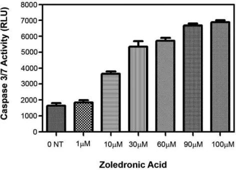
time- and dose-dependent apoptosis in both cell lines. Data for PC-3 cell line regarding apoptosis is given in Figure-1. For PC-3 cell line, Caspase 3/7 activity was
VLJQL¿FDQWO\LQFUHDVHGLQ=$WUHDWHGFHOOVFRPSDUHG WRXQWUHDWHGFRQWUROVS)LJXUH
IL-6 secretion as detected in the supernatants of PC-3 and DU145 cells - Incubation of PC-3 and DU145 cells with increasing concentrations of ZA
IRUDQGKRXUVUHVXOWHGLQDVLJQL¿FDQWGRVH GHSHQGHQWGHFUHDVHLQ,/VHFUHWLRQS)LJ -ure-3 and 4). This effect was detected with the lowest dose and at the earliest time points. A difference in terms of dose-dependent inhibition of IL-6 secretion between two cell lines could only be observed at 72 hours. The lowest level of IL-6 secretion was achieved at 24 hours for PC-3 cells.
For DU-145 cells, a four-fold decrease in IL-6 secretion was found in ZA treated cells with 60
ȝ0DQGKLJKHUFRQFHQWUDWLRQVFRPSDUHGWRXQWUHDWHG
controls. However, for PC-3 cells, IL-6 secretion was only halved with the same concentrations and IL-6
VHFUHWLRQZDVVLJQL¿FDQWO\KLJKHULQ'8FHOOVWKDQ 3&FHOOVS
Figure 1 – Concentration dependent relative apoptosis in PC-3 cell line following exposure to zoledronic acid. PC-3 cell line was treated with increasing concentrations of zoledronic acid for 72 hours and then the levels of mono-oligo nucleosome fragments was TXDQWL¿HGXVLQJ&HOO'HDWK'HWHFWLRQ.LW&ROXPQVWKHPHDQVRIWZRLQGHSHQGHQWH[SHULPHQWVEDUV6'17XQWUHDWHGFRQWUROV 3YVFRQWUROV
0HDVXUHPHQWRI,/P51$OHYHOVLQ=$ WUHDWHG'8FHOOVE\573&5573&5ZDVSHU -formed in DU-145 cells, in order to examine whether the reduction of IL-6 levels is associated with a
de-FUHDVHGH[SUHVVLRQRI,/P51$7KHQXPEHURI F'1$FRSLHVZDVVLJQL¿FDQWO\ORZHULQ'8FHOOV
treated with 30 and 90μM ZA, compared to untreated
FRQWUROVS)LJXUH
Figure 3 –(IIHFWRI]ROHGURQLFDFLGWUHDWPHQWRQ,/VHFUHWLRQLQ3&FHOOOLQH viable cell/well was treated with increasing con-centrations of zoledronic acid. IL-6 levels were measured after 48 and 72 hours by ELISA in the supernatants of zoledronic acid treated FHOOV3RLQWVWKHPHDQRIDWOHDVWWKUHHLQGHSHQGHQWH[SHULPHQWVEDUV6'XQWUHDWHGFRQWUROV3YVFRQWUROV
COMMENTS
7KHSUHVHQWVWXG\FRQ¿UPHGWKDW=$LQGXFHV
apoptosis in PC-3 and DU145 prostate cancer cell lines in a dose- and time-dependent manner. However,
WKHH[WHQWRIWKLVHIIHFWZDVVLJQL¿FDQWO\GLIIHUHQWIRU
PC-3 and DU145 cell lines. In addition, two cell lines differed in terms of IL-6 secretion. The degree of apoptosis was not related to the level of the inhibition of IL-6 secretion for PC-3 cells, which also secrete
ORZOHYHOVRI,/FRPSDUHGWR'8FHOOV2QWKH
other hand, the level of reduction in IL-6 secretion was correlated with the degree of ZA induced apoptosis in DU145 cell lines. Based on this data, it might be speculated that anti-tumoral effects of ZA could also
be mediated by IL-6 and related signaling pathways in prostate cancer cells.
It has been well-documented that IL-6 is a multifunctional cytokine that plays an important role in the regulation of hematopoiesis, immune response,
LQÀDPPDWLRQERQHPHWDEROLVPDQGQHXUDOGHYHORS -ment (12). It is produced by different cells including lymphoid or non-lymphoid cells and malignant tis-sues (13). All prostate cells including normal pros-tate epithelia, cells originated from benign prostatic hyperplasia and malignant prostate cancer are shown to be capable of secreting IL-6 in cell cultures (14). Furthermore, increased secretion of IL-6 ligand and its receptors in serum has been reported for all stages of prostate cancer including hormone refractory
tients (14,15). Also, clinical prognosis of prostate cancer is directly affected by serum IL-6 levels (15) and IL-6 plays an important role for the development of resistance to chemotherapeutics used in prostate cancer (16). Moreover, exogenous administration of IL-6 has been shown to inhibit doxorubicin-induced apoptosis in PC-3 cells (17).
In vitro studies demonstrated an increase in the proliferation of prostate cancer cells with IL-6 stimulation (18,19) and a decrease in growth rate of androgen insensitive PC-3 and DU145 cell lines treated with anti-IL-6 antibodies (16). These results suggest that the combined use of anticancer agents with drugs resulting in an inhibition of IL-6
expres-VLRQFRXOGLQFUHDVHWKHHI¿FDF\RIFKHPRWKHUDS\SDU -ticularly in patients with hormone refractory prostate cancer.
+RZHYHUFRQÀLFWLQJUHVXOWVKDYHEHHQUHSRUW -ed regarding the stimulatory/inhibitory effects of IL-6 on the proliferation on various prostate cancer cell lines (11,20,21). These differences may be attributed to a several reasons related to IL-6 signaling pathway. Firstly, there are membranous and soluble forms of IL-6 ligand and its receptor, which are strictly regulated. Secondly, gp130, the signal transducer of IL-6 on the membrane, can be activated by various growth fac-tors. Thirdly, activated gp130, either simultaneously or preferentially, triggers three intracellular pathways by the alteration of intracellular domain. IL-6 signal-ing is mediated by JAK-STAT, ras-raf-MAPK and PI3K-Akt signaling. It has been suggested that one or two alternative pathways are preferentially more active in different cell lines. IL-6 can also be up- and down regulated by autocrine or paracrine effects and feed-back mechanism (11,20,22,23). Its expression is regulated by several transcription factors such as
$31)N%&5(%DQGF(%3,WLVFRQVLGHUHGWKDW
intracellular signaling pathways of IL-6 also regulate these transcription factors.
Zoledronic acid may affect a some molecules in signal transduction pathways including cell prolif-eration process (ras-raf-MAPK), tumor suppressor genes, apoptotic pathways, cell cycle proteins and posttranslational processes. Since ZA affects the binding of ras proteins to the membrane via protein prenylation (10), it might indirectly inhibit cell pro-liferation. In a recent study by Cavarretta et al., the
effect of IL-6 was shown to be mediated by oncogene Mcl-1 (myeloid cell leukemia-1), an anti-apoptotic member of the Bcl-2 family in prostate cell line (24). The association between ZA treatment and IL-6 secre-tion may also be regulated by Mcl-1 expression.
Several authors have previously suggested that ZA cannot induce apoptosis (9,25). Such an inconsistency might be explained by the differences in ZA concentrations (25) and treatment durations (9). The present study indicates that a longer treat-ment period with higher concentrations of ZA is necessary to induce apoptosis. Interestingly, when bisphosphonates are combined with other common
DQWLQHRSODVWLFGUXJVDVLJQL¿FDQWV\QHUJ\RFFXUV
The synergic cytotoxic effect of ZA has previously been detected on prostate cancer cells (26,27).
Few studies investigated the relation between IL-6 expression/secretion and ZA treatment. A de-creased IL-6 expression has been reported after ZA treatment in bone marrow stromal cells under in vitro
FRQGLWLRQV2QWKHFRQWUDU\DWUDQVLHQWLQGXF
-WLRQRIDQLQFUHDVHLQ71)DOSKDDQG,/OHYHOVZLWK
ZA infusion has been demonstrated in cancer patients with fever (30). Although the disagreements between the studies may be explained by the variations of in vivo and in vitro conditions, all of these observations clearly points out that IL-6 has an important role in the processes related to both bone microenvironment and metastases in prostate cancer (31). The present study shows a correlation between the degree of ZA induced apoptosis and the inhibition of IL-6 secretion, imply-ing that the apoptotic effect of ZA is associated with IL-6 and related pathways. Exogenous administration of IL-6 do not interfere the anticancer actions of ZA on PC-3 cells, which supports the above-mentioned association (17).
7KHVH¿QGLQJVUDLVHWZRSRVVLEOHLQWHUSUH -tations: either the reduction of IL-6 secretion itself induces the apoptotic process or it may be the out-come of ZA induced apoptosis in a dose dependent
PDQQHU,IQRVLJQL¿FDQWFRUUHODWLRQKDGEHHQIRXQG
between the decrease in IL-6 expression and the de-gree of apoptotic process, it could be suggested that ZA directly inhibits the autocrine mechanisms of IL-6 expression.
affecting signal transduction pathways on the up-stream region of apoptotic pathway. This probability may explain the reduction of IL-6 secretion with increased apoptosis, which was observed after ZA treatment in our study. Therefore, it can be suggested that ZA not only directly induces apoptotic pathways, but also indirectly affects one or more signal trans-duction molecules located on upstream region, which cause the apoptosis in PC-3 and DU145 cell lines. For these reasons, it is necessary to determine the target molecules that play key roles on the effects of ZA.
CONCLUSION
The present in vitro study shows a time- and dose-dependent apoptotic effect of ZA on both PC-3 and DU145 prostate cancer cell lines, which correlates with an inhibitory effect on IL-6 expression in DU145 cells. Additional research is required to further eluci-date the activity of IL-6 and its role in the pathogenesis of advanced prostate cancer at cellular and molecular levels. Also, further studies are required to investigate the down regulation of oncogene Mcl-1 (myeloid cell leukemia-1), an anti-apoptotic member of the Bcl-2 family, which is regulated directly by IL-6 in ZA treated cells (24). The inhibition of IL-6 with anti-IL-6 antibody sensitizes androgen-independent prostate cancer cells to chemotherapeutic agents in vitro (32); thus, treatment modalities targeting IL-6 may have multiple advantages in prostate cancer patients who
UHFHLYHOLPLWHGWKHUDSHXWLFDQGVXUYLYDOEHQH¿WIURP
conventional treatment alternatives (18).
CONFLICT OF INTEREST
1RQHGHFODUHG
REFERENCES
-HPDO$6LHJHO5:DUG(0XUUD\7;X-7KXQ0-Cancer statistics, 2007. CA -HPDO$6LHJHO5:DUG(0XUUD\7;X-7KXQ0-Cancer J Clin. 2007; 57: 43-66.
2. Culig Z, Steiner H, Bartsch G, Hobisch A: Interleu-kin-6 regulation of prostate cancer cell growth. J Cell Biochem. 2005; 95: 497-505.
1DNDVKLPD -7DFKLEDQD 0 +RULJXFKL< 2\D 0 2KLJDVKL7$VDNXUD+HWDO6HUXPLQWHUOHXNLQDV a prognostic factor in patients with prostate cancer. &OLQ&DQFHU5HV
*LUL'2]HQ0,WWPDQQ0,QWHUOHXNLQLVDQDX -tocrine growth factor in human prostate cancer. Am J Pathol. 2001; 159: 2159-65.
6PDOO(-6PLWK056HDPDQ--3HWURQH6.RZDOVNL 02&RPELQHGDQDO\VLVRIWZRPXOWLFHQWHUUDQGRP -ized, placebo-controlled studies of pamidronate disodium for the palliation of bone pain in men with PHWDVWDWLFSURVWDWHFDQFHU-&OLQ2QFRO 4277-84.
6DDG)*OHDVRQ'00XUUD\57FKHNPHG\LDQ6 9HQQHU3/DFRPEH/(7$/$UDQGRPL]HGSOD -cebo-controlled trial of zoledronic acid in patients with hormone-refractory metastatic prostate carcinoma. J 1DWO&DQFHU,QVW
6KLSPDQ&05RJHUV0-$SSHUOH\-)5XVVHOO5* Croucher PI: Bisphosphonates induce apoptosis in hu-man myeloma cell lines: a novel anti-tumour activity. Br J Haematol. 1997; 98: 665-72.
)URPLJXH2/DJQHDX[/%RG\--%LVSKRVSKRQDWHV induce breast cancer cell death in vitro. J Bone Miner 5HV
/HH09)RQJ(06LQJHU)5*XHQHWWH56%LVSKRV -phonate treatment inhibits the growth of prostate FDQFHUFHOOV&DQFHU5HV
2DGHV *0 6HQDUDWQH 6* &ODUNH ,$ .LUE\ 56 &ROVWRQ.:1LWURJHQFRQWDLQLQJELVSKRVSKRQDWHV induce apoptosis and inhibit the mevalonate pathway, LPSDLULQJ5DVPHPEUDQHORFDOL]DWLRQLQSURVWDWHFDQ -cer cells. J Urol. 2003; 170: 246-52.
&KXQJ7'<X--6SLRWWR07%DUWNRZVNL06L -mons JW: Characterization of the role of IL-6 in the progression of prostate cancer. Prostate. 1999; 38: 199-207.
12. Kishimoto T, Akira S, Taga T: Interleukin-6 and its receptor: a paradigm for cytokines. Science. 1992; 258: 593-7.
13. Kishimoto T: The biology of interleukin-6. Blood. 1989; 74: 1-10.
14. Twillie DA, Eisenberger MA, Carducci MA, Hseih :6.LP:<6LPRQV-:,QWHUOHXNLQDFDQGLGDWH mediator of human prostate cancer morbidity. Urology. 1995; 45: 542-9.
%RUVHOOLQR1%HOOGHJUXQ$%RQDYLGD%(QGRJHQRXV interleukin 6 is a resistance factor for cis-diamminedi-chloroplatinum and etoposide-mediated cytotoxicity RIKXPDQSURVWDWHFDUFLQRPDFHOOOLQHV&DQFHU5HV 1995; 55: 4633-9.
7HQWD57LEODOH[L'6RWLULRX(/HPEHVVLV30D -noussakis M, Koutsilieris M: Bone microenviron-ment-related growth factors modulate differentially the anticancer actions of zoledronic acid and doxorubicin on PC-3 prostate cancer cells. Prostate. 2004; 59: 120-31.
/RX:1L='\HU.7ZHDUG\'-*DR$&,QWHUOHX -kin-6 induces prostate cancer cell growth accompanied by activation of stat3 signaling pathway. Prostate. 2000; 42: 239-42.
%RUVHOOLQR 1 %RQDYLGD % &LOLEHUWR * 7RQLDWWL &7UDYDOL 6 '¶$OHVVDQGUR 1 %ORFNLQJ VLJQDOLQJ through the Gp130 receptor chain by interleukin-6 and oncostatin M inhibits PC-3 cell growth and sensitizes the tumor cells to etoposide and cisplatin-mediated cytotoxicity. Cancer. 1999; 85: 134-44.
2NDPRWR0/HH&2\DVX5,QWHUOHXNLQDVDSDUD -crine and auto-crine growth factor in human prostatic FDUFLQRPDFHOOVLQYLWUR&DQFHU5HV 6. 0RUL60XUDNDPL0RUL.%RQDYLGD%2QFRVWDWLQ 020SURPRWHVWKHJURZWKRI'8KXPDQSURV -WDWHFDQFHUFHOOVEXWQRW3&RU/1&D3WKURXJKWKH VLJQDOLQJRIWKH20VSHFL¿FUHFHSWRU$QWLFDQFHU5HV 1999; 19: 1011-5.
22. Klein B, Zhang XG, Jourdan M, Content J, Houssiau F, Aarden L, et al.: Paracrine rather than autocrine regulation of myeloma-cell growth and differentiation by interleukin-6. Blood. 1989; 73: 517-26.
0LNL6,ZDQR00LNL<<DPDPRWR07DQJ%<R -kokawa K, et al.: Interleukin-6 (IL-6) functions as an in vitro autocrine growth factor in renal cell carcinomas. FEBS Lett. 1989; 250: 607-10.
&DYDUUHWWD,71HXZLUW+8QWHUJDVVHU*0RVHU3/ Zaki MH, Steiner H, et al.: The antiapoptotic effect of
IL-6 autocrine loop in a cellular model of advanced SURVWDWHFDQFHULVPHGLDWHGE\0FO2QFRJHQH 26: 2822-32.
%RLVVLHU6)HUUHUDV03H\UXFKDXG20DJQHWWR6 Ebetino FH, Colombel M, et al.: Bisphosphonates inhibit breast and prostate carcinoma cell invasion, an early event in the formation of bone metastases. &DQFHU5HV
1HYLOOH:HEEH+/5RVWDPL+RGMHJDQ$(YDQV&$ &ROHPDQ5(+ROHQ,6HTXHQFHDQGVFKHGXOHGHSHQ -dent enhancement of zoledronic acid induced apoptosis by doxorubicin in breast and prostate cancer cells. Int J Cancer. 2005; 113: 364-71.
27. Ullen A, Lennartsson L, Harmenberg U, Hjelm-Er-iksson M, Kalkner KM, Lennernas B, et al.: Addi-tive/synergistic antitumoral effects on prostate cancer cells in vitro following treatment with a combination RIGRFHWD[HODQG]ROHGURQLFDFLG$FWD2QFRO 44: 644-50.
'HUHQQH6$PLRW0%DULOOp6&ROOHWWH05RELOODUG 1%HUWKDXG3HWDO=ROHGURQDWHLVDSRWHQWLQKLEL -tor of myeloma cell growth and secretion of IL-6 and MMP-1 by the tumoral environment. J Bone Miner 5HV
29. Corso A, Ferretti E, Lunghi M, Zappasodi P, Mangia-cavalli S, De Amici M, et al.: Zoledronic acid down-regulates adhesion molecules of bone marrow stromal cells in multiple myeloma: a possible mechanism for its antitumor effect. Cancer. 2005; 104: 118-25. 'LFXRQ]R*9LQFHQ]L%6DQWLQL'$YYLVDWL*5RFFL
L, Battistoni F, et al.: Fever after zoledronic acid ad-PLQLVWUDWLRQLVGXHWRLQFUHDVHLQ71)DOSKDDQG,/ -,QWHUIHURQ&\WRNLQH5HV
(DWRQ &/ &ROHPDQ 5( 3DWKRSK\VLRORJ\ RI ERQH metastases from prostate cancer and the role of ELVSKRVSKRQDWHVLQWUHDWPHQW&DQFHU7UHDW5HY 29: 189-98.
32. Smith PC, Hobisch A, Lin DL, Culig Z, Keller ET: In-terleukin-6 and prostate cancer progression. Cytokine *URZWK)DFWRU5HY
Accepted after revision: $SULO Correspondence address:
Dr. Cag Cal
Department of Urology Ege University Tip Fakultesi Izmir, Turkey
EDITORIAL COMMENT
Interleukin-6 (IL-6) is an important regula-tor of cellular events in human prostate cancer. It has multifunctional effects on proliferation, apoptosis, and angiogenesis and is a target for novel therapies. Most studies were performed with the anti-IL-6
an-WLERG\&172LQYLWURDQGLQYLYR7KH\
have demonstrated differences in responsiveness to the antibody between these two different cell lines. The authors of the present paper show that zoledronic acid, that is used for late stage prostate cancer treat-ment, has a negative effect on IL-6 expression. This is a novel important aspect of action of that drug in human prostate cancer therapy. Since IL-6 is con-sidered a survival factor in some but not all human prostate cancers, this therapy may increase rate of cell death. However, growth-inhibitory effects of IL-6 in selected cell lines were also observed. For that reason, it is important to determine who are the patients who
ZLOOEHQH¿WIURPDQWL,/WKHUDS\LQWKHIXWXUH,Q
summary, the manuscript by Asbagh et al. is transla-tionally relevant and may stimulate research on IL-6 regulatory effects in prostate cancer in the future.
REFERENCES
1. Smith PC, Keller ET: Anti-interleukin-6 monoclonal antibody induces regression of human prostate cancer xenografts in nude mice. Prostate. 2001; 48: 47-53. =DNL0+1HPHWK-$7ULNKD0&172DPRQR
-clonal antibody to IL-6, inhibits human tumor-induced cachexia in nude mice. Int J Cancer. 2004; 111: 592-5.
3. Steiner H, Cavarretta IT, Moser PL, Berger AP, Bektic -'LHWULFK+HWDO5HJXODWLRQRIJURZWKRISURVWDWH cancer cells selected in the presence of interleukin-6 E\WKHDQWLLQWHUOHXNLQDQWLERG\&1723URVWDWH 2006; 66: 1744-52.
Dr. Zoran Culig
![Figure 3 –(IIHFWRI]ROHGURQLFDFLGWUHDWPHQWRQ,/VHFUHWLRQLQ3&FHOOOLQH viable cell/well was treated with increasing con- con-centrations of zoledronic acid](https://thumb-eu.123doks.com/thumbv2/123dok_br/15143522.518421/5.918.196.744.183.468/figure-iihfwri-rohgurqlfdflgwuhdwphqwrq-vhfuhwlrqlq-fhooolqh-increasing-centrations-zoledronic.webp)
![Figure 5 – ,/P51$H[SUHVVLRQOHYHOVRI'8FHOOOLQHDIWHU]ROHGURQLFDFLGWUHDWPHQW$'8FHOOOLQHZDVWUHDWHGZLWK0 DQG0FRQFHQWUDWLRQVRI]ROHGURQLFDFLGIRUKRXUVDQGWKHQ,/P51$H[SUHVVLRQOHYHOVDVF'1$FRS\QXPEHUZHUHPHDVXUHG E\573&55RFKH/LJKW&\FOHU6\VWHP17XQWUHDWHGFRQWUR](https://thumb-eu.123doks.com/thumbv2/123dok_br/15143522.518421/6.918.197.719.177.625/suhvvlrqohyhovri-rohgurqlfdflgwuhdwphqw-fhooolqhzdvwuhdwhgzlwk-frqfhqwudwlrqvri-rohgurqlfdflgirukrxuvdqgwkhq-suhvvlrqohyhovdvf-qxpehuzhuhphdvxuhg-xqwuhdwhgfrqwur.webp)
