The article was published by Academy of Chemistry of Globe Publications www.acgpubs.org/RNP © Published 01/01/2016 EISSN:1307-6167
Rec. Nat. Prod
. 10:4 (2016) 441-451
Antioxidative and Antitumor Effects of Isoflavones Isolated from
the Leaves of
Maackia fauriei
.
Ki Hoon Yoon
1*, Kwang Jun Park
1, Jun Yin
1, Kyu Hyung Yoon
1,
Ji Yeon Lee
1, Yoon Jeong Hwang
1, Do Ik Lee
2, Young Wook Choi
3and Min Won Lee
1*1
Laboratory of Pharmacognosy and Natural Product Derived Medicine, College of Pharmacy, Chung-Ang University, Seoul 156-756, Republic of Korea
2
Laboratory of immunology, College of Pharmacy, Chung-Ang University, Seoul 156-756, Republic of Korea
3
Laboratory of Drug Delivery Research, College of Pharmacy, Chung-Ang University, Seoul 156-756, Republic of Korea
(Received June 21, 2015; Revised August 30, 2015; Accepted August 30, 2015)
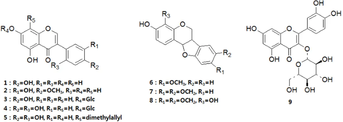
Abstract: The flowers of Maackia fauriei have traditionally been used to treat hypertension, apoplexy, hemostasis, vaginal bleeding, and dystocia; moreover, the bark of this plant has been used as a natural dye. In the present study, activity-guided isolation of the leaves of M. fauriei yielded five isoflavones [genistein (1), pratensein (2), genistin (3), 2'-hydroxygenistein-7-O-β-D-glucopyranoside (4), and 2,3-dehydrokievitone (5)]; three pterocarpans [medicarpin (6), maackiain (7), and 4-hydroxy maackiain (8)]; and one flavonol [isoquercitrin (9)]. To evaluate the anti-oxidative effects of these compounds, their 1,1-diphenyl-2-picryl-hydrazyl (DPPH) radical scavenging assays and nitrotetrazolium blue chloride (NBT) superoxide scavenging assays were measured. And the anti-tumor activity against human cancer cell lines in genital system, LNCaP, PC-3,HeLa and OVCAR-3 cells were evaluated by MTT method. Furthermore, the apoptosis of the PC-3 and HeLa cells were determined by by annexin V-FITC and PI their fluorescence was analyzed by flow cytometry. The flavonol (9, isoquercitrin) and pterocarpan (8, 4-hydroxymaackiain) showed strong anti-oxidative activities. Besides, the isoflavones (1-5) did not showed anti-oxidative activity and the isoflavones (1-5) and pterocarpans (6-8) generally showed the potent cytotoxic activity against all of four human genital cancer cells. Especially, 2,3-dehydrokievitone (5) which had a prenyl group at C-8 position of the A-ring exhibited strong cytotoxic activity and induced apoptosis efficiently in cancer cells.
Keywords: Maackia fauriei; flavonoids; pterocarpans; anti-oxidant; anti-tumor; cytotoxicity. © 2016 ACG Publications. All rights reserved.
1. Introduction
Maackia fauriei (Leguminosae) is native to Korea and endemic to Halla Mountain on Jeju
Island (Nakai T, 1952). The flowers of M. fauriei have traditionally been used to treat hypertension,
apoplexy, hemostasis, vaginal bleeding, and dystocia, and the bark of this plant has been used as a natural dye (Song JT, 1989). Several flavonoids and lectin were isolated from the stems and
heartwood of M. fauriei (Hwang MH et al., 1998; Ko SH. et al., 1999; Kim BS. et al., 2004; Kim JM.
*
et al., 2010). And the tyrosinase inhibition of isoflavonoid and cytotoxicity against cancer cells and hemagglutination activity of lectin were reported (Kim JM. et al., 2010; Kim BS. et al., 2004).
Phenolic compounds, especially flavonoids, have been increasingly investigated due to their potential benefits for the treatment and prevention of cancer and other pathological disorders (Schroeter C. et al. 2002; Rice-Evans C. 2001).
Prostate cancer (PCa) is a typically observed in men who are middle-aged or older; the probability of having a prostate tumor increases with age and an extremely common male-specific malignancy and is the most common cancer among men worldwide (Siegel R, Naishadham, D. and Jemal A. 2012). During the early state of prostate cancer, tumor growth and development depend on androgens. However, in the advanced stages of PCa, tumors develop in an androgen-independent manner and cell growth becomes unregulated (Kampa. et al., 1997). Because they share characteristics typical of the different stages of PCa progression, the LNCaP and PC-3 cell lines are widely used to study prostate cancer. The LNCaP cell line was established from a human lymph node metastatic lesion of a prostatic adenocarcinoma, whereas the PC-3 cell line was established from a human prostatic adenocarcinoma metastatic to bone. LNCaP cells express the androgen receptor and exhibit androgen-dependent proliferation; in contrast, PC-3 cells express very little to androgen receptor and are androgen-insensitive.
Gynecologic cancer which implies cancers of the female reproductive system is the fourth most common type of cancer in women, affecting approximately 1 in 20 women (Siegel, R., Naishadham, D. and Jemal A. 2012). It includes ovarian cancer, uterine cancer, vaginal cancer, cervical cancer and vulvar cancer. HeLa and OVCAR-3 are widely known as cervical cancer cell lines and ovarian cancer cell lines, respectively. HeLa cancer cell line was established from human cervical adenocarcinoma. These cells have been reported to contain human papilloma virus 18 (HPV-18) sequences. OVCAR-3 cell line was established from the malignant ascites of a patient with progressive adenocarcinoma of the ovary. These cells express both androgen and estrogen receptor.
This paper describes the isolation and the identification of isoflavonoid from the leaves of M. fauriei and evaluations of their anti-oxidative and anti-proliferative activities against human genital cancer cell lines.
2. Materials and Methods
2.1. Plant material
The leaves of M. fauriei (3.4 kg) were collected from Halla Arboretum (Jeju Island, Republic of Korea) in June 2012 and its identity was confirmed by Prof. Lee. M. W. (Pharmacognosy Lab, College of Pharmacy, Chung-Ang University) and Lee. C. I. (Kwang-Leung Korean National Arboretum in Pochoen, Korea). A voucher specimen was deposited at the herbarium of the College of Pharmacy, Chung-Ang University.
2.2. General experimental procedures
Column chromatography was performed with Sephadex LH-20 resin (10-25 μm, GE Healthcare Bio-Science AB, Uppsala, Sweden) and MCI-gel CHP 20P (75-150 μm, Mitsubishi Chemical, Tokyo, Japan). ODS-B gel (40-60 μm, Daiso, Osaka, Japan) was used as the stationary phase on a middle pressure liquid chromatography (MPLC) system equipped with an injector (Waters 650E), a detector (110 UV/VIS detector, Gilson, Middleton, WI, USA) and a pump (TBP5002, Tauto Biotech, Shanghai, China).
ratios). Spots were detected under UV radiation (254 nm) after spraying the plate with FeCl3 and either 10% H2SO4 or anisaldehyde-H2SO4, followed by heating.
Chemical structures were elucidated by several different methods. 1D NMR techniques such as 1H-(300 or 600 MHz) and 13C-(150 MHz) nuclear magnetic resonance (NMR), in addition to 2D-NMR techniques such as heteronuclear single quantum coherence (HSQC) and heteronuclear multiple bond coherence (HMBC) were recorded on a Gemini 2000 and a VNS (Varian, Palo Alto, CA, USA) at the research facilities of Chung-Ang University. Low resolution fast atom bombardment mass spectra (LRFAB-MS) were recorded with a JMSAX505WA instrument (JEOL, Tokyo, Japan) at the National Center for Inter-University Research Facilities at Seoul National University.
2.3. Extraction and Isolation
The leaves of M. fauriei (3.4 kg) were extracted with 80% acetone (40 L) at room temperature. The acetone extract was concentrated under a vacuum, yielding 270 g of extract. After acetone evaporation, the extract was partitioned by solvent fractionation with hexane, ethyl acetate, and water. Of the three solvent fractions, the ethyl acetate layer (48 g) exhibited moderate antioxidative activity and was highly cytotoxic to LNCaP prostate cancer cells. Thus, the ethyl acetate layer was subjected to column chromatography (CC) with Sephadex LH-20 resin. Fractions were eluted with an H2O:MeOH gradient (from 4:6 to 0:10), yielding 9 subfractions (MF-1 to MF-9). Of these subfractions, MF-5 to MF-8 showed antioxidative activity and were cytotoxic. Repeated CC of fraction MF-5 (4.2 g) using an MCI gel column with a gradient solvent system of H2O:MeOH (from 4:6 to 0:10) yielded three compounds, 3 (104 mg), 4 (16 mg), and 9 (116 mg). Similarly, CC of fraction MF-6 (1.5 g) was performed using MCI gel in an open column with a gradient solvent system of H2O:MeOH (from 4:6 to 0:10) and an ODS gel with a gradient solvent system of H2O:MeOH (from 6:4 to 0:10) on an MPLC system (10 mL/min). Column chromatography was followed by recrystallization, thus yielding compounds 6 (62 mg) and 8 (60 mg). Fraction MF-7 (2.1 g) was separated using an MCI gel column and an ODS gel column with an H2O:MeOH gradient (from 4:6 to 0:10), yielding compound 2 (54 mg). Further recrystallization of subfraction MF-7 yielded compound
7 (72 mg). Fraction MF-8 (1.7 g) was applied to an MCI gel in an open column with a gradient solvent system of H2O:MeOH (from 4:6 to 0:10). The subfractions of MF-8 were passed through an ODS gel with a gradient solvent system of H2O:MeOH (from 6:4 to 0:10) on an MPLC system (10 mL/min), thus yielding compounds 1 (20 mg) and 5 (27 mg).
2.3.1. Genistein: 5,7,4'-trihydroxyisoflavone (
1
).
White amorphous powder; LRFAB-MS m/z: 270 [M-H]-
1H-NMR (300 MHz, Acetone-d6 + D2O) : δ 8.11 (1H, s, H-2), 7.41 (2H, d, J=8.7 Hz, H-2', 6'), 6.87 (2H, d, J=8.7 Hz, H-3', 5'), 6.38 (1H, d, J=2.1 Hz, H-8), 6.25 (1H, d, J=2.1 Hz, H-6)
13C-NMR (150 MHz, Acetone-d6 + D2O) : δ 180.7 (C-4), 164.3 (C-7), 162.3 (C-5), 158.1 (C-9), 157.6 (C-4'), 153.3 (C-2), 130.2 (C-2', 6'), 123.1 (C-3), 122.0 (C-1'), 115.0 (C-3', 5'), 105.1 (C-10), 98.9 (C-6), 93.6 (C-8)
2.3.2. Pratensein: 5,7,3'-trihydroxy-4'-methoxy isoflavone (
2
).
White amorphous powder; LRFAB-MS m/z : 300 [M-H]-
1H-NMR (300 MHz, Acetone-d6 + D2O) : δ 8.18 (1H, s, H-2), 7.24 (1H, d, J=2.1 Hz, H-2'), 7.05 (1H, dd, J=8.1, 2.1 Hz, H-6'), 6.88 (1H, d, J=8.1Hz, H-5’), 6.40 (1H, d, J=2.1 Hz, H-8), 6.28 (1H, d, J =2.1 Hz, H-6), 3.87 (3H, s, -OCH3)
2.3.3. Genistin: genistein-7-O-
β
-D-glucopyranoside (
3
).
White amorphous powder; LRFAB-MS m/z : 432 [M-H]-
1H-NMR (300 MHz, DMSO-d6 + D2O) : δ 8.34 (1H, s, H-2), 7.39 (2H, d, J=8.7 Hz, H-2', 6'), 6.83 (2H, d, J=8.7 Hz, H-3', 5'), 6.71 (1H, d, J=2.1 Hz, H-8), 6.47 (1H, d, J=2.1 Hz, H-6), 5.05 (1H, d, J = 7.2 Hz, H-1''), 3.71 (1H, d, J = 10.2 Hz, H-6''b), 3.42 (overlapped, m, H-5'', 6''a), 3.32 (1H, t, J = 8.6 Hz, H-3''), 3.27 (1H, t, J = 8.6 Hz, H-2''), 3.17 (1H, t, J = 8.6 Hz, H-4'')
13C-NMR (150 MHz, DMSO-d6 + D2O) : δ 180.9 (C-4), 163.4 (C-7), 162.0 (C-5), 161.7 (C-9), 157.6 4'), 155.0 2), 130.6 2', 6'), 123.0 3), 121.5 1'), 115.5 3', 5'), 106.5 10), 100.3 (C-6), 100.0 (C-1''), 95.0 (C-8), 77.5 (C-5''), 76.6 (C-3''), 73.4 (C-2''), 69.9 (C-4''), 60.9 (C-6'')
2.3.4. 2'-Hydroxygenistein-7-O-
β
-D-glucopyranoside :
5,7,2',4'-tetrahydroxyisoflavone-7-O-β
-D-glucopyranoside (
4
).
White amorphous powder; LRFAB-MS m/z : 448 [M-H]-
1H-NMR (300 MHz, DMSO-d6 + D2O) : δ 8.22 (1H, s, H-2), 6.98 (1H, d, J=8.1 Hz, H-6'), 6.70 (1H, d, J=2.1 Hz, H-8), 6.46 (1H, d, J=2.1 Hz, H-6), 6.36 (1H, d, J=2.4 Hz, H-3'), 6.28 (1H, dd, J=8.4, 2.4 Hz, H-5'), 5.04 (1H, d, J = 7.2 Hz, H-1''), 3.69 (1H, d, J = 10.2 Hz, H-6''b), 3.42 (overlapped, m, H-5'', 6''a), 3.32 (1H, t, J = 8.6 Hz, H-3''), 3.27 (1H, t, J = 8.6 Hz, H-2''), 3.17 (1H, t, J = 8.6 Hz, H-4'')
13
C-NMR (150 MHz, DMSO-d6 + D2O) : δ 181.0 (C-4), 163.3 (C-7), 161.9 (C-5), 159.0 (C-4'), 157.7 (C-9), 156.7 (C-2'), 156.2 (C-2), 132.6(C-6'), 121.2 (C-3), 108.9 (C-1'), 106.7 (C-5'), 106.5 (C-10), 103.0 (C-3'), 100.3 (C-1''), 99.9 (C-6), 95.0 (C-8), 77.5 (C-5''), 76.6 (C-2''), 73.4 (C-3''), 69.9 (C-4''), 60.9 (C-6'')
2.3.5. 2,3-dehydrokievitone: 5,7,2',4'-tetrahydroxy-8-(3,3-dimethylallyl) isoflavone (
5
).
White amorphous powder; LRFAB-MS m/z : 354 [M-H]-1H-NMR (600 MHz, Acetone-d6 + D2O) : δ 8.20 (1H, s, H-2), 7.11 (1H, d, J=8.4 Hz, H-6'), 6.48 (1H, d, J=2.4 Hz, H-3'), 6.43 (1H, dd, J=8.4, 2.4 Hz, H5'), 6.41 (1H, s, H6), 5.22 (1H, t, J=7.2 Hz, H2''), 3.42 (2H, d, J=7.2 Hz, H1''), 1.78 (3H, s, -CH3), 1.64 (3H, s, -CH3)
13C-NMR (150 MHz, Acetone-d6 + D2O) : δ 181.6 (C-4), 161.76 (C-7), 159.9 (C-5), 159.1 (C-4'), 156.7 2'), 155.5 2), 155.4 9), 132.0 6'), 131.2 3''), 122.2 2''), 121.0 3), 109.8 1'), 107.1 5'), 106.4 10), 104.9 8), 103.6 3'), 98.6 6), 25.0 4''), 21.1 1''), 17.0 (C-5'')
2.3.6. Medicarpin: 3-hydroxy-9-methoxy pterocarpan (
6
).
Pale yellow needle crystals; LRFAB-MS m/z : 270 [M-H]-
1H-NMR (300 MHz, Acetone-d6 + D2O) : δ 7.29 (1H, d, J=8.4 Hz, H-1), 7.20 (1H, d, J=8.4 Hz, H-7), 6.55 (1H, dd, J=8.4, 2.4 Hz, H-2), 6.43 (1H, dd, J=8.4, 2.4 Hz, H-8), 6.37 (1H, d , J=2.4 Hz, H-10), 6.36 (1H, d , J=2.4 Hz, H-4), 5.49 (1H, d, J=6.0 Hz, H-11a), 4.24 (1H, m, H-6), 3.72 (3H, s, -OCH3), 3.57(2H, m, H-6, 6a)
13C-NMR (150 MHz, Acetone-d6 + D2O) : δ 161.1 (C-9), 160.8 (C-10a), 158.9 (C-3), 156.7 (C-4a), 132.1 1), 125.0 7), 119.6 6b), 111.7 11b), 109.7 2), 106.0 8), 103.0 4), 96.3 (C-10), 78.6 (C-11a), 66.2 (C-6), 54.9 (-OCH3), 39.5 (C-6a)
2.3.7. Maackiain: 3-hydroxy-8,9-methylendioxy pterocarpan (
7
).
Pale yellow needle crystals; LRFAB-MS m/z : 284 [M-H]-
13C-NMR (150 MHz, Acetone-d6 + D2O) : δ 158.9 (C-3), 156.7 (C-4a), 154.3 (C-10a), 147.9 (C-9), 141.5 (C-8), 132.0 (C-1), 118.6 (C-6b), 111.7 (C-11b), 109.6 (C-2), 105.0 (C-7), 102.9 (C-4), 101.2 (C-12), 93.1 (C-10), 78.5 (C-11a), 66.1 (C-6), 40.1 (C-6a)
2.3.8. 4-Hydroxymaackiain: 3,4-hydroxy-8,9-methylendioxy pterocarpan (
8
).
Pale yellow needle crystals; LRFAB-MS m/z : 300 [M-H]-
1H-NMR (300 MHz, Acetone-d6 + D2O) : δ 6.87 (1H, s, H-7), 6.83 (1H, d, J=8.4 Hz, H-1), 6.58 (1H, d, J=8.4 Hz, H-2), 6.38 (1H, s, H-10), 5.91 and 5.89 (each 1H, d, J=1.2 Hz, H-12), 5.50 (1H, d, J=6.9 Hz, H-11a), 4.30 (1H, m, H-6), 3.64 (1H, t, J=10.2 Hz, H-6), 3.55 (1H, m, H-6a)
13C-NMR (150 MHz, Acetone-d6 + D2O) : δ 154.3 (C-10a), 147.9 (C-9), 145.8 (C-3), 144.5 (C-4a), 141.5 (C-8), 133.0 (C-4), 120.9 (C-1), 118.5 (C-6b), 112.7 (C-11b), 109.3 (C-2), 105.1 (C-7), 101.2 (C-12), 93.1 (C-10), 78.8 (C-11a), 66.4 (C-6), 40.3 (C-6a)
2.3.9. Isoquercitrin: quercetin-3-O-
β
-D-glucopyranoside (
9
).
Yellow amorphous powder; LRFAB-MS m/z : 464 [M-H]-
1H-NMR (300 MHz, DMSO-d6 + D2O) : δ 7.57 (1H, d, J=2.3 Hz, H-2'), 7.57 (1H, dd, J=9.0, 2.3 Hz, H-6'), 6.85 (1H, d, J=9.0 Hz, H-5'), 6.41 (1H, d, J=2.0 Hz, H-8), 6.20 (1H, d, J=2.0 Hz, H-6), 5.45 (1H, d, J= 7.8 Hz, H-1''), 3.59-3.09 (6H in total, m, glycoside protons)
13C-NMR (150 MHz, DMSO-d6 + D2O) : δ 177.8 (C-4), 164.4 (C-7), 161.6 (C-5), 161.3 (C-9), 156.8 (C-2), 148.7 (C-4'), 145.1 (C-3'), 133.7 (C-3), 122.0 (C-6'), 121.6 (C-1'), 116.6 (C-2'), 115.6 (C-5'), 104.4 (C-10), 101.3 (C-1''), 99.0 (C-6), 93.9 (C-8), 77.9 (C-5''), 76.8 (C-3''), 74.4 (C-2''), 70.2 (C-4''), 61.3 (C-6'')
2.4. Measurement of DPPH radical scavenging activity
Anti-oxidant activity was determined by measuring the ability of each compound to scavenge the
stable DPPH free radical (Sigma, St. Louis, USA). For this assay, 20 μL of each sample (in absolute ethanol) was added to 180 μL of DPPH (0.2 mM in absolute ethanol). After mixing gently and incubating for 30 min, the optical density was measured at 540 nm using an ELISA reader (TECAN, Salzburg, Austria). The free radical scavenging activity was calculated as the inhibition rate (%) = [1-(sample O.D. / control O.D.)] × 100. The IC50 values were defined as the concentrations at which 50% of the DPPH free radicals were scavenged. L-ascorbic acid was used as a positive control.
2.5. Measurement of NBT/superoxide scavenging activity
To measure NBT reduction, reactions containing 50 mM phosphate buffer (pH 7.5), EDTA (0.05
mM), hypoxanthine (0.2 mM), 63 μL NBT (1 mM) (Sigma, St. Louis, MO, USA), 63 μL of either aqueous or ethanolic extract (or distilled water as a control), and 63 μL of xanthine oxidase (1.2 U/μL;
2.6. Cell culture
LNCaP, PC-3, HeLa, and OVCAR-3 cells were purchased from the Korean Cell Line Bank. Cells
were grown at 37 °C in a humidified atmosphere (5 % CO2) in RPMI medium (Sigma) containing 10
% fetal bovine serum and 100 IU/ml penicillin G (Gibco BRL, Grand Island, NY, USA).
2.7. MTT assay
PC-3, HeLa, and OVCAR-3 cells were seeded onto 24-well plates at a density of 105 cells/well,
whereas LNCaP cells were seeded at 104 cells/well. Cells were grown for 24 h in 5% CO2 at 37 °C
and then incubated for 48 h in medium supplemented with 0.5% FBS. Next, the medium was replaced with PBS containing 0.5 mg/ml MTT [3-(4,5-dimethylthiazol-2-yl)-2,5-diphenyltetrazolium bromide]. The supernatants were then removed after 4 h and the MTT-formazan reaction products were
dissolved in 500 μl DMSO. The extent of reduction of MTT to formazan within the cells was assessed by measuring the absorbance at 540 nm with a microplate reader (TECAN, Salzburg, Austria). Cell
viability was calculated as follows: sample O.D. / blank O.D. × 100 (%).
2.8. Flow cytometry analysis
Cells were harvested, sequentially washed in PBS and annexin V binding buffer, and resuspended in binding buffer. Cells were then stained with annexin V-FITC and propidium iodide (PI) for 15 min
in the dark at 4 °C. The resultant fluorescence was analyzed on a flow cytometer (BD-LSR II, San Jose, CA, USA) using Cell Quest 2.0 software. The percentages of cells in the upper left (necrotic cells), upper right (late apoptotic cells), lower right (early apoptotic cells), and lower left (viable cells) quadrants of the histogram were calculated for comparison.
2.9. Statistical analysis
All data are expressed as means ± SDs. Values were analyzed by one-way analysis of variance (ANOVA) followed by the Student-Newman-Keuls (S-N-K) test. Calculations were performed using the Statistical Package for the Social Sciences (SPSS) software pack; differences were considered significant for p values less than 0.05 (designated by different superscripts in the same column).
3. Results and Discussion
Figure 1. Structures of the compounds isolated from M. fauriei (1-9).
The radical scavenging activities were determined against DPPH and NBT superoxide of the M. fauriei leaf extract and the three fractions (hexane, ethyl acetate, and water) generated by solvent partitioning. And the ethyl acetate layer showed moderate anti-oxidative activity (DPPH: IC50 =
76.82±1.59 μg/mL, NBT: IC50 = 20.84±4.09 μg/mL) (Table 1-2).
Table 1. IC50 values of the extract of M. fauriei, the fractions and and isolated compounds against DPPH radicals scavenging activity.
Fraction IC50 (μg/mL) Compound IC50 (μM) Whole leaf extract >100b) 1 >100d)
Water >100b) 2 >100d) Ethyl acetate 76.82±1.59a) 3 >100d) Hexane >100b) 4 >100d) 5 >100d) 6 >100d) 7 >100d)
8 48.05±1.65c)
9 42.64±2.55b)
L-ascorbic acid 35.29±1.23a)
Values represent means ± SDs of three independent experiments. In the IC50 column, values that are not significantly different from one another are designated with the same letter, whereas values that are significantly different from one another (p < 0.05) are designated with different letters.
Table 2. IC50 values of the extract of M. fauriei, the fractions and and isolated compounds against NBT superoxide scavenging activity.
Fraction IC50(μg/mL) Compound IC50(μM)
Whole leaf extract 32.40±2.40b) 1 >100c) Water 35.89±0.83b) 2 >100c) Ethyl acetate 20.84±4.09a) 3 >100c) Hexane >100c) 4 >100c) 5 >100c) 6 >100c) 7 >100c) 8 4.55±0.21a) 9 10.40±1.25b) Allopurinol 9.90±0.05b)
The isoflavonoid (1-5) which were isolated from ethylacetate fraction did not showed anti-oxidative activity but the pterocarpan, 8 and the flavone, 9 showed potent DPPH radical scavenging
activity (IC50 = 48.05±1.65 and 42.64±2.55 μM, respectively) compared with the positive control, L
-ascorbic acid (IC50 = 35.29±1.23 μM) (Table 1).
As shown in Table 2, 4-hydroxymaackiain (8) and isoquercitrin (9) also showed potent NBT superoxide scavenging activity. In particular, 8 (IC50 = 4.55±0.21 μM) showed potent antioxidant
activity compared with the positive control, allopurinol (IC50 = 9.90±0.05 μM). The only structural
difference between 7 and 8 is the hydroxyl group at the C-4 position of the A-ring. Thus, the ortho-hydroxyl group at A-ring may help stabilize the radicals.
To determine the cytotoxic activities against four different human genital cancer cell lines, LNCaP, PC-3, HeLa and OVCAR-3 cells, MTT assays were performed with telimagrandin II and EGCG as positive controls which have been reported to inhibit proliferation of prostate cancer cells and other cancer cells (Yokoyama M. et al. 2008; Gupta S. et al. 2003; Kim MH. et al. 2013) (Table 3 and 4). The extract of M. fauriei exhibited moderate cytotoxic activity and the ethyl acetate fraction showed potent cytotoxicity against all the cancer cell lines (Table 3).
Table 3. IC50 values of extract of M. fauriei and the fractions against cancer cells.
Sample IC50(μg/mL)
LNCaP PC-3 HeLa OVCAR-3
Whole leaf extract 81.46±2.13c) 86.32±5.02c) 31.60±0.33c) 71.05±0.85c) Water >100d) >100d) >100d) >100d) Ethyl Acetate 31.08±0.06a) 23.01±0.09a) 7.82±0.19a) 19.88±0.05a)
Hexane 74.85±1.42b) 42.03±3.26b) 19.66±0.56b) 36.73±4.28b)
Values represent means ± SDs of three independent experiments. In the IC50 column, values that are not significantly different from one another are designated with the same letter, whereas values that are significantly different from one another (p < 0.05) are designated with different letters.
Table 4. IC50 values of the compounds isolated from M. fauriei against cancer cells.
Sample IC50(μM)
LNCaP PC-3 HeLa OVCAR-3 1 61.24±1.28e) 90.27±1.28d) 14.02±1.10b) 21.39±0.36d) 2 >100f) >100e) 21.45±0.22d) 22.69±0.16e) 3 >100f) >100e) 60.30±2.44f) >100i) 4 >100f) >100e) >100g) >100i) 5 20.20±0.12b) 11.06±0.09a) 8.58±0.01a) 8.92±0.03a) 6 98.8±0.43f) 85.25±1.00c) 46.28±0.70e) 49.21±0.91h) 7 >100f) >100e) 21.33±0.04d) 36.12±0.43f) 8 58.24±2.06d) 25.44±0.11b) 18.88±0.36c) 16.47±0.26c) 9 >100f) >100e) >100g) >100i) Tell II 5.93±0.26a) 27.47±3.11b) 7.35+0.71a) 12.23±0.01b) EGCG 33.30±1.62c) >100e) >100g) 38.66±0.38g)
Values represent means ± SDs of three independent experiments. Within a given column, values that are not significantly different from one another are designated with the same letter, whereas values that are significantly different from one another (p < 0.05) are designated with different letters.
2,3-Dehydrokievitone (5), which has a prenyl group at the C-8 position of the A-ring showed the most potent cytotoxicity against the cancer cell lines (Table 4).
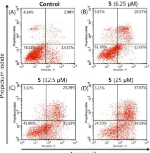
Flowcytometry analysis of 5 which showed strong cytotoxic activity was tested. With increasing concentrations of the 5, the percentages of normal cells were reduced and the percentages of cells in early and late apoptosis were increased. The result showed that 2,3-dehydrokievitone (5) induces apoptosis more effectively than necrosis (Figure 2-3).
Figure 2. Flow cytometry analysis of apoptosis as assessed by annexin V/PI staining of PC-3 cells. Cells were treated with compound 5(6.25, 12.5, or 25 μM) and incubated for 2 days in low serum (0.5% FBS) medium. The
percentages of cells in each quadrant are indicated (lower left, normal; lower right, early apoptosis; upper right, late apoptosis; upper left, necrosis).
Figure 3. Flow cytometry analysis of apoptosis as assessed by annexin V/PI staining of HeLa cells. Cells were treated with compound 5(6.25, 12.5, or 25 μM) and incubated for 2 days in low serum (0.5% FBS) medium. The
3. Conclusions
The activity-guided isolation of M.fauriei yielded five isoflavones (1-5), three pterocarpans (6-8), and one flavonol (9).
Among the phenolic compounds, 4-hydroxymaackiain (8), which has hydroxyl groups at the C-3 and C-4 positions of its A-ring, showed exceptionally strong antioxidant activity. Besides, the phenolic compounds (1-8) generally showed potent cytotoxic activity against four human genital cancer cell lines, LNCaP, PC-3, HeLa and OVCAR-3. In particular, 2,3-dehydrokievitone (5), which has a prenyl group at the C-8 position of its A-ring, exhibited strong cytotoxic activity against all cancer cell lines and efficiently induced apoptosis in PC-3 and HeLa cells.
The results of the present study suggest that the leaves of M. fauriei, and its isolated phenolic compounds are a promising source of natural products that can be developed as a new candidate for treatment of human genital cancer in the future.
Acknowledgments
This work was supported by the Basic Science Research Program through the National Research Foundation of Korea (NRF), which is funded by the Ministry of Education, Science, and Technology (2014R1A1A2056899) and was also supported by a Chung-Ang University Research Scholarship Grant in 2014.
Supporting Information
Supporting Information accompanies this paper on http://www.acgpubs.org/RNP
References
[1] T.S. Chang, H.Y. Ding, S.K. Tai and C.Y. Wu (2007). Mushroom tyrosinase inhibitory effects of isoflavones isolated from soygerm koji fermented with Aspergillus oryzae BCRC 32288, Food Chemistry105, 1430-1438.
[2] S. Gupta, T. Hussain and H. Mukhtar (2003). Molecular pathway for (− )-epigallocatechin-3-gallate-induced cell cycle arrest and apoptosis of human prostate carcinoma cells, Archives of Biochemistry and Biophysics410, 177–185.
[3] [3] N. Hasan, H. Osman, S. Mohamad, W.K. Chong, K. Awang and A.S.M. Zahariluddin (2012). The chemical components of Sesbania grandiflora root and their antituberculosis activity, Pharmaceuticals
5, 882-889.
[4] [4] T. Hatano, R. Edamatsu, M. Hiramatsu, A. Mori, Y. Fujita, T. Yasuhara, T. Yoshi-da and T. Okuda (1989). Effects of the interaction of tannins with co-exist substances. Ⅳ Effects of tannins and related polyphenols on superoxide anion radical, and on 1,1-diphenyl-2- picrylhydrazyl radical, Chemical and Pharmaceutical Bulletin37, 2016.
[5] [5] M.H. Hwang, Y.S. Kwon and C.M. Kim (1998). A new isoflavone glycoside from heartwood of
Maackia fauriei, Natural Medicines52, 527–528.
[6] [6] M. Ichige, E. Fukuda, S. Miida, J. Hattan, N. Misawa, S. Saito, T. Fujimaki, M. Imoto and K. Shindo (2013). Novel isoflavone glucosides in groundnut (Apios americana Medik) and their antiandrogenic activities, Journal of Agricultural and Food Chemistry 61, 2183−7.
[7] [7] X. Du, Y. Bai, H. Liang, Z. Wang, Y. Zhao, Q. Zhang and L. Huang (2006). Solvent effect in 1H
NMR spectra of 3′-hydroxy-4′-methoxy isoflavonoids from Astragalus membranaceus var.
mongholicus, Magnetic Resonance in Chemistry44 (7), 708-712.
[8] [8] M. Iinuma, M. Ohyama, T. Tanaka, M. Mizuno and S.K. Hong (1992). Three 2’,4’,6’-trioxygenated flavonoes in roots of Echinosophora koreensis, Phytochemistry31, 665-669.
PC3 and DU145) through a partial interaction with opioid receptors, European Journal of Pharmacology 335, 255–265.
[10] [10] B.S. Kim, K.T. Oh, D.H. Cho, Y.J. Kim, W.M. Koo, K.H. Kong and H.H. Kim (2004). A sialic acid-binding lectin from the legume Maackia fauriei: comparison with lectins from, M. Amurensis.
Plant Science 167, 1315-1321.
[11] [11] J.M. Kim, R.K. Ko, D.S. Jung, S.S. Kim and N.H. Lee (2010). Tyrosinase inhibitory constituents from the stems of Maackia fauriei, Phytotherapy Research24, 70–75.
[12] [12] M.H. Kim, S.Y. Ha, M.H. Oh, H.H. Kim, S.R. Kim and M.W. Lee (2013). Anti-oxidative and anti-proliferative activity on human prostate cancer cells lines of the phenolic compounds from Corylopsis coreana Uyeki, Molecules 18, 4876-4886.
[13] [13] S.H. Ko, Y.S. Kwon and S.H. Do (1999). Flavonoids from the heartwood of Maackia fauriei, Yakhak Hoeji43, 553–558.
[14] [14] Y.N. Lee (2006). New flora of Korea (I), Kyohaksa, Seoul, Korea. 599.
[15] [15] Y. Lu and L.Y. Foo (1997). Identification and quantification of major polyphenols in apple pomace, Food Chemistry59, 187-194.
[16] [16] X.Q. Ma, C.J. Zheng, Y. Zhang, C.L. Hu, B. Lin, X.Y. Fu, L.Y. Han, L.S. Xu, K. Rahman and L.P. Qin (2013). Antiosteoporotic flavonoids from Podocarpium podocarpum, Phytochemistry Letters 6, 118-122.
[17] [17] T. Nakai (1952). A synoptical sketch of Korean flora. Bull Natl Sci Mus Tokyo31, 1–152.
[18] [18] I. Parejo, F. Viladomat, J. Bastida, A. Rosas-Romero, N. Flerlage, J. Burillo and C. Codina (2002). Comparison between the radical scavenging activity and antioxidant activity of six distilled and nondistilled mediterranean herbs and aromatic plants, Journal of Agricultural and Food Chemistry50, 6882-6890.
[19] [19] J.A. Park, H.J. Kim, C. Jin, K.T. Lee and Y.S. Lee (2003). A new pterocarpan, (-)-Maackiain sulfate, from the roots of Sophora subprostrata, Archives of pharmacal research26, 1009-1013. [20] [20] G. Poli, G. Leonarduzzi, F. Biasi and E. Chiarpotto (2004). Oxidative stress and cell signaling,
Current Medicinal Chemistry11, 1163–1182.
[21] [21] C. Rice-Evans (2001). Flavonoid antioxidants. Current Medicinal Chemistry8, 797–807.
[22] [22] H. Schroeter, C. Boyd, J.P.E. Spencer, R.J. Williams, E. Cadenas and C. Rice-Evans (2002). MAPK signaling in neurodegeneration: influences of flavonoids and of nitric oxide, Neurobiology of Aging23, 861–880.
[23] [23] R. Siegel, D. Naishadham and A. Jemal (2012). Cancer statistics, CA: A Cancer Journal for Clinicians62, 10–29.
[24] [24] A.J.M. Silva, C.D. Netto and P.R.R. Costa (2004). The first synthesis of (±) -3,4-Dihydroxy-8,9-methylenedioxypterocarpan, an antitumoral agent and its coumestan derivative, Journal of the Brazilian Chemical Society 15, 979-981.
[25] [25] J.T. Song (1989). Handbook of Korean flora, Korea Institute of Plant Ressourcen, Seoul, Korea. p.524.
[26] [26] M. Yokoyama, M. Noguchi, Y. Nakao, M. Ysunaga, F. Yamasaki and T. Iwasaka (2008). Antiproliferative effects of the major tea polyphenol, (−)-epigallocatechingallate and retinoic acid in cervical adenocarcinoma, Gynecologic Oncology108, 326–331.
[27] [27] M. Valko, M. Izakovic, M. Mazur, C.J. Rhodes and J. Telser (2004). Role of oxygen radicals in DNA damage and cancer incidence, Molecular and Cellular Biochemistry266, 37–56.