In vivo evaluation of a new vena cava filter
Avaliação in vivo de um novo filtro de veia cava
Gilberto do Nascimento Galego1
*
, Pierre Galvagni Silveira1, Júlia Jochen Broering1, Eduardo da Silva Eli1,
Marcelo Peixer Corbellini2, Amir Antônio Martins de Oliveira Junior2
Abstract
Background: Pulmonary embolism is an important cause of cardiovascular death. Inferior vena cava ilters have been shown to be efective for prevention of this condition. Objectives: To determine the safety, performance and eicacy of a new inferior vena cava ilter in an ovine model. Methods: BKone1 ilters are self-centering with over-the-wire deployment, have three iltering regions and are made from nickel-titanium alloy. Eight of these ilters were implanted in 8 sheep. he sheep were divided into 4 groups of two animals (A and B) and the number of clots injected difered by group. Two clots were injected in group 2, four in group 3, eight in group 4 and zero clots in group 1. A animals underwent euthanasia soon after the procedure and B animals were observed for 30 days and then euthanized after a control cavography. All inferior vena cavas were processed for histological examination. Clots were prepared in a metal mold, sectioned and then radiopaque markers were inserted. Clot capture was analyzed by identifying the radiopaque marker on luoroscopy. Results: No clot migration was observed during follow-up. Control cavographies showed patent inferior vena cavas. Pathological examination indicated little inlammatory tissue response. All clots were captured in the condition with 2 clots, only one clot was missed in the group injected with 4 clots and in the condition of 8 clots, they were partly captured. Conclusions: he ilters were deployed safely. here was a reduction in eicacy as the number of blood clots increased.
Keywords: vena cava ilter; pulmonary embolism; in vivo model; ovine model.
Resumo
Contexto: Embolia pulmonar é uma importante causa de morte cardiovascular. Filtros de veia cava inferior têm se mostrado efetivos na sua prevenção. Objetivos: Determinar a segurança, o desempenho e a eicácia de um novo iltro de veia cava inferior em estudo experimental utilizando modelos ovinos. Métodos: Filtros BKone1 são autocentrantes,
over-the-wire (OTW), compostos por três regiões de iltragem e construídos em liga de níquel-titânio. Oito iltros foram implantados em oito ovelhas. As ovelhas foram divididas em quatro grupos, de acordo com o número de êmbolos injetados, com dois animais em cada grupo (A e B). Foram injetados dois êmbolos no grupo 2, quatro no grupo 3, oito no grupo 4 e nenhum êmbolo no grupo 1. Os animais denominados A foram submetidos a eutanásia logo após o procedimento e os animais B foram observados por 30 dias, sendo submetidos a eutanásia após a realização de uma cavograia de controle. Após a eutanásia, todos os animais foram submetidos a explante do segmento de veia cava inferior contendo o iltro para análise anatomopatológica. Os êmbolos foram preparados em molde metálico e seccionados, adicionando-se marcadores radiopacos. A retenção dos êmbolos foi constatada através da identiicação da marca radiopaca na seção de captura do iltro, via luoroscopia. Resultados: Não foi observada migração do iltro após o período de 30 dias. As cavograias de controle mostraram perviedade das veias cava inferior. Os resultados dos exames anatomopatológicos indicaram pouca resposta inlamatória dos tecidos. Os êmbolos foram capturados totalmente na condição com dois êmbolos, apenas um êmbolo não foi capturado no grupo com quatro êmbolos e na condição de oito êmbolos, eles foram parcialmente capturados. Conclusões: Os iltros foram entregues com segurança. Há uma queda na eicácia de captura com o aumento da quantidade de êmbolos.
Palavras-chave: iltro de veia cava; embolia pulmonar; modelo in vivo; modelo ovino.
1 Universidade Federal de Santa Catarina – UFSC, Departamento de Cirurgia, Florianópolis, SC, Brazil.
2 Universidade Federal de Santa Catarina – UFSC, Departamento de Engenharia Mecânica, Florianópolis, SC, Brazil.
Financial support: None.
Conlicts of interest: No conlicts of interest declared concerning the publication of this article. Submitted: September 10, 2015. Accepted: February 15, 2016.
INTRODUCTION
Pulmonary embolism (PE) is an important cause of cardiovascular death and is responsible for an estimated 120,000 to 150,000 deaths annually in the United States.1,2 These clots commonly arise from deep
venous thrombosis and often present in patients with risk factors such as recent surgery, hospitalization, infection or malignant neoplasm.3 Anticoagulation
remains the gold standard of care, but many patients have contraindications against this treatment or do not respond to it.4 In these cases surgical prophylaxis
with vena cava ilters5 is necessary.
Vena cava ilters (VCF) are endovascular devices
indicated for prevention of PE and death in patients for whom anticoagulation is contraindicated or ineffective.6
The search for a VCF with good clinical outcomes and
low complication rates has prompted an increase in the number of models available on the market. An ideal device would have the following characteristics: high
clot capture eficacy, non‑thrombogenic properties,
retrievability and absence of change to local venous
low. However, a perfect VCF has not yet been
developed.7
This study investigates a VCF prototype manufactured
by Biokyra Research and Development (BKone1).
The company initially designed many VCFs with
different capture areas. These different designs were evaluated in tests performed on an experimental bench using a pressurized system designed to reproduced
human hemodynamic conditions. The model with the best performance was selected on the basis
of results for capture eficacy, change in pressure
gradient with accumulation of clots and clot sizes captured (Figure 1). The ilter thus selected exhibited
the same capture eficacy as commercially available
ilters.8 The purpose of this study is to evaluate the
safety, performance and eficacy of this ilter in an
ovine model.
METHODS
Device description
The BKone1 ilter is designed for permanent placement. This ilter is made of nitinol elements and is a self‑centering, over‑the‑wire (OTW) ilter with three distinct iltering regions, a proximal region, an
intermediate region and a distal region.
The distal region is conical in shape and is made up of 0.25 mm wire rods with limited penetration.
Its primary function is to capture clots (iltering),
and its secondary function is to provide anchorage
to prevent ilter migration. When the ilter is in the
maximum diameter of 35 mm, it is 45 mm height. The intermediate region has an inverted cone shape and is connected to the proximal region, which is also conical in shape. The main function of these
regions is to centralize the ilter, but they are also
designed to retain clots that have not been captured by the distal region.
The rods are joined mechanically by deformation of a stainless steel sheet 3 mm in length, creating an
interference it at the junction between the distal and
the intermediate region, and another similar 3 mm piece joining the proximal ends of the rods, also by interference (Figure 2).
The ilter can be delivered via either femoral or jugular access. Femoral access was chosen for these tests.
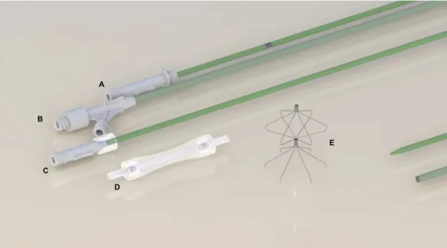
The device is delivered through a catheter introducer
sheath (12Fr internal diameter) with an introducer
catheter (600 mm), containing an hemostatic valve dilator catheter (670 mm), a pusher catheter (700mm) and a guide wire (0.035” × 1500mm).
BKone1 comes inside a transparent polycarbonate cartridge. There is a tube within the guide wire path that directs the passage of the guide wire. There is also a security lock at each end of the cartridge to hold the guide wire tube in the correct position. The position, femoral or jugular, is indicated on the cartridge (Figure 3).
Figure 2. BKone1 ilter in the upper oblique view, from the side and from below, respectively. Central picture: arrows show the
three capture regions.
Figure 3. VCF and delivery system shown from top to bottom: (A) pusher catheter; (B) catheter introducer sheath; (C) dilator
Animal groups
This study was approved by the Ethics Committee
at the Albert Einstein medical school. Eight Santa Ines sheep, each weighing between 42.5 and 75 kg and with vena cava diameters ranging from 1.3 to 4.0 cm, were distributed randomly into 4 groups (1, 2, 3 and 4), two animals per group (A and B), and the number
of clots injected after VCF placement differed by
group. Group 1 was a control group and so zero clots were injected in the animals in this group. Two clots were injected in group 2, four in group 3 and eight in group 4. Animals designated A were euthanized shortly after the procedure and animals designated B were observed for at least 30 days before euthanasia.
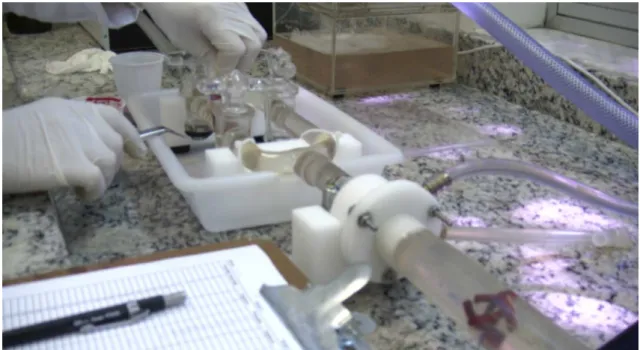
Clot preparation
Around 10‑20 ml of blood was collected by
intravenous puncture of sheep from groups 2, 3 and 4
on the day before VCF placement. The blood collected was then used to ill a metal mold built speciically
for this study and sterilized in advance. The mold has 2 parts, each 9 cm long, and was designed with furrows that create a space 5 mm in diameter when the two parts are joined together. After 24 hours, the two parts of the mold were separated (Figure 4).
Clots were removed from the mold and cut to a
standard length of 35 mm. Two pieces of platinum wire, each 5 mm long and with a diameter of 0.2 mm, were inserted into the extremities of each clot (Figure 5).
The clots were then drawn up into a 14 Fr sheath one
by one to serve as a source of emboli.
Filter placement
All the procedures were performed in a Hemodynamic Laboratory in the Training and Research Center at the
Instituto Israelita de Ensino e Pesquisa Albert Einstein, São Paulo, SP, Brazil. The anesthetic procedure was initiated by a veterinarian using intramuscular ketamine
(10 mg/kg dose) and midazolam (0.25 mg/kg). One of
the animal’s ear veins was punctured and propofol was administered (7 mg/kg) to aid positioning into a supine position, and then tracheal intubation was performed. A capnograph was connected between the endotracheal tube and the ventilation circuit. Anesthesia
was maintained using isolurane in concentrations of
2 to 4% in a closed system with oxygen and compressed
air low set to 1.5 to 2 L / min.
An orogastric tube was inserted to facilitate decompression of the rumen and a pulse oximetry probe was placed on the tongue, ear, lip or nipple for monitoring. Temperature, heart rate and respiratory rate were recorded throughout anesthesia.
After induction of anesthesia, the right common
femoral vein was identiied and punctured with the
aid of Doppler ultrasonography. Using the Seldinger technique, a 0.035 guide wire was progressed up to
the VCI and then the (12Fr) introducer system (also
developed by Biokyra Research and Development) was inserted. At this point, the veterinarian injected the animal with 1ml (50mg) of sodium heparin to prevent coagulation. A cavography was performed with injection of 20 ml of contrast (Telebrix 30 Meglunina) for anatomical study and to locate the renal veins.
The distal extremity of the sheath was positioned below the renal vein and the dilator was removed.
Next, the ilter was docked at the distal extremity of
Figure 4. Mold built speciically for this study, illed with blood.
Figure 5. Insertion of the platinum wire into one extremity of
the sheath and, with the aid of a pusher catheter, was placed and released into the infrarenal segment by a
pull‑back mechanism. After deployment, a second cavography was performed to conirm that the VCF was correctly placed and the IVC was patent.
For animals in groups 2, 3 and 4, once the VCF had been delivered, the 12 Fr system was removed and an 18 Fr introducer was inserted and positioned
in line with the iliac bone and the guide wire was
removed. The 14Fr sheath containing the clots was then introduced into this 18Fr introducer.
Clots were injected into the iliac vein with 20 ml
of 0.9% saline solution, slowly and progressively,
using direct luoroscopy visualization to monitor their trajectory. Fluoroscopies were conducted of the thoracic region in order to locate non‑captured
clots (Figure 6).
For B animals, 1ml of protamine hydrochloride
diluted in 20 ml of 0.9% saline solution was injected over a period of 1 minute in order to neutralize the heparin. These animals were brought out of anesthesia and were observed for a period of at least 30 days.
Group A animals were subjected to induction of deep anesthesia and painless death was effected by
anesthetic overdose and hypovolemia. Hypovolemia
was achieved by puncturing the femoral artery and connecting it to a vacuum system. After euthanasia,
a laparotomy was performed and an IVC segment of approximately 7cm containing the VCF was removed.
This specimen was stored in 10% formaldehyde and sent for histopathological analysis.
Group B animals were observed to evaluate their
post‑operative condition and to detect possible
complications resulting from the procedure. After
the follow‑up period, these animals were transported
to the hemodynamic laboratory and subjected to the
same anesthetic protocol as in the irst procedure. A cavography was performed to evaluate IVC patency and ilter position and compared to the cavography
from day 1.
After this procedure, the animals were euthanized
and the IVC segment containing the ilter was removed,
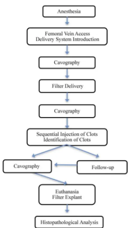
as had previously been performed with A animals. This specimen was stored in 10% formaldehyde and sent for histopathological analysis. Figure 7 summarizes the methods.
RESULTS
Delivery of the VCFs using the dedicated delivery system, developed by Biokyra Company, proved
to be feasible, with good accuracy and free from
complications or tilts. All VCFs were deployed in the
infra renal portion of the IVC, via femoral vein access, with easy navigation and luoroscopic observation.
The efficacy of clot capture, which was also
observed with luoroscopy, can be seen in Table 1.
Figure 7. In vivo test summary: Group A animals were subjected
to euthanasia after cavography and VCF delivery; Group B animals were followed for at least 30 days, subjected to a second cavography and subsequently euthanized. he VCI segment containing the VCF was sent for histopathological analysis after explantation.
Figure 6. Time luoroscopy image showing clots captured by
Group 2 capture eficacy was 100%. In group 3, the
capture rate was 100% for one animal and 75% for
the other. In group 4, capture eficacy was 50%. Clots that had not been captured by the VCF were located in pulmonary artery branches by luoroscopy. Although
clot capture was not 100% in some experiments, there were no clinical consequences for the animals during the procedure or during the observation period.
During follow‑up, which varied from 30 to 78 days,
B animals remained healthy and exhibited no
complications related to implantation of the VCF or
other adverse effects, such as bruising at the insertion site, infection or hemodynamic instability.
Cavographies were performed before IVC explantation. They showed patent IVCs in all animals
and there was no evidence of damage to the blood
vessel, device migration or VCF angle changes. No adhesion of the VCF to the vessel was observed
during histopathological analysis. The inner layer was composed of typical endothelial cells with presence of
discrete hemorrhage and there was no inlammation or tissue calciication. The VCFs had not suffered
deformation, deterioration, fracture or migration.
DISCUSSION
The process of bringing a new VCF to the market requires a well‑deined set of performance criteria
and mathematical models to identify factors affecting device performance. Procedures should be designed to accurately simulate the results of a biological event.9
Therefore, a comprehensive evaluation of the device’s behavior in both in vitro scenarios and using animal experiments is necessary.
At a time in which there are concerns about animal experiments, it is essential that in vivo and in vitro
studies be designed to obtain maximum beneit from
limited resources. An accurate in vitro evaluation of
VCF prototypes provides us with data showing that
this product is, at least, comparable to designs already on the market, limiting use of animal resources and
preventing exposure of patients to devices with low
eficacy or important adverse effects.9
The device used in this study has previously been evaluated on an experimental bench where tests were
performed in an artiicial system. The ilter selected exhibited the same capture eficacy as commercially
available ilters.8
Although the predictive value of in vitro models has
signiicantly reduced the number of animal required
for testing, certain features such as navigation, behavior upon release, thrombogenicity, endothelial reaction and migration can only be evaluated using in vivo tests. These tests allow us to judge safety and
eficacy as well as preventing premature adoption of
unsuitable products. Successful in vivo and in vitro tests are generally associated with successful clinical performance.9
Sheep are good animals to use in these experimental
tests because the diameter of the ovine VCI is similar
to the diameter in humans.10 Sheep were chosen for
this study because of this similarity and because of the high correlations between in vivo studies with these animals and clinical trial results.
The BKone1 ilter was designed to offer a high clot capture rate, to preserve IVC patency and to remain
centered. Minimal changes in metal elasticity and the length or angle of rods or their components can
signiicantly affect ilter eficacy.
The experimental method allowed us to evaluate clot retention as well as other desired parameters. Using a metal mold to make standardized clots enabled better comparison between groups. The radiopaque
markers inserted into these clots enabled luoroscopic analysis of VCF capture eficacy.
In this study, all devices were positioned accurately and correctly oriented and the animals did not have any complications related to implementation of the
VCFs. The same results have also been observed
in other studies of commercially available VCFs.1,9
Table 1. Capture eicacy and complication rates.
Group 1 2 3 4
Conditions 1A 1B 2A 2B 3A 3B 4A 4B
Number of clots injected 0 0 2 2 4 4 8 8
Number of clots captured 0 0 2 2 4 3 4 4
Eicacy - - 100% 100% 100% 75% 50% 50%
Migration No No No No No No No No
Cava perforation No No No No No No No No
Inlammatory response No No No No No No No No
The results indicate that ilter eficacy decreases with
a higher embolic load, which has also been observed in other studies.11 However, these results can not be
compared or extrapolated from one study to another because a standardized experimental protocol has
not been deined. Despite the lower capture eficacy
in groups with a higher embolic load, no sheep died
during experiments or follow‑up.
Use of a guide wire to deploy VCFs (over‑the‑wire
method) ensures correct device positioning. In vitro studies have shown that changes in angle relative to the original position are associated with loss of clots and consequently lost capture capability.12 In this
study, the devices remained centralized according to both control cavography and histopathological examination. Moreover, there was no deformation, fracture or migration, showing good material quality and good device design. Proctor et al.9 tested the
GreenieldTM vena cava ilter in ovine models and
compared it with other commercially available ilters
and reported similar results for these variables.
As was observed with the GreenieldTM VCF and
modiied Greenield ilter,9,10 histopathological analysis
of the BKone1 ilter revealed good biocompatibility and incorporation of the VCF with limited vessel
reaction, which is important for future development of a temporary device. Another important factor is
IVC patency and this was observed in all animals, conirming low device material thrombogenicity.
CONCLUSION
Overall, the experimental method used in this
study proved adequate for assessing clot retention as
well as other desired parameters. The BKone1 VCF
demonstrated ease of deployment, resistance to migration
and durability in an ovine model. Histopathological
analysis showed good biocompatibility and incorporation.
There was a reduction in eficacy with larger numbers
of clots, but no clinical repercussions were observed.
REFERENCES
1. Murphy HE, White AR, Rosenthal D, et al. Evaluation of the Crux IVC filter in an animal model. J Endovasc Ther. 2008;15(3):292-9. http://dx.doi.org/10.1583/08-2374.1. PMid:18540703.
2. Arcelus JI, Caprini JA, Monreal M, Suarez C, González-Fajardo J. The management and outcome of acute venous thromboembolism: a prospective registry including 4011 patients. J Vasc Surg. 2003;38(5):916-22. http://dx.doi.org/10.1016/S0741-5214(03)00789-4. PMid:1460319http://dx.doi.org/10.1016/S0741-5214(03)00789-4.
3. McRae S. Treatment options for venous thromboembolism: lessons learnt from clinical trials. Thromb J. 2014;12(1):27. http://dx.doi. org/10.1186/s12959-014-0027-8. PMid:25506267.
4. Kearon C, Akl EA, Comerota AJ, et al. Antithrombotic therapy for VTE disease: antithrombotic therapy and prevention of thrombosis,
9th ed: American College of Chest Physicians evidence-based Clinical practice guidelines. Chest Journal. 2012;141(2 Suppl):e419S-94S.
5. Hoppe H. Optional vena cava filters: indications, management, and results. Dtsch Arztebl Int. 2009;106(24):395-402. PMid:19623306.
6. Weichman K, Ansell JE. Inferior vena cava filters in venous thromboembolism. Prog Cardiovasc Dis. 2006;49(2):98-105. http:// dx.doi.org/10.1016/j.pcad.2006.04.002. PMid:17046435.
7. Kinney TB. Update on inferior vena cava filters. J Vasc Interv Radiol. 2003;14(4):425-40. http://dx.doi.org/10.1097/01. RVI.0000064860.87207.77. PMid:12682199.
8. Corbellini MP. Estudo experimental do desempenho de um novo filtro de veia cava inferior [dissertation]. Florianópolis: Universidade Federal de Santa Catarina; 2012.
9. Proctor MC, Cho KJ, Greenfield LJ. Development and evaluation of investigational vena caval filters: the complementary roles of in vitro and in vivo studies. J Surg Res. 2003;110(1):241-54. http:// dx.doi.org/10.1016/S0022-4804(03)00002-7. PMid:12697408.
10. Yoshida WB, Sequera J, De Abreu Maffei FH. Long-term histopathologic evaluation of inferior vena cava after modified Greenfield filter implantation. Experimental study in sheep. Int Angiol. 2004;23(2):170-6. PMid:15507896.
11. Xian ZY, Roy S, Hosaka J, Kvernebo K, Laerum F. Multiple emboli and filter function: an in vitro comparison of three vena cava filters. J Vasc Interv Radiol. 1995;6(6):887-93. http://dx.doi.org/10.1016/ S1051-0443(95)71208-8. PMid:8850665.
12. Katsamouris AA, Waltman AC, Delichatsios MA, Athanasoulis CA. Inferior vena cava filters: in vitro comparison of clot trapping and flow dynamics. Radiology. 1988;166(2):361-6. http://dx.doi. org/10.1148/radiology.166.2.3336712. PMid:3336712.
*
Correspondence
Gilberto do Nascimento Galego Rua Menino Deus, 63, Baia Sul Medical Center, Bloco C, 2º andar - Centro CEP 88020-210 - Florianópolis (SC), Brazil Tel.: +55 (48) 3322-1043 E-mail: [email protected]
Author information
GNG and PGS - PhDs, Professors of Vascular Surgery, Departamento de Cirurgia, Universidade Federal de Santa Catarina (UFSC). JJB and ESE - Medical students, Universidade Federal de Santa Catarina (UFSC). MPC - MSc, Materials Engineer, Departamento de Engenharia Mecânica, Universidade Federal de Santa Catarina (UFSC). AAMOJ - PhD, Professor of Mechanical Engineering, Departamento de Engenharia Mecânica, Universidade Federal de Santa Catarina (UFSC).
Author contributions
Conception and design: GNG, PGS, JJB, ESE, MPC, AAMOJ Analysis and interpretation: GNG, PGS, JJB, ESE, MPC, AAMOJ Data collection: GNG, PGS, JJB, ESE, MPC, AAMOJ Writing the article: GNG, PGS, JJB, ESE, MPC, AAMOJ Critical revision of the article: GNG, PGS, JJB, ESE, MPC, AAMOJ Final approval of the article*: GNG, PGS, JJB, ESE, MPC, AAMOJ Statistical analysis: GNG, PGS, JJB, ESE, MPC, AAMOJ Overall responsibility: GNG, PGS, JJB, ESE, MPC, AAMOJ