Matrix-Assisted Laser Desorption/Ionization
Time of Flight Mass-Spectrometry
(MALDI-TOF MS) Based Typing of Extended-Spectrum
β-Lactamase Producing
E
.
coli
–
A Novel Tool
for Real-Time Outbreak Investigation
Adrian Egli1,2*, Sarah Tschudin-Sutter3, Michael Oberle4, Daniel Goldenberger1, Reno Frei1, Andreas F. Widmer3
1Division of Clinical Microbiology, University Hospital Basel, Basel, Switzerland,2Infection Biology Lab, Department Biomedicine, University of Basel, Basel, Switzerland,3Division of Infectious Diseases and Hospital Epidemiology, University Hospital Basel, Basel, Switzerland,4Clinical Microbiology, Cantonal Hospital Aarau, Aarau, Switzerland
Abstract
Epidemiologically linked clusters are confirmed by typing strains with molecular typing such as pulsed-field gel electrophoresis (PFGE). We compared six extended-spectrumβ -lacta-mase producingE.coliof a PFGE-related cluster with Matrix-assisted laser desorption/ioni-zation-time of flight mass-spectrometry based typing that confirmed relatedness faster and more cost-effective, but as reliable as PFGE.
Introduction
Rapid and specific identification of strain-identity are integral components of outbreak investi-gation and subsequent implementation of effective infection control measures [1,2]. Conven-tional molecular typing methods such as pulsed-field gel electrophoresis (PFGE) or MLST-typing are labor-intensive and require up to one week from sample collection to delivery of typ-ing results [3–5]. Matrix-assisted laser desorption/ionization time of flight mass-spectrometry (MALDI-TOF MS) is able to identify bacterial species via determination of highly specific pro-tein mass-spectra profiles within minutes and has revolutionized the identification of bacteria [6]. MALDI-TOF MS has a high resolution within the mass-range of 2 to 20kDa, covering pre-dominantly ribosomal proteins demonstrating important differences at a species level [7–9]. These features may be used to rapidly assess strain relatedness. We hypothesize that (i) vari-ance at a species level may be rapidly determined by using MALDI-TOF MS and that (ii) re-sults reveal similar clustering when compared to conventional PFGE based typing. Therefore, we aimed to assess the validity of MALDI-TOF MS based typing in a pilot study in a previously OPEN ACCESS
Citation:Egli A, Tschudin-Sutter S, Oberle M, Goldenberger D, Frei R, Widmer AF (2015) Matrix-Assisted Laser Desorption/Ionization Time of Flight Mass-Spectrometry (MALDI-TOF MS) Based Typing of Extended-Spectrumβ-Lactamase ProducingE.
coli–A Novel Tool for Real-Time Outbreak Investigation. PLoS ONE 10(4): e0120624. doi:10.1371/journal.pone.0120624
Academic Editor:Paul Proost, University of Leuven, BELGIUM
Received:June 22, 2014
Accepted:February 5, 2015
Published:April 10, 2015
Copyright:© 2015 Egli et al. This is an open access article distributed under the terms of theCreative Commons Attribution License, which permits unrestricted use, distribution, and reproduction in any medium, provided the original author and source are credited.
Data Availability Statement:All MALDI-TOF raw data files are available at:figshare.com/s/ 3820a176a72f11e4906406ec4bbcf141.
published nosocomial outbreak of extended-spectrumβ-lactamase (ESBL)-producing Escheri-chia coliwith PFGE to confirm relatedness of strains [10].
Material and Methods
We re-analysed six isolates of ESBL-producingE.colicollected during an outbreak at the Uni-versity hospital Basel, Switzerland, [10] by performing MALDI-TOF MS based typing and comparing the results to those obtained by pulsed-field gel electrophoresis. Two non-related independent control ESBLE.colistrains were included.
Pulsed-field gel electrophoresis (PFGE) typing
The PFGE method has been performed as previously published [11]. Briefly, DNA restriction fragments were separated by PFGE afterXbaI digestion and dendrograms were drawn with use of the software GelCompar, version 4.5 (Applied Maths, Belgium).
MALDI-TOF MS based typing
A detailed standard operating procedure is provided inS1 Text. Briefly, all bacterial isolates were stored at−80°C. These were thawed and sub-cultivated for re-analysis at standard
condi-tions on a blood agar plate in an aerobic atmosphere at 37°C for 18h. All isolates were of same age to control for senescence-associated changes in the mass peak spectrum. Full protein ex-traction using ethanol washes and formic acid (70%) was used to increase the spectrum quality and was repeated three times independently to assess reproducibility. For each isolate, four sep-arate spectra were recorded using the Flex Control software (Bruker Daltonics, Bremen, Ger-many). The measurements were performed on Microflex MALDI-TOF (Bruker Daltonics). Voltage settings were: digitizer 1000V, detector gain voltage offset linear at 2650V, and reflec-tor at 1400V. Spectrometer ion source 1 was set at 19.98kV, source 2 at 18.08kV, and lens at 6kV. Spectra were recorded within the range of 2 to 20 kDa. Species were confirmed in com-parison with the mass-spectrum library using the MALDI Biotyper 3 software (OC 3.1, Bruker Daltonics) at standard conditions. Time for performance of MALDI-TOF MS based typing was recorded.
Bioinformatic analysis
Recorded profiles were analysed first to confirm the bacterial species using the library database including mass spectrometry profiles of 4623 pathogens. The profiles were then smoothed and baseline peak shifts were subtracted using the Biotyper 3 software. Principal component analy-sis (PCA) was used to determine clusters with similar protein expression by applying euclidean distance measures and single linkage algorithms. Lower bound was set at 3000 arbitrary units, upper bound at 15000 arbitrary units, and resolution was 2.
Flex Analysis software (Bruker Daltonics) was employed to identify single peaks of each iso-late (seeS1 Table). In addition, in an overlay of all isolates‘peak shifts’were identified between outbreak and non-related clusters. A significant peak has to be above 1000 arbitrary units and a signal-to-noise ratio of>10. Peaks between different isolates had to be separated by at least 5
m/z, but less than 50m/z, as this can reflect an unrelated peak.
Results
Species identification
All strains previously phenotypically identified by VITEK 2 were re-confirmed by MALDI--TOF MS (S1A Fig). Highly reproducible peaks were identified for each isolate by repetition of full protein extraction in three independent experiments.
High resolution clustering of outbreak and non-related isolates with
MALDI-TOF MS in comparison to PFGE based typing
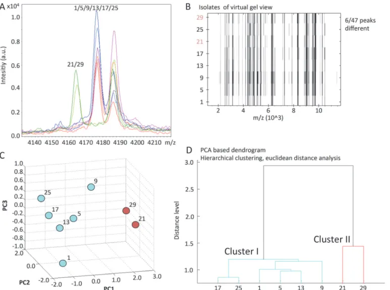
All spectra were magnified and mass peak profiles were screened to determine typical peaks of theE.coliisolates. In total 47 peaks were identified. We then screened for differences corre-sponding to changes within the mass-spectrum of an individual isolate (correcorre-sponding to changes in the amino acid sequence). Typical shifts in the mass peak spectrum observed in ESBL-producingE.coliisolates are illustrated inFig 1AandS1B Fig. Mass peaks, which were significantly shifted (>10 and<100m/z) allowing the separation of outbreak and non-related
clusters are summarized inS1 Table.A virtual gel-view of all isolates is represented inFig 1B. Principal component analysis revealed PC1 as the strongest denominator explaining the clus-tering with 70% (Fig 1C), while PC2 and PC3 explained 20% and 10% of the cluster distribu-tion, respectively.S2A-C Figsummarize the results of a two-dimensional cluster analysis. Plotting PC1 and PC2 showed a high resolution to discriminate the outbreak and non-related clusters. Similarly a PCA-based dendrogram accurately delineates clusters (Fig 1D), identifying six highly related (outbreak), and two unrelated ESBLE.coliisolates. All results from MALDI--TOF MS based typing including the detailed analysis of peak frame shifts were obtained within less than one day.
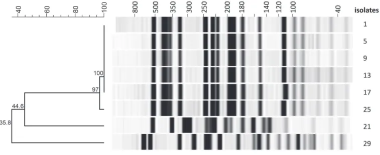
The PFGE and MALDI-TOF MS based dendrograms in direct comparison are highly simi-lar (Fig 2). In spite of divergent methodology—dice correlation analysis and PCA—each ana-lytical method reliably identified the non-related cluster.
Discussion
This MALDI-TOF MS based typing approach can reliably identify clusters of ESBLE.coli iso-lates arising during a hospital outbreak in a timely manner. In our study, we could obtain all re-sults including a detailed frame shift analysis of MS peaks in less than one day compared to one week with PFGE. Typing results (in particular the PCA-based dendrogram) become avail-able at the same time as susceptibility results, at a speed that allows adequate and efficient im-plementation of infection control measures.
Key steps for successful MALDI-TOF MS based typing are the same age of bacterial subcul-tures, a full protein extraction protocol, and inclusion of non-related isolates. Subculture of the isolates requires overnight incubation, however the full protein extraction and data analysis using the algorithms presented can be accomplished within three hours. All methods are pro-vided in full detail in the supplementary material.
Our study has a number of limitations. First, this was a small outbreak focusing on one pathogen, which may be more conducive to taking a MALDI-TOF based approach. However, even with more samples the processing time is significantly shorter in comparison to PFGE. An important second limitation is that, the small sample size may limit generalizability of our results to other settings. However, we would like to point out that the results were highly repro-ducible in three independent experiments. Nevertheless, further studies comparing PFGE and MALDI-TOF on larger sample sizes are needed [14]. To the best of our knowledge, no software is currently available, which would allow the direct comparison between MALDI-TOF (protein peak data) and PFGE (DNA restriction enzyme pattern) typing data.
Overall, our pilot study highlights an impressive time gain in identifying similarities and dif-ferences between ESBLE.colistrains. This novel approach to outbreak investigation may allow
Fig 1. MALDI-TOF MS based typing. A. Representative example of mass spectrum differences between ESBLE.coli.Isolates are indicated by color code. Isolates 1, 5, 9, 13, 17 and 25 (outbreak cluster) show a clear distinguished peak at position 4175 m/z, whereas isolates 21 and 29 show a shift to position 4165 m/z (non-related cluster). This corresponds to change of about 10Da. Various other areas of peak shifts have been identified between outbreak and non-related ESBLE.coliclusters.B. MALDI-TOF MS based virtual gel view.MS generated peaks of the protein expression profile are shown for every bacterial isolate. Red labels highlight the non-related isolates.C. Three-dimensional principal component analysis.Based on the Euclidean distance analysis algorithm a three dimensional plot was generated. PC1 and PC2 showed the highest discriminatory potential and indicated a cluster for the outbreak associated isolates, whereas the non-related isolates are less similar.D. PCA-based dendrogram generated by MALDI-TOF MS.The PCA based dendrogram uses a hierarchical clustering algorithm with a Euclidean distance analysis.
real-time typing and revolutionize outbreak investigations if further larger studies are able to confirm our results. Definitions on what constitutes a significant peak change in MALDI-TOF MS for typing should be approached.
Supporting Information
S1 Fig. A. Identification ofE. colicompared to reference library spectra.Blue lines show MS
peaks of an individual isolate. Red lines indicate the reference peaks.B. Representative exam-ple of mass spectrum differences between ESBLE.coli.Isolates are indicated by color code.
Isolates 1, 5, 9, 13, 17 and 25 (outbreak cluster) show a clear distinguished peak at position 4859 m/z, whereas isolates 21 and 29 show a shift to position 4872 m/z (non-related cluster). This corresponds to change of about 13Da.
(TIF)
S2 Fig. (A-C) Two-dimensional principal component analysis (PCA) of the protein expres-sion profiles.The influence on the data distribution by PC1, PC2 and PC3 is 70%, 20%, and 10% respectively.
(TIF)
S1 Text. A detailed step-by-step protocol is provided for performing MALDI-TOF based typing.
(DOCX)
S1 Table. List of all identified peaks.
(DOCX)
Acknowledgments
We wish to thank Clarisse Straub, Magdalena Schneider, Elisabeth Schultheiss, and Sabrina Stammler (University Hospital Basel, Switzerland) for excellent technical assistance with the
Fig 2. PFGE based typing with gel-view and dendrogram.Dice analysis indicates a very high relatedness in the outbreak-associated cluster. Isolates 21 and 29 are clearly separated.
validation of the typing protocol. We thank Dr. Daire O’Shea (University of Alberta, Canada) for critically reading the manuscript.
Author Contributions
Conceived and designed the experiments: AE ST DG RF. Performed the experiments: AE. Ana-lyzed the data: AE ST MO. Contributed reagents/materials/analysis tools: RF AW. Wrote the paper: AE ST MO DG RF AW.
References
1. Eppinger M, Mammel MK, Leclerc JE, Ravel J, Cebula TA. Genomic anatomy of Escherichia coli O157: H7 outbreaks. Proc Natl Acad Sci U S A. 2011 Dec 13; 108(50):20142–7. PMID:22135463. Pubmed
Central PMCID: 3250189. doi:10.1073/pnas.1107176108
2. Rasko DA, Webster DR, Sahl JW, Bashir A, Boisen N, Scheutz F, et al. Origins of the E. coli strain caus-ing an outbreak of hemolytic-uremic syndrome in Germany. N Engl J Med. 2011 Aug 25; 365(8):709–
17. PMID:21793740. Pubmed Central PMCID: 3168948. doi:10.1056/NEJMoa1106920
3. Foley SL, Lynne AM, Nayak R. Molecular typing methodologies for microbial source tracking and epide-miological investigations of Gram-negative bacterial foodborne pathogens. Infection, genetics and evo-lution: journal of molecular epidemiology and evolutionary genetics in infectious diseases. 2009 Jul; 9 (4):430–40. PMID:19460308. doi:10.1016/j.meegid.2009.03.004
4. Woodford N, Turton JF, Livermore DM. Multiresistant Gram-negative bacteria: the role of high-risk clones in the dissemination of antibiotic resistance. FEMS microbiology reviews. 2011 Sep; 35(5):736–
55. PMID:21303394. doi:10.1111/j.1574-6976.2011.00268.x
5. Goering RV. Pulsed field gel electrophoresis: a review of application and interpretation in the molecular epidemiology of infectious disease. Infection, genetics and evolution: journal of molecular epidemiology and evolutionary genetics in infectious diseases. 2010 Oct; 10(7):866–75. PMID:20692376. doi:10.
1016/j.meegid.2010.07.023
6. Bizzini A, Greub G. Matrix-assisted laser desorption ionization time-of-flight mass spectrometry, a revo-lution in clinical microbial identification. Clinical microbiology and infection: the official publication of the European Society of Clinical Microbiology and Infectious Diseases. 2010 Nov; 16(11):1614–9. PMID:
20636422.
7. Josten M, Reif M, Szekat C, Al-Sabti N, Roemer T, Sparbier K, et al. Analysis of the matrix-assisted laser desorption ionization-time of flight mass spectrum of Staphylococcus aureus identifies mutations that allow differentiation of the main clonal lineages. Journal of clinical microbiology. 2013 Jun; 51 (6):1809–17. PMID:23554199. Pubmed Central PMCID: 3716067. doi:10.1128/JCM.00518-13
8. Suarez S, Ferroni A, Lotz A, Jolley KA, Guerin P, Leto J, et al. Ribosomal proteins as biomarkers for bacterial identification by mass spectrometry in the clinical microbiology laboratory. Journal of microbio-logical methods. 2013 Sep; 94(3):390–6. PMID:23916798. doi:10.1016/j.mimet.2013.07.021
9. Xiao D, Zhang H, He L, Peng X, Wang Y, Xue G, et al. High natural variability bacteria identification and typing: Helicobacter pylori analysis based on peptide mass fingerprinting. Journal of proteomics. 2014 Feb 26; 98:112–22. PMID:24382553. doi:10.1016/j.jprot.2013.11.021
10. Tschudin-Sutter S, Frei R, Battegay M, Hoesli I, Widmer AF. Extended spectrum ss-lactamase-produc-ing Escherichia coli in neonatal care unit. Emergss-lactamase-produc-ing infectious diseases. 2010 Nov; 16(11):1758–60.
PMID:21029537. Pubmed Central PMCID: 3294509. doi:10.3201/eid1611.100366
11. Stranden A, Frei R, Widmer AF. Molecular typing of methicillin-resistant Staphylococcus aureus: can PCR replace pulsed-field gel electrophoresis? Journal of clinical microbiology. 2003 Jul; 41(7):3181–6.
PMID:12843061. Pubmed Central PMCID: 165370.
12. Tenover FC, Arbeit RD, Goering RV, Mickelsen PA, Murray BE, Persing DH, et al. Interpreting chromo-somal DNA restriction patterns produced by pulsed-field gel electrophoresis: criteria for bacterial strain typing. Journal of clinical microbiology. 1995 Sep; 33(9):2233–9. PMID:7494007. Pubmed Central
PMCID: 228385.
13. Lasch P, Fleige C, Stammler M, Layer F, Nubel U, Witte W, et al. Insufficient discriminatory power of MALDI-TOF mass spectrometry for typing of Enterococcus faecium and Staphylococcus aureus iso-lates. Journal of microbiological methods. 2014 May; 100:58–69. PMID:24614010. doi:10.1016/j.
mimet.2014.02.015