Mechlorethamine DNA Crosslink at a Cytosine-Cytosine
Mismatch Pair
Pornchai Rojsitthisak1*, Nutthapon Jongaroonngamsang1, Rebecca M. Romero2, Ian S. Haworth2
1Department of Food and Pharmaceutical Chemistry, Faculty of Pharmaceutical Sciences, Chulalongkorn University, Bangkok, Thailand,2Department of Pharmacology and Pharmaceutical Sciences, University of Southern California, Los Angeles, California, United States of America
Abstract
Background:Mechlorethamine [ClCH2CH2N(CH3)CH2CH2Cl], a nitrogen mustard alkylating agent, has been proven to form a
DNA interstrand crosslink at a cytosine-cytosine (C-C) mismatch pair using gel electrophoresis. However, the atomic connectivity of this unusual crosslink is unknown.
Methodology/Principal Findings: HPLC-UV, MALDI-TOF-MS, and ESI-MS/MS were used to determine the atomic connectivity of the DNA C-C crosslink formed by mechlorethamine, MALDI-TOF-MS of the HPLC-purified reaction product of mechlorethamine with the DNA duplex d[CTCACACCGTGGTTC]Nd[GAACCACCGTGTGAG] (underlined bases are a C-C
mismatch pair) indicated formation of an interstrand crosslink at m/z 9222.088 [M22H+Na]+. Following enzymatic digestion of the crosslinked duplex by snake venom phosphodiesterase and calf intestinal phosphatase, ESI-MS/MS indicated the presence of dC-mech-dC [mech = CH2CH2N(CH3)CH2CH2] at m/z 269.2 [M]2+(expected m/z 269.6, exact mass 539.27) and its
hydrolytic product dC-mech-OH at m/z 329.6 [M]+(expected m/z 329.2). Fragmentation of dC-mech-dC gave product ions at
m/z 294.3 and 236.9 [M]+, which are both due to loss of the 4-amino group of cytosine (as ammonia), in addition to dC and
dC+HN(CH3)CH = CH2, respectively. The presence of m/z 269.2 [M]2+and loss of ammonia exclude crosslink formation at
cytosine N4or O2and indicate crosslinking through cytosine N3with formation of two quaternary ammonium ions.
Conclusions:Our results provide an important addition to the literature, as the first example of the use of HPLC and MS for analysis of a DNA adduct at the N3position of cytosine.
Citation:Rojsitthisak P, Jongaroonngamsang N, Romero RM, Haworth IS (2011) HPLC-UV, MALDI-TOF-MS and ESI-MS/MS Analysis of the Mechlorethamine DNA Crosslink at a Cytosine-Cytosine Mismatch Pair. PLoS ONE 6(6): e20745. doi:10.1371/journal.pone.0020745
Editor:Sue Cotterill, St. Georges University of London, United Kingdom
ReceivedNovember 1, 2010;AcceptedMay 12, 2011;PublishedJune 6, 2011
Copyright:ß2011 Rojsitthisak et al. This is an open-access article distributed under the terms of the Creative Commons Attribution License, which permits
unrestricted use, distribution, and reproduction in any medium, provided the original author and source are credited.
Funding:The authors have no support or funding to report.
Competing Interests:The authors have declared that no competing interests exist. * E-mail: [email protected]
Introduction
DNA damage and mutation can have major effects on genetic information that may alter the function of essential proteins and cause disease. Mismatching of paired bases is a common type of DNA damage that can result in harmful mutations and a specific DNA mismatch repair mechanism is required for this damage [1]. Sources of mismatch base pairs include replication errors due to direct misincorporation of bases, lesions in the parent strand, and formation of a heteroduplex between two homologous DNA molecules during recombination [2–4]. Mismatches may also be generated in hairpins formed by trinucleotide repeat sequences [5–7]. Mismatch base pairs cause thermodynamic instability of DNA duplexes [8], but most retain an intrahelical conformation with one or more hydrogen bonds between the bases: examples include A-A [9], G-G [9], A-C [10], G-T [11], C-T [12] and C-C [12–14] pairs. Establishment of DNA structures containing mismatch base pairs is important for understanding their involvement in replication, repair, and recombination.
The antiparallel C-C pair is one of the least stable mismatch pairs [8]. This instability causes the C-C pair to adopt different conformations, including both intra- and extrahelical positions
[12–18]. Gao et al. first showed that a C-C mismatch pair could adopt an extrahelical conformation in a DNA duplex [15], whereas Boulard et al. described an intrahelical C-C mismatch pair with a single hydrogen bond, but also noted the flexibility and apparent pH dependence of the conformation [12]. In the d[CCG]ntriplet repeat
hairpin, a C-C mismatch pair is present as every third base pair of the stem [16–18]. In such hairpins and equivalent duplexes, the C-C mismatch seems to be mainly intrahelical and hydrogen bonded [13,14]. However, the d[CCG]15 hairpin shows pH-dependent
electrophoretic mobility, which led to the proposal that multiple C-C mismatch pairs in this hairpin might partly adopt extrahelical locations [18]. The structure of this hairpin may also be of interest from a disease perspective, since the d[CCG]n triplet repeat
sequence is present in the d[CGG]n?d[CCG]ngenomic region that
is expanded in Fragile X syndrome [6,7]. It has been suggested that both strands of this sequence form hairpins, and that these hairpins are important in the mechanism of repeat expansion and consequently in development of the disease [13–21].
The dynamic properties of d[CCG]nhairpins and their potential
[ClCH2CH2N(CH3)CH2CH2Cl], a nitrogen mustard alkylating
agent, is able to form a DNA interstrand crosslink at a C-C mismatch pair [19–21], with this crosslink forming in preference to the better known 1,3 G-G interstrand crosslink of mechlorethamine [22,23]. We proposed that the mechlorethamine C-C crosslink forms in the DNA minor groove through the N3of cytosine [19], but at the time we were unable to prove the atomic connectivity of the crosslink.
High performance liquid chromatography (HPLC) has been used widely for detection and purification of DNA adducts and crosslinks [22–26], and characterization of DNA adducts and crosslinks has been achieved using HPLC coupled with mass spectrometry (MS) [27–43]. Several of these studies have used MALDI-TOF-MS for determination of the molecular weight of the crosslinked duplex, followed by enzymatic digestion to obtain a fragment containing the specific crosslinked nucleosides for comparison with a synthetic standard [31–33]. Noll et al. used this approach for a duplex containing a synthetic N4dC-ethyl-N4dC crosslink [31] and these techniques have similarly been applied for N3dT-alkyl-N3dT and O6dG-heptyl-O6dG crosslinks [32,33].
In ESI-MS/MS analysis using the product ion scan mode, a molecular ion detected from the initial electrospray ionization is fragmented by collision-induced dissociation (CID)-MS [28–30]. In this mode, molecular ions with m/z corresponding to the expected molecular weight of a crosslinked DNA species can be selected for fragmentation. The resulting product ions are then used to interpret the structure of the molecular ion [34,35]. ESI-MS/MS has been used to determine the atomic connectivity of crosslinks formed by mechlorethamine at A and G [34] and to study G-G crosslinking by 1,2,3,4-diepoxybutane [36,37] and 1, 3-butadiene [38]. In this study, we used HPLC purification,
MALDI-TOF-MS, enzymatic digestion and ESI-MS/MS to determine the connectivity of the DNA C-C crosslink formed by mechlorethamine.
Results
Characterization of the mechlorethamine-crosslinked DNA duplex by HPLC and MALDI-TOF-MS
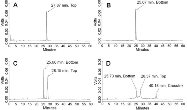
HPLC chromatograms of top- and bottom-strand DNA gave single peaks at 27.87 and 25.07 min, respectively (Figures 1A & 1B). The chromatogram of the annealed duplex gave two peaks at 25.60 and 28.15 min (Figure 1C) due to denaturation of the duplex on the column. The chromatogram of the crosslinked DNA (Figure 1D) showed three peaks at retention times of 25.73, 28.37 and 40.18 min. The first two of these peaks correspond to bottom-strand and top-bottom-strand DNA, respectively. The third peak was tentatively assigned to the mechlorethamine-DNA crosslinked duplex and comprised 25% of the total peak area.
Following purification and desalting of the eluent collected at 40.18 min, the eluted sample was analyzed by MALDI-TOF-MS and a signal with [M22H+Na]+
m/z 9222.088 (expected [M22H+Na]+
m/z 9214.2) was detected (Figure 2). The difference between the observed and expected m/z was about 7.9 amu, giving a mass difference of 0.08% or a mass accuracy of 99.92%. Mass-accuracy measurements of large oligonucleotides can have errors of 0.3% [44–45] due to the instability of mixed-base oligodeoxynu-cleotides and metal ion contamination [30,46–48]. DNA analysis by MALDI-TOF-MS in the presence of metal contaminants such as sodium and potassium may show degraded performance including peak broadening and reduced mass resolution, sensitivity and accuracy due to interacion between cations and the negatively
Figure 1. HPLC-UV chromatograms of DNA and the mechlorethamine-crosslinked DNA duplex. A:Top-strand DNA.B:Bottom-strand DNA.C:DNA duplex (denatured on the column).D:The mechlorethamine-crosslinked DNA duplex.
charged sugar-phosphate backbone [30]. Therefore, the broad signal and the slight discrepancy between the observed and expected m/z may be due to interaction of cations with the DNA. The cations could be contaminants from salts and buffers used in the preparation and purification of the crosslinked duplexes.
Enzymatic digestion of the mechlorethamine-crosslinked DNA duplex
HPLC chromatograms of the SVPD and CIP-digested products of poly-dC, poly-dG, poly-dT and poly-dA showed peaks at 3.83,
8.15, 9.32 and 11.13 min (Figure 3A). These corresponded to the standard monodeoxynucleosides dC, dG, dT and dA, which had retention times of 3.81, 8.18, 9.25 and 11.08 min, respectively (Figure 3B). Enzymatic digestion of top-strand DNA, bottom-strand DNA and the DNA duplex gave similar chromatograms with four peaks at retention times of about 3.8, 8.3, 9.3 and 11.2 min (Figure 3C–E). These chromatograms also showed an additional peak at a retention time of 7.9 min, which is a deoxyinosine peak resulting from deamination of dA due to the contamination of adenine deaminase in the snake venom phosphodiesterase, as previously reported by Wilds et al [33]. This conclusion was also supported by the observation that this peak appeared only on the chromatogram of the SVPD and CIP-digested products of poly-dA, but not for poly-dC, poly-dG and poly-dT (Figure 3A). The deoxyinosine peak and the four mononucleoside peaks were also observed after digestion of the mechlorethamine-crosslinked duplex, together with two additional peaks at retention times of 10.72 and 11.45 min (Figure 3F).
ESI-MS/MS analysis of the atomic connectivity of the mechlorethamine C-C crosslink
The products of digestion of the mechlorethamine-crosslinked duplex were analyzed by ESI-MS/MS. Molecular ions with m/z corresponding to possible crosslink species were selected for fragmentation. In this process, mechlorethamine crosslinks forming through the O2, N3 and N4 atoms of cytosine were considered (Figure 4). The O2dC-mech-O2dC and N4
dC-mech-N4dC crosslinks [mech = CH2CH2N(CH3)CH2CH2] are neutral
and should give a [M+H]+
molecular ion of m/z 538.2. In contrast, the N3dC-mech-N3dC crosslink has a double positive charge (Figure 4) and should give a [M]2+ molecular ion with
m/z 269.6.
No signals were present in a range of m/z 53865, but a signal with m/z 269.2 was detected. Fragmentation of [M]2+
m/z 269.2 gave product ions with m/z 177.1, 189.5, 209.4, 236.9 and 294.3
Figure 2. MALDI-TOF-MS spectrum of the mechlorethamine-crosslinked DNA duplex.
doi:10.1371/journal.pone.0020745.g002
Figure 3. HPLC-UV chromatograms of monodeoxynucleosides. A: SVPD and CIP-digested poly-dC, poly-dG, poly-dT and poly-dA. B: Standard dC, dG, dT and dA.C:SVPD- and CIP-digested products of top-strand DNA.D:SVPD- and CIP-digested products of bottom-strand DNA.E: SVPD- and CIP-digested products of DNA duplex.F:SVPD- and CIP-digested products of the mechlorethamine-crosslinked DNA duplex.
(Figure 5). The ion at m/z 294.3 occurs due to loss of deoxycytidine (dC) and ammonia [M2(dC+NH3)]+. Loss of dC
and ammonia combined with loss of HN(CH3)CH = CH2results
in the ion at m/z 236.9 [M2(dC+NH3+HN(CH3)CH = CH2)]+.
Loss of dC, ammonia and deoxyribose gives the ion at m/z 177.1 [M2(dC+NH3+deoxyribose)]+. The ion at m/z 177.1 can also be
formed through homolytic cleavage of the glycosidic bond in the ion at m/z 294.3.
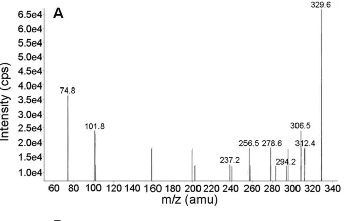
Based on possible hydrolysis of the N3dC-mech-N3dC crosslink, we also considered the presence of N3dC-mech-OH (Figure 6) in the ESI-MS/MS analysis. This species has an expected m/z of 329.2, and a signal at m/z 329.6 was detected. Fragmentation of [M]+
m/z 329.6 gave product ions with m/z 312.4 [M2NH3]+, m/z 294.2
[M2(NH3+H2O)]+, m/z 306.5 [M2(H2O+CH2= CH2)+Na)]+,
and m/z 237.2 [M2(NH3+H2O+HN(CH3)CH = CH2)]+. The
product ions with m/z 237.2 and 294.2 are identical to the respective ions with m/z 236.9 and 294.3 obtained in fragmentation of the N3dC-mech-N3dC molecular ion. Therefore, the presence of N3dC-mech-OH and its fragmentation pattern supports the con-clusion that the dC-mech-dC crosslink forms through cytosine N3.
Discussion
The key results in the ESI-MS/MS analysis of the mechloreth-amine-crosslinked DNA were the appearance of a doubly charged molecular ion and fragmentation of this ion with neutral loss of ammonia, consistent with the presence of an unreacted exocyclic amino group at N4. This fragmentation is in contrast to that observed for benzo[a]pyrene-7,8-dihydrodiol-9,10-epoxide DNA adducts formed at amino groups on guanine, cytosine and adenine,
which gave no product ions with loss of ammonia [35]. Our observed loss of ammonia is also inconsistent with the imine that would be formed at N4 following crosslinking through O2. The absence of a molecular ion at m/z 538 further indicates that the mechlorethamine C-C crosslink does not form through N4or O2. In addition, the heterolytic cleavage to give [dC2CH = CH2]+m/z
236.9 is consistent with similar cleavage of mechlorethamine crosslinks from guanine to adenine [34]. Collectively, these results show that mechlorethamine crosslinks a C-C mismatch pair in a DNA duplex through cytosine N3.
The absence or presence of loss of ammonia (17 mass units) in fragmentation of the ESI-MS/MS product ion has been used in several previous studies as evidence for base modification at a ring N atom (N3 in cytosine) or an exocyclic amino group (N4 in cytosine). Loss of ammonia is taken to indicate that the original base had an intact amino group, and thus the adduct formed at the ring N. On the other hand, the absence of loss of ammonia suggests that the amino group was modified in the adduct. Singh et al. showed that ethylation of guanine occurred at the ring N7atom and not at exocyclic N2 based on observation of the loss of ammonia from ethylguanine [41]. Similarly, Chao et al. showed that methylation and ethylation of guanine occurred at the ring N7 atom [42]. In contrast, Gaskell et al. showed that the reaction of benzo[a]pyrene-7,8-dihydrodiol-9,10-epoxide (B[a]PDE) with 29-deoxynucleoside-39-monophosphates occurred at the exocyclic amino group of each base, based on the observation of no loss of ammonia in MS fragmentation of the reaction products [35]. These findings should also be viewed in the context of the results obtained by Cao and Wang for fragmentation of the protonated ions of 29-deoxycytidine and 5-substituted 29-deoxycytidine selectively labeled with15N atoms [43]. Substitution at position 5 influenced the fragmentation pattern and ammonia was lost from either the exocyclic N4 or ring N3 due to exchange of nitrogen between these positions [43]. These results suggest that care is required with identification of modifications on N4or N3of cytosine by MS/MS. This is an important caveat in the current work, but we note that the nitrogen exchange occurred with cytosine modified at position 5 and without modifications on the exocyclic N4or ring N3atoms [43]. Loss of ammonia from a cyto-sine base with modification at N3gave fragmented ions containing a heteroaryne skeleton. The presence of six-membered rings with a triple bond such as heteroarynes and arynes (e.g. benzyne) is commonly found in ESI-MS/MS fragmentation [49–52].
It is possible that the crosslink formed through N4or O2could undergo spontaneous double protonation in the mass spectrom-eter to form a doubly charged molecular ion. However, it is likely that the formation of a molecular ion with a single charge would occur more easily than that with a double charge, and the MS spectra should then show both [M+H]+
and [M+2H]2+
signals at m/z 538.2 and 269.6, respectively. However, the presence of an ion close to m/z 269.6 and the absence of an ion close to m/z 538.2 make crosslink formation through N4or O2 unlikely. On this basis, we also exclude a possible asymmetric mechloreth-amine crosslink formed through N3and O2, N3and N4or N4and O2. In addition, the positive ion ESI-MS/MS product ion scan mode did not detect any m/z units that corresponded to other possible mechlorethamine-crosslinked dinucleosides, such as dG-mech-dG ([M+H]+
, m/z 619.28; [M]2+
, m/z 309.6) and dA-mech-dA ([M+H]+
, m/z 587.29; [M]2+
, m/z 293.64). This is due to the lack of suitable sites for these crosslinking reactions in our designed duplex. For example, the duplex lacks a 1,3 G-G interstrand crosslinking site, which is a favorable site for the mechlorethamine crosslinking reaction [22,23]. The signal at
m/z 329.6 was assigned to N3dC-mech-OH, which may have
Figure 4. Structures of possible mechlorethamine crosslinks at a cytosine-cytosine mismatch pair. A:A mechlorethamine crosslink through O2of cytosine.B:A mechlorethamine crosslink through N3of cytosine.C:A mechlorethamine crosslink through N4of cytosine. Note that the O2and N4crosslinks are neutral species that form with loss of 2H+
from the nucleosides, whereas the N3 crosslink has a double positive charge.
formed by aqueous hydrolysis of the N3C-mech-N3dC crosslink either during or after enzymatic digestion of the crosslinked duplex.
In the HPLC purification of the crosslink, use of a column temperature of 33uC and a slow increase in the amount of acetonitrile over 60 min was able to resolve the dC-mech-dC crosslink from single-stranded DNA. These chromatographic conditions were optimized in an earlier study [53]. The conditions are also sufficient to denature the DNA duplex because the presence of the C-C mismatch pair results in a low melting temperature (Tm). This is advantageous in the HPLC separation, since the duplex peak could have a similar retention time to that of the crosslink peak. The percentage of mechlorethamine-cross-linked C-C mismatch DNA detected by this method (about 25%) was in good agreement with our previous results for this reaction based on detection by gel electrophoresis [20,21].
Enzymatic hydrolysis of the mechlorethamine-crosslinked DNA by SVPD and CIP was monitored using HPLC coupled with UV detection. The use of HPLC-UV for monitoring the progress of enzymatic digestion is useful for optimization of the reaction conditions (e.g. reaction time and temperature), including the suitable amount of SVPD and CIP. The crosslinked DNA was degraded to single nucleosides within 48 h, based on the similarity of the chromatogram with those for control sequences and the free DNA duplex, suggesting the complete hydrolysis of crosslinked DNA. The appearance of additional peaks on the chromatogram of the digested mixture indicated the presence of reaction products of mechlorethamine and DNA duplex. The HPLC chromatogram for the enzymatic digestion mixture of the crosslinked duplex showed two new peaks at 10.72 and 11.45 min. However, we detected only one crosslink species that gave an ion of m/z 269.2, corresponding to N3dC-mech-N3dC. Therefore, we are currently unable to define which of the peaks at 10.72 or 11.45 min corresponds to the ion of m/z 269.2.
Applications of mass spectrometry to analysis of DNA adducts are becoming more common. Enzymatic digestion coupled with HPLC-ESI-MS/MS analysis provides a basis for quantitative detection and characterization of small amounts of DNA adducts in vitro and in vivo [38]. Synthesis of an authentic standard for comparison of the fragmentation pattern is also useful. We have attempted synthesis of the N3dC-mech-N3dC crosslink for this purpose and for spectroscopic analysis, but formation of the species has proven to be difficult. This may be due to the requirement for multiple reactions (i.e. the need for reactions of two cytosine bases with one mechlorethamine) and the rapid hydrolysis of mechlorethamine. In addition, small-molecule synthetic methods may not appropriately mimic oligonucleotide secondary structure. Some nucleophilic groups in oligonucleotide bases may be hydrogen bonded, making them less available for reaction in comparison with individual bases with less hydrogen bonding. This may be a common problem for other adducts, and the growing number of DNA crosslinks for which the fragmen-tation pattern has been established should improve the reliability of future analyses of unknown adducts. Therefore, the fragmen-tation pattern of the mechlorethamine C-C crosslink reported here provides an important addition to the literature, as the first
example of ESI-MS/MS analysis of a DNA adduct at the N3
position of cytosine.
Materials and Methods
Chemicals
Synthetic 15-mer oligodeoxynucleotides were purchased from Sigma-Proligo (St. Louis, MO). Mechlorethamine hydrochloride and monodeoxynucleosides (dA, dT, dG and dC) were also purchased from Sigma. Snake venom phosphodiesterase (SVPD,
fromCrotalus adamanteus) and calf intestinal phosphatase (CIP) were
purchased from Sigma and Finnzyme (Espoo, Finland), respec-tively. Other reagents were of AR or HPLC grade.
Preparation of the mechlorethamine-DNA crosslink
A DNA duplex solution (10mM) in 0.1 M sodium chloride and 0.05 M Tris buffer pH 7.5 was prepared using stock solutions of
d[CTCACACCGTGGTTC] (top strand, 100mM, MW = 4505)
and d[GAACCACCGTGTGAG] (bottom strand, 100mM,
MW = 4603). The solution was heated at 90uC for 2 min and then slowly cooled to room temperature to allow annealing of the DNA to form a duplex (MW = 9108) containing a C-C mismatch pair (the underlined bases in the two strands form this pair). The DNA duplex was also designed to avoid the presence of a site for formation of a mechlorethamine 1,3 G-G crosslink [22,23]. The duplex solution (100ml) was then incubated with 0.1 M mechlor-ethamine in dimethylsulfoxide (1ml) for 2 h at room temperature
to allow the reaction to go to completion [19–21].
Purification and characterization of the mechlorethamine-DNA crosslinked duplex
The crosslinked duplex was purified by HPLC (Shimadzu-VP, Kyoto, Japan) using a Biobasic-C4 column (4.66250 mm, 5m)
(Thermo Electron, Waltham, MA) with a column temperature of 33uC. Optimization of the conditions of HPLC purification for the crosslink is described elsewhere [49]. Gradient elution was performed with 5–15% acetonitrile (ACN) in 100 mM TEAA and 0.1 mM EDTA over 60 min at a flow rate of 1 ml/min with UV detection at 260 nm. The injection volume was 20ml. Control experiments were performed under the same HPLC conditions by injecting top-strand DNA, bottom-strand DNA, or annealed DNA duplex.
The percentage of DNA crosslinked by mechlorethamine was calculated from the peak area of the crosslink divided by the sum of all peaks in the chromatogram multiplied by 100. The HPLC-purified crosslinked duplex was obtained by collecting the appropriate eluting peak and desalting by ultracentrifugal filtration (MicroconH YM-3; Millipore, Billerica, MA) at 5000 rpm for 45 minutes. The purified crosslink solution was then freeze-dried and subjected to molecular weight determination by Autoflex II MALDI-TOF-MS (Bruker Daltonics, Billerica, MA) using the linear positive mode. Data processing was performed with Flex Analysis software. The sample was prepared by dispersing the purified crosslink in a matrix consisting of 3-hydroxypicolinic acid in acetonitrile/H2O (1/1, v/v) and ammonium acetate solution
(12 mg/ml) at the volume ratio of 1:1.
Enzymatic digestion of the mechlorethamine-crosslinked DNA duplex
The purified crosslinked duplex was dissolved in water at 10mM and 6.6ml of this solution was combined with magnesium chloride
Figure 5. ESI-MS/MS analysis of N3dC-mech-N3dC. A:ESI-MS/MS spectrum of N3
dC-mech-N3dC.B:Fragmentation pattern of N3dC-mech-N3dC. The IUPAC name for N3dC-mech-N3dC is [4-amino-3-[2-[2-[6-amino-3-[4-hydroxy-5-(hydroxymethyl)tetrahydrofuran-2-yl]-2-oxo-pyrimidin-1-ium-1-yl]ethyl-methyl-amino]ethyl]-1-[4-hydroxy-5-(hydroxymethyl)tetrahydrofuran-2-yl]pyrimidin-3-ium-2-one].
Figure 6. ESI-MS/MS analysis of N3dC-mech-OH. A:ESI-MS/MS spectrum of N3dC-mech-OH.B:Fragmentation pattern of N3dC-mech-OH. The IUPAC name for N3dC-mech-OH is [4-amino-3-[2-(2-hydroxyethyl(methyl)amino)ethyl]-1-[4-hydroxy-5-(hydroxymethyl)tetrahydrofuran-2-yl]pyrimidin-3-ium-2-one].
(10 mM, 0.67ml), Tris buffer pH 8.1 (50 mM, 0.67ml), SVPD (0.4 units), and CIP (20 units). The resulting solution was adjusted to 100ml with distilled water and incubated at 37uC for 48 h.
After incubation, the enzymes were removed by ultracentrifugal filtration (MicroconHYM-3) at 5000 rpm for 45 minutes. Control experiments were performed using the same conditions with 6.6ml
of 10mM top-strand DNA, 10mM bottom-strand DNA, 10mM DNA duplex, or 100mM polydeoxynucleotides (15mer sequences of poly-dA, poly-dT, poly-dG and poly-dC).
Formation of the products of digestion of the mechlorethamine-crosslinked duplex and the control DNAs was monitored by injecting 20ml the digested products after ultracentrifugal filtration
into HPLC (Shimadzu-VP) using a Rainin Microsorb-C18 column (4.66150 mm, 5m) (Varian, Lake Forest, CA) with the column temperature at 25uC. Gradient elution was performed with 2–20% ACN in 50 mM sodium phosphate buffer (pH 5.8) over 20 min at a flow rate of 1 ml/min with UV detection at 260 nm.
ESI-MS/MS analysis of the mechlorethamine C-C crosslink
The crosslink species produced by enzymatic digestion of the mechlorethamine-crosslinked duplex was analyzed using an API 4000 triple quadrupole instrument (Applied Biosystems, Foster City, CA) with a turbo-ion spray source using electrospray ionization in positive ion mode. The ion source conditions were an ion spray voltage of 4500 V. Nitrogen was used as nebulizing
gas (20 psi) and curtain gas (10 psi). The filtrate after ultracentrif-ugal filtration of the enzymatic digest of the mechlorethamine-crosslinked duplex was diluted with purified water and directly infused into the mass spectrometer at a flow rate of 10ml/min. The scan range was m/z 50–600. The selected ion was isolated and fragmented by collisions with nitrogen (4 psi). Data processing was performed using Analyst software (version 4.4.2). Signals with appropriate values of m/z corresponding to a mechlorethamine C-C crosslink or hydrolytic products of the crosslink were selected and fragmented. The resulting MS spectra were analyzed to determine the connectivity. IUPAC names for the fragments were obtained using SymyxHDraw Vervion 3.2 (Symyx Technologies, CA, USA).
Acknowledgments
We thank Pharma Nueva Co. (Bangkok, Thailand) for assistance with ESI-MS/MS. We also thank Drs. Tim Gallaher and Patrick Callery for helpful discussions and advice.
Author Contributions
Conceived and designed the experiments: PR. Performed the experiments: PR NJ. Analyzed the data: PR NJ RMR ISH. Contributed reagents/ materials/analysis tools: PR. Wrote the paper: PR NJ RMR ISH.
References
1. Modrich P, Lahue R (1996) Mismatch repair in replication fidelity, genetic recombination, and cancer biology. Annu Rev Biochem 65: 101–133. 2. Dalhus B, Laerdahl JK, Backe PH, Bjøra˚s M (2009) DNA base repair -
reco-gnition and initiation of catalysis. FEMS Microbiol Rev 33: 1044–1078. 3. Lee AM, Xiao J, Singleton SF (2006) Origins of sequence selectivity in
homologous genetic recombination: insights from rapid kinetic probing of RecA-mediated DNA strand exchange. J Mol Biol 360: 343–359.
4. Goodman MF (1997) Hydrogen bonding revisited: geometric selection as a principal determinant of DNA replication fidelity. Proc Natl Acad Sci 94: 10493–10495.
5. Gacy AM, Goellner G, Juranic N, Macura S, McMurray CT (1995) Trinucleotide repeats that expand in human disease form hairpin structures in vitro. Cell 81: 533–540.
6. Mitas M (1997) Trinucleotide repeats associated with human disease. Nucleic Acids Res 25: 2245–2253.
7. Nakayabu M, Miwa S, Suzuki M, Izuta S, Sobue G, et al. (1998) Mismatched nucleotides may facilitate expansion of trinucleotide repeats in genetic diseases. Nucleic Acids Res 26: 1980–1984.
8. Peyret N, Ananda Seneviratne P, Allawi HT, SantaLucia Jr. J (1999) Nearest-neighbor thermodynamics and NMR of DNA sequences with internal ANA, CNC, GNG and TNT Mismaiches. Biochemistry 38: 3468–3477.
9. Chou SH, Zhu L, Gao Z, Cheng JW, Reid BR (1996) Hairpin loops consisting of single adenine residues closed by sheared A?A and G?G Pairs formed by the DNA triplets AAA and GAG: solution structure of the d(GTACAAAGTAC) hairpin. J Mol Biol 264: 981–1001.
10. Sarma MH, Gupta G, Sarma RH (1987) DNA structure in which an adenine-cytosine mismatch pair forms an integral part of the double helix. Biochemistry 26: 7707–7715.
11. Hunter WN, Brown T, Knealee G, Anand NN, Rabinovich D, et al. (1987) The structure of guanosine-thymidine mismatches in B-DNA at 2.5 resolution. J Biol Chem 262: 9962–9970.
12. Boulard Y, Cognet JAH, Fazakerley GV (1997) Solution structure as a function of pH of two central mismatches, CNT and CNC, in the 29 to 39 K-ras gene
sequence, by nuclear magnetic resonance and molecular dynamics. J Mol Biol 268: 331–347.
13. Mariappan SV, Catasti P, Chen X, Ratliff R, Moyzis RK, et al. (1996) Solution structures of the individual single strands of the Fragile X DNA triplets (GCC)nN(GGC)n. Nucleic Acids Res 24: 784–792.
14. Mariappan SV, Silks III LA, Bradbury EM, Gupta G (1998) Fragile X DNA triplet repeats, (GCC)n, form hairpins with single hydrogen-bonded cytosineN cy-tosine mispairs at the CpG sites: isotope-edited nuclear magnetic resonance spectroscopy on (GCC)nwith selective15
N4-labeled cytosine bases. J Mol Biol 283: 111–120.
15. Gao X, Huang X, Smith GK, Zheng M, Liu H (1995) New antiparallel duplex motif of DNA CCG repeats that is stabilized by extrahelical bases symmetrically located in the minor groove. J Am Chem Soc 117: 8883–8884.
16. Darlow JM, Leach DRF (1998) Secondary structures in d(CGG) and d(CCG) repeat tracts. J Mol Biol 275: 3–16.
17. Darlow JM, Leach DRF (1998) Evidence for two preferred hairpin folding patterns in d(CGG)Nd(CCG) repeat tracts in vivo. J Mol Biol 275: 17–23.
18. Yu A, Barron MD, Romero RM, Christy M, Gold B, et al. (1997) At physiological pH, d[CCG]nforms a hairpin containing protonated cytosines and a distorted helix. Biochemistry 36: 3687–3699.
19. Romero RM, Mitas M, Haworth IS (1999) Anomalous cross-linking by mechlorethamine of DNA duplexes containing C-C mismatch pairs. Biochem-istry 38: 3641–3648.
20. Rojsitthisak P, Romero RM, Haworth IS (2001) Extrahelical cytosine bases in DNA duplexes containing d[GCC]nNd[GCC]nrepeats: detection by a
mechlor-ethamine crosslinking reaction. Nucleic Acids Res 29: 4716–4723.
21. Romero RM, Rojsitthisak P, Haworth IS (2001) DNA interstrand crosslink formation by mechlorethamine at a cytosine-cytosine mismatch pair: kinetics and sequence dependence. Arch Biochem Biophys 386: 143–153.
22. Rink SM, Solomon MS, Taylor MJ, Raja SB, Mclaughlin LW, et al. (1993) Covalent structure of a nitrogen mustard-induced DNA interstrand cross-link, an N7-to-N7linkage of deoxyguanosine residues at the duplex sequence 59-d(CNG). J Am Chem Soc 115: 2551–2557.
23. Rink SM, Hopkins PB (1995) A mechlorethamine-induced DNA interstrand cross-link bends duplex DNA. Biochemistry 34: 1439–1445.
24. Hecker KL, Green SM, Kobayashi K (2001) Analysis and purification of nucleic acids by ion-pair reversed-phase high-performance liquid chromatography. J Biochem Biophys Methods 46: 83–93.
25. Hartley JA, Souhami RL, Berardini MD (1993) Electrophoretic and chromato-graphic separation methods used to reveal interstrand crosslinking of nucleic acids. J Chromatogr 618: 277–288.
26. Cummings J, French RC, Smyth JF (1993) Application of high-performance liquid chromatography for recognition of covalent nucleic acid modification with anticancer drugs. J Chromatogr 618: 251–276.
27. Gupta R, Beck JL, Sheil MM, Ralph SF (2005) Identification of bifunctional GA and AG intrastrand crosslinks formed between cisplatin and DNA. J Inorg Biochem 99: 552–559.
28. Singh R, Farmer PB (2006) Liquid chromatography-electrospray ionization-mass spectrometry: the future of DNA adduct detection. Carcinogenesis 27: 178–196.
29. Gut IG (2004) DNA analysis by MALDI-TOF mass spectrometry. Hum Mutat 23: 437–441.
30. Tost J, Gut IG (2006) DNA analysis by mass spectrometry - past, present and future. J Mass Spectrom 41: 981–995.
31. Noll DM, Noronha AM, Miller PS (2001) Synthesis and characterization of DNA duplexes containing an N4
C-Ethyl-N4
C interstrand cross-link. J Am Chem Soc 123: 3405–3411.
32. Wilds CJ, Noronha AM, Robidoux S, Miller PS (2004) Mispair-aligned N3 T-alkyl-N3
duplexes with interstrand cross-links of variable lengths. J Am Chem Soc 126: 9257–9265.
33. Wilds CJ, Booth JD, Noronha AM (2006) Synthesis of oligonucleotides containing an O6
-G-alkyl-O6
-G interstrand cross-link. Tetrahedron Lett 47: 9125–9128.
34. Balcome S, Park S, Quirk Dorr DR, Hafner L, Phillips L, et al. (2004) Anedine-containing DNA-DNA cross-links of antitumor nitrogen mustards. Chem Res Toxicol 17: 950–962.
35. Gaskell M, Kaur B, Farmer PB, Singh R (2007) Detection of phosphodiester adducts formed by the reaction of benzo[a]pyrene diol epoxide with 29-deoxynucleotide using collision-induced dissociation electrospray ionization tandem mass spectrometry. Nucleic Acids Res 35: 5014–5027.
36. Tretyakova N, Livshits A, Park S, Bisht B, Goggin M (2007) Structural elucidation of a novel DNA-DNA cross-link of 1,2,3,4-diepoxybutane. Chem Res Toxicol 20: 284–289.
37. Park S, Anderson C, Loeber R, Seetharaman M, Jones R, et al. (2005) Interstrand and intrastrand DNA-DNA cross-linking by 1,2,3,4-diepoxybutane: role of stereochemistry. J Am Chem Soc 127: 14355–14365.
38. Goggin M, Loeber R, Park S, Walker V, Wickliffe J, et al. (2007) HPLC-ESI+
-MS/MS analysis of N7-guanine-N7-guanine DNA cross-links in tissues of mice exposed to 1,3-butadiene. Chem Res Toxicol 20: 839–847.
39. Wang Y, Wang Y (2003) Structure elucidation of DNA interstrand cross-link by a combination of nuclease P1 digestion with mass spectrometry. Anal Chem 75: 6306–6313.
40. Wang Y, Zhang Q, Wang Y (2004) Tandem mass spectrometry for the determination of the sites of DNA interstrand cross-link. J Am Soc Mass Spectrom 15: 1565–1571.
41. Singh R, Kaur B, Farmer PB (2005) Detection of DNA damage derived from a direct acting ethylating agent present in cigarette smoke by a use of liquid chromatography-tandem mass spectrometry. Chem Res Toxicol 18: 249–256. 42. Chao MR, Wang CJ, Yen CC, Yang HH, Lu YC, et al. (2007) Simultaneous
determination of N7-alkylguanines in DNA by isotope-dilution LC-tandem MS coupled with automated solid phase extraction and its application to small fish model. Biochem J 402: 483–490.
43. Cao H, Wang Y (2006) Collisionally activated dissociation of protonated 29-deoxycytidine, 29-deoxyuridine and their oxidatively damaged derivatives. J Am Soc Mass Spectrom 17: 1335–1341.
44. Miketova P, Schram KH (1997) Mass spectrometry of nucleotides and oligonucleotides. Mol Biotechnol 8: 249–253.
45. Nordhoff E, lngendoh A, Cramer R, Overberg A, Stahl B, et al. (1992) Matrix-assisted laser desorption/ionization mass spectrometry of nucleic acids with wavelengths in the ultraviolet and infrared. Rapid Commun Mass Spectrom 6: 771–776.
46. Wu KJ, Shaler TA, Becket CH (1994) Time of flight mass spectrometry of underivatized single-stranded DNA oligomers by matrix-assisted laser desorp-tion. Anal Chem 66: 1637–1645.
47. Nordhoff E, Kirpekar F, Karas M, Cramer R, Hahner S, et al. (1994) Comparison of IR- and UV-matrix assisted laser desorption/ionization mass spectrometry of oligodeoxynucleotides. Nucleic Acids Res 22: 2460–2465. 48. Hillenkamp F, Karas M, Ingendoh A, Stahl B (1990) Matrix-assisted UV laser
desorption/ionization: a new approach to mass spectrometry of large biomolecules, in Biological Mass Spectrometry Burlingame AL, McCloskey JA, eds. Elsevier Science Publishers, Amsterdam. pp 49–60.
49. Fields EK, Meyerson S (1968) Formation and Interaction of Arynes, in Advances in Physical Organic Chemistry Gold V, ed. Academic Press, London, Vol 6. pp 2–68.
50. Widicus Weaver SL, Remijan AJ, McMahon RJ, McCall1 BJ (2007) A search forortho-benzyne (o-C6H4) in CRL 618. Astrophys J 671: L153–L156.
51. Monsandl T, Macfarlane G, Flammang R, Wentrup C (2010) Mass spectrometry of benzyne and cyclopentadienylideneketene. Aust J Chem 63: 1076–1083.
52. Tu YP, Yuan HS, Liu SY (1996) Mass spectrometric evidence for the generation of benzyne from benzoyl peroxide in thermolysis. Rapid Commun Mass Spectrom 10: 1093–1095.