Utilizing Chemical Genomics to Identify
Cytochrome
b
as a Novel Drug Target for
Chagas Disease
Shilpi Khare1, Steven L. Roach2¤a, S. Whitney Barnes1, Dominic Hoepfner3, John R. Walker1, Arnab K. Chatterjee2¤b, R. Jeffrey Neitz4, Michelle R. Arkin4, Case
W. McNamara1¤b, Jaime Ballard1, Yin Lai1, Yue Fu1, Valentina Molteni2, Vince Yeh2, James H. McKerrow5, Richard J. Glynne1, Frantisek Supek1*
1Department of Genetics and Neglected Diseases, Genomics Institute of the Novartis Research Foundation, San Diego, California, United States of America,2Department of Medicinal Chemistry, Genomics Institute of the Novartis Research Foundation, San Diego, California, United States of America, 3Novartis Institutes for BioMedical Research, Novartis Campus, Basel, Switzerland,4Small Molecule Discovery Center and Department of Pharmaceutical Chemistry, University of California, San Francisco, San Francisco, California United States of America,5Skaggs School of Pharmacy and Pharmaceutical Sciences, University of California, San Diego, La Jolla, California, United States of America
¤a Current Address: Dart NeuroScience, San Diego, California, United States of America
¤b Current Address: California Institute for Biomedical Research (Calibr), La Jolla, California, United States of America
Abstract
Unbiased phenotypic screens enable identification of small molecules that inhibit pathogen growth by unanticipated mechanisms. These small molecules can be used as starting points for drug discovery programs that target such mechanisms. A major challenge of the approach is the identification of the cellular targets. Here we report GNF7686, a small mole-cule inhibitor ofTrypanosoma cruzi, the causative agent of Chagas disease, and identifica-tion of cytochromebas its target. Following discovery of GNF7686 in a parasite growth inhibition high throughput screen, we were able to evolve a GNF7686-resistant culture of
T.cruziepimastigotes. Clones from this culture bore a mutation coding for a substitution of
leucine by phenylalanine at amino acid position 197 in cytochromeb. Cytochromebis a component of complex III (cytochromebc1) in the mitochondrial electron transport chain and catalyzes the transfer of electrons from ubiquinol to cytochromecby a mechanism that utilizes two distinct catalytic sites, QNand QP. The L197F mutation is located in the QNsite and confers resistance to GNF7686 in both parasite cell growth and biochemical cyto-chromebassays. Additionally, the mutant cytochromebconfers resistance to antimycin A, another QNsite inhibitor, but not to strobilurin or myxothiazol, which target the QPsite. GNF7686 represents a promising starting point for Chagas disease drug discovery as it potently inhibits growth of intracellularT.cruziamastigotes with a half maximal effective concentration (EC50) of 0.15µM, and is highly specific forT.cruzicytochromeb. No effect on the mammalian respiratory chain or mammalian cell proliferation was observed with up to 25µM of GNF7686. Our approach, which combinesT.cruzichemical genetics with OPEN ACCESS
Citation:Khare S, Roach SL, Barnes SW, Hoepfner D, Walker JR, Chatterjee AK, et al. (2015) Utilizing Chemical Genomics to Identify Cytochromebas a Novel Drug Target for Chagas Disease. PLoS Pathog 11(7): e1005058. doi:10.1371/journal.ppat.1005058
Editor:Margaret A Phillips, University of Texas Southwestern Medical Center, UNITED STATES
Received:February 17, 2015
Accepted:June 30, 2015
Published:July 17, 2015
Copyright:© 2015 Khare et al. This is an open access article distributed under the terms of the
Creative Commons Attribution License, which permits unrestricted use, distribution, and reproduction in any medium, provided the original author and source are credited.
Data Availability Statement:All relevant data are within the paper and its Supporting Information files.
Funding:This work was partly funded from the 1R01AI090605 grant awarded to JHM by National Institute of Allergy and Infectious Diseases (http:// www.niaid.nih.gov/Pages/default.aspx). The funders had no role in study design, data collection and analysis, decision to publish, or preparation of the manuscript.
biochemical target validation, can be broadly applied to the discovery of additional novel drug targets and drug leads for Chagas disease.
Author Summary
Chagas Disease, or American trypanosomiasis, is caused by the kinetoplastid protozoan Trypanosoma cruziand is primarily transmitted to a mammalian host via a triatomine
insect vector (the“kissing bug”) infected withT.cruziparasites. Although discovered in
1909 by the physician Dr. Carlos Chagas, the disease gained recognition by the public health community only following a major outbreak in Brazil during the 1960s.
Approxi-mately eight million people (primarily in Central and South America) are infected withT.
cruziand cases are becoming more widespread due to migration out of the endemic regions. Current treatment options have severe problems with toxicity, limited efficacy, and long administration. Hence, discovery of new drugs for treatment of Chagas disease has become of prime interest to the biomedical research community. In this study, we
report identification of a potent inhibitor ofT.cruzigrowth and use a chemical
genetics-based approach to elucidate the associated mechanism of action. We found that this
com-pound, GNF7686, targets cytochromeb, a component of the mitochondrial electron
trans-port chain crucial for ATP generation. Our study provides new insights into the use of phenotypic screening to identify novel targets for kinetoplastid drug discovery.
Introduction
Chagas disease, or American trypanosomiasis, is a neglected disease caused by the kinetoplastid
protozoanTrypanosoma cruzi(T.cruzi). Endemic to Latin America, Chagas disease is
increas-ingly globalized due to population migration from endemic regions into developed countries, and the U.S. in particular. About eight million people are estimated to harbor the infection
with 40,000 new cases added annually [1,2].
In the 100+ years that have passed since the first description of Chagas disease by Carlos Chagas, only two drugs have been developed to treat this infection: nifurtimox and
benznida-zole [2,3]. While both these drugs can clearT.cruzifrom the infected mammalian hosts, they
are both inadequate to address the medical need of millions of patients chronically infected today. The drug shortcomings include toxicity, prolonged treatment time, and high rate of
treatment failure [2,3].
T.cruziis transmitted to mammalian hosts primarily via hematophagous triatomine bugs
[4]. While in the vector insect,T.cruzicells propagate as flagellated epimastigotes that
trans-form into non-dividing infective trypomastigotes. As theT.cruzi-infected bug takes a blood
meal from a host, it deposits motileT.cruzitrypomastigotes near the wound site. Following
invasion of host cells in the bite wound or at mucous membranes, intracellular trypomastigotes
undergo a morphological transformation into amastigotes and start to replicate [1,4,5]. After
completing several rounds of intracellular division, the amastigotes transform into trypomasti-gotes that then leave the infected cell and initiate a new cycle of infection.
The acute phase of Chagas disease is often asymptomatic, characterized by readily detect-able parasitemia, and usually resolves within a few weeks through control of parasite
prolifera-tion by the adaptive immune system [4]. In the chronic stage, infected individuals rarely
display symptoms or evidence of the disease for decades. However, ~30% of these patients
eventually go on to develop a severe cardiomyopathy and ~10% of patients progress with
gas-trointestinal complications [1,4].
New drug discovery for Chagas disease is hampered by very limited number of validatedT.
cruzidrug targets. Drug discovery efforts have focused on the trypanosome ergosterol
biosyn-thesis pathway and cruzain, aT.cruzicysteine protease [6]. During the last decade, much
attention has been paid to inhibitors of sterol 14α-demethylase (CYP51), an essential enzyme
in the ergosterol biosynthesis pathway [6,7]. To a large degree, this interest has been fueled by
the availability of drugs targeting fungal sterol 14α-demethylase, such as posaconazole or
ravu-conazole [8,9]. Both these drugs are exceptionally potent onT.cruzi in vitroand have been
shown to effect radical parasitological cure in mouse models of Chagas disease [10]. Also,
treat-ment with posaconazole curedT.cruziinfection in an immunosuppressed patient following
benznidazole treatment failure [11]. However, a 60-day treatment with posaconazole, while
transiently clearing the parasite from Chagas patients, did not prevent recrudescence of infec-tion in a majority of patients (81%) as determined by PCR. A similar trial testing the efficacy of ravuconazole prodrug E1224 has recently reported failure to cure infection in a majority
(~70%) of the treated Chagas patients [12,13]. These failures in clinical phase 2 trials have
been attributed to insufficient drug exposure or dosing duration [14].
In addition to inhibitors of the parasite ergosterol biosynthesis, several inhibitors of cruzain were reported as promising candidates for treating Chagas disease. Of these, the most advanced
is K777, a vinyl sulfone peptidomimetic inhibitor [15,16]. K777 has been shown to be safe and
efficacious in animal models of acute and chronic Chagas disease [17,18] and is currently
undergoing preclinical development evaluation.
Identification ofT.cruzigrowth inhibitors by phenotypic screening represents a viable
alter-native to target-based Chagas disease drug discovery. The approach allows efficient discovery of small molecules that perturb new molecular targets. Limitations of this approach stem from ignorance of the molecular mechanism, which precludes the use of structure-assisted drug design and prevents early predictions of toxicity through inhibition of homologous host enzymes. Chemical optimization of hit molecules in the absence of target-based activity mea-surements can be confounded by complex structure-activity-relationships, as biochemical
activity and cellular permeability cannot be distinguished [19,20].
To overcome these limitations, we have established a chemical genetics approach to the
determination of the mechanism of action of small molecule growth inhibitors inT.cruzi.
Starting with a novelT.cruziinhibitor GNF7686, we evolved resistantT.cruzimutantsin
vitro, and then identified the resistance-conferring mutation by whole genome sequencing. Finally, we demonstrated inhibition of the putative target in a biochemical assay. An expansion
of this approach to otherT.cruzigrowth inhibitors could lead to identification of many
addi-tional drug targets and associated lead inhibitors for Chagas disease, and is already underway
in our laboratory. As with the case of theT.cruzicytochromebtarget reported in this study,
such an approach could point to drugs and drug targets from other fields, and substantially accelerate the introduction of novel Chagas disease treatments into the clinic.
Methods
Chemical reagents
T
.
cruzi
culture
T.cruziCL strain was propagated in NIH/3T3 fibroblast cells. NIH/3T3 cells were grown in RPMI-1640 media supplemented with 10% heat-inactivated fetal bovine serum (FBS, Hyclone)
and 100 IU penicillin / 100μg streptomycin (Hyclone) per mL at 37°C / 5% CO2, and passaged
every three to four days. To establish infection, 6.25 × 10⁵of 3T3 cells were seeded in a T-175
flask. After attachment, cells were infected with 20–40 × 106T.cruzitrypomastigotes.
Follow-ing cell infection, parasites cycled between the trypomastigote and the intracellular proliferative amastigote forms and medium was changed biweekly.
T.cruziepimastigotes were maintained in liver infusion tryptose (LIT) medium (9 g / L liver
infusion broth, 5 g / L tryptose, 1 g / L NaCl, 8 g / L Na2HPO4, 0.4 g / L KCl, and 1 g / L glucose,
pH = 7.2) supplemented with 10% heat-inactivated FBS and 15μM hemin, and passaged every
five days during middle to late logarithmic growth phase (maintained at 26°C / 0% CO2).
T
.
cruzi in vitro
metacyclogenesis
For differentiation of epimastigotes into trypomastigotes, saturated cultures ofT.cruziCL
epi-mastigotes were harvested by centrifugation (1,000 ×gfor 10 min at 10°C), resuspended in
arti-ficial triatomine urine medium (TAU; 190 mM NaCl, 17 mM KCl, 2 mM MgCl2, 2 mM CaCl2,
0.035% NaHCO3, 8 mM phosphate buffer, pH 6.9) at a density of 5 x 10⁸cells / mL, and
incu-bated at 26°C. Two hours later, the parasites were transferred to TAU 3AAG medium (TAU supplemented with 10 mM L-proline, 50 mM sodium L-glutamate, 2 mM sodium L-aspartate and 10 mM D-glucose) in T-25 culture flasks with a layer of culture medium approximately 1 cm in depth. This cell density was previously shown to be the optimal density for epimastigote
differentiation [21,22]. After 72 hour incubation, the mixture of epimastigotes and newly
dif-ferentiated trypomastigotes was used for infection of mammalian host cells (NIH/3T3 mouse embryonic fibroblast line).
Briefly, NIH/3T3 cells were plated at a density of 0.025 million cells / mL into T-175 flasks and infected with 5 mL of pelleted epimastigote / trypomastigote mixture (collected from 25 mL of transformed culture) resuspended in RPMI-1640 medium supplemented with 10%
FBS and 100 IU penicillin / 100μg streptomycin per mL. After 24 hour incubation,
non-inter-nalized extracellular epimastigotes and trypomastigotes were removed, and infected NIH/3T3 cells were cultured for additional seven days. By then, newly formed trypomastigotes released from infected NIH/3T3 host cells were present in the medium and further used as described in the“T.cruzi in vitroefficacy assays”section.
T
.
cruzi in vitro
efficacy assays
To determine compound efficacy onT.cruziintracellular amastigotes, NIH/3T3 cells were
seeded (1,000 cells / well, 40μL) into microscopy-grade, clear bottom, 384-well plates (Greiner)
in RPMI-1640 medium containing 5% heat-inactivated fetal bovine serum and 100 IU
penicil-lin / 100μg streptomycin per mL. Plates were incubated overnight at 37°C / 5% CO2. Cells
were infected withT.cruzitrypomastigotes at a multiplicity of infection (MOI) of 10 for the
wild-type strain, and 20 for the GNF7686-resistant mutant strain. After six hours of infection
(at 37°C / 5% CO2), the plates were washed by aspirating the medium and replacing with fresh
screening medium to remove remaining extracellular trypomastigotes. The plates with infected
cells were incubated overnight (37°C / 5% CO2) and compounds dissolved in DMSO were
added to plate wells on the following day (0.2% DMSO final concentration). After 48-hour compound treatment, infected cells were fixed (4% paraformaldehyde in phosphate-buffered
saline containing 0.5 mM CaCl2and 0.5 mM MgCl2), permeabilized (0.1% Triton X-100 in
Technologies). The plates were then scanned using the Evotec Opera High Content Screening System (Perkin Elmer) and amastigote proliferation was assessed by counting parasites within
the 3T3 cells using CellProfiler version 2.1.0 cell image analysis software [23]. In some
experi-ments, an alternative protocol for measurement of compound activity on intracellularT.cruzi
was used [24].
To determine compound activity on theT.cruziepimastigote form, epimastigotes (20μL;
2.5 × 10⁵parasites / mL) were added to 384-well assay plates containing 20
μL of LIT media
with pre-dispensed compounds (0.2% DMSO final concentration) and incubated for seven
days at 26°C / 0% CO2. Parasite viability was assessed at the end of this incubation period using
the CellTiter-Glo Luminescent Cell Viability Assay (Promega). Luminescence as a measure of parasite viability was measured on the EnVision plate reader.
To assess compound efficacy on trypomastigotes, parasites (30μL; 2 × 10⁶trypomastigotes / mL)
were added to 384-well plates containing 10μL of RPMI-1640 without phenol red (Invitrogen)
and supplemented with 10% FBS and 100 IU penicillin / 100 µg streptomycin per mL, and then treated with compounds (0.2% DMSO final concentration). Following a 48-hour incuba-tion period, viability was assessed using the CellTiter-Glo Luminescent Cell Viability Assay (Promega).
Leishmania donovani in vitro
efficacy assay
Leishmania donovani(L.donovani) axenic amastigotes (strain MHOM/SD/62/1S-CL2D) were
maintained at 37°C / 5% CO2in RPMI-1640 medium containing 4 mM L-glutamine, 20% heat
inactivated FBS, 100 IU penicillin / 100μg streptomycin per mL, 23μM folic acid, 100 M
aden-osine, 22 mM D-glucose, and 25 mM 2-(N-morpholino)ethanesulfonic acid (pH 5.5 adjusted at 37°C using 1M HCl).
For compound screening, axenic amastigotes were seeded into 384-well plates containing axenic amastigote medium with pre-dispensed compounds (0.25% final DMSO concentration)
at a density of 9,600 cells / well. Plates were incubated for 48 hours at 37°C / 5% CO2. Cell
via-bility was assessed using the CellTiter-Glo Luminescent Cell Viavia-bility Assay (Promega).
Trypanosoma brucei in vitro
efficacy assay
Trypanosoma brucei brucei(T.b.brucei) bloodstream form (strain Lister 427) was maintained in HMI-9 medium: IMDM (Iscove's Modified Dulbecco's Media), 10% heat-inactivated FBS,
10% Serum Plus medium supplement (SAFC biosciences), 1 mM hypoxanthine, 50μM
batho-cuproine disulfonate.Na2, 1.5 mM cysteine, 1 mM pyruvate, 39μg/mL thymidine, and 0.2 mM
2-mercapthoethanol) at 37°C / 5% CO2.
For compound screening, parasites were seeded into 384-well assay plates containing HMI-9 with pre-dispensed compounds (0.25% final DMSO concentration) at a density of
6,000 cells / well, and plates were then incubated for 48 hours at 37°C / 5% CO2. Cell viability
was assessed using the CellTiter-Glo Luminescent Cell Viability Assay (Promega).
For conversion from the bloodstream form to the procyclic form, bloodstream form para-sites were transferred from HMI-9 medium to Differentiating Trypanosome Medium (DTM,
pH = 7.2), consisting of: 6.8 g / L NaCl, 400 mg / L KCl, 200 mg / L CaCl2, 140 mg / L
NaH2PO4.H2O, 200 mg / L MgSO4.7H2O, 7.94 g / L HEPES, 2.2 g / L NaHCO3, 110 mg / L
sodium pyruvate, 10 mg / L phenol red, 14 mg / L hypoxanthine, 1 mg / L biotin, 760 mg / L glycerol, 640 mg / L proline, 236 mg / L glutamic acid, 1.34 g / L glutamine, 7.5 mg / L hemin (in 50 mM sodium hydroxide), 1X MEM amino acid solution (Invitrogen),1X MEM
non-essential amino acids solution (Invitrogen), 28.2 mg / L bathocuproine disulfonate.Na2,
citrate and cis-aconitate [21,22]. Following medium exchange, parasites were incubated at a
lower temperature (27°C / 5% CO2), monitored for change in morphology to procyclic
para-sites, and sub-cultured for long-term cultivation.
For compound screening, 20μliters (5,000 parasites) ofT.b.bruceiprocyclic culture were
added to 384-well plate wells filled with 20μliters of DTM medium and pre-dispensed
com-pounds (0.25% final DMSO concentration). Plates were then incubated for 72 hours at 27°C /
5% CO2. Cell viability was assessed using the CellTiter-Glo Luminescent Cell Viability Assay
(Promega).
Plasmodium falciparum in vitro
efficacy assay
GNF7686 and cytochromebinhibitors were assayed for activity on twoPlasmodium
falcipa-rum(P.falciparum) lines: D10attB and yDHODH-D10attB. The D10attB line is reliant on the coenzyme Q-dependent malarial dihydroorotate dehydrogenase (PfDHODH), whereas the
yDHODH-D10attB line expresses also the fumarate-utilizingSaccharomyces cerevisiae(S.
cere-visiae) DHODH, which circumvents reliance onPfDHODH and renders this line fully resistant
to cytochromebinhibitors [25]. The use of these two lines allows for distinction of selection of
potent cytochromebinhibitors as described in detail in the Results section [26,27]. Growth
and viability ofP.falciparumcell lines (in the presence or absence of drug) in infected
erythro-cytes were assessed using a SYBR Green-based proliferation assay exactly as described previ-ously [28].
Mammalian cell cytotoxicity assay
NIH/3T3 fibroblast cells were maintained in RPMI-1640 medium supplemented with 10%
heat-inactivated FBS and 100 IU penicillin / 100μg streptomycin per mL at 37°C / 5% CO2.
For compound screening, cells were diluted to 4 × 10⁴cells / mL in assay medium
(RPMI-1640, 5% FBS, and 100 IU penicillin / 100μg streptomycin per mL) and seeded at 50μL / well
into 384-well plates. Following overnight incubation, compounds were added to each well (0.2% DMSO final concentration) and plates were further incubated for 96 hours. Cell viability was assessed using the CellTiter-Glo Luminescent Cell Viability Assay (Promega). Measured luminescence values were normalized to the value obtained for 0.2% DMSO, and plotted against the corresponding compound concentration for half maximal cytotoxic concentration
(CC50) value determination.
S
.
cerevisiae
susceptibility assay
The minimal inhibitory concentrations (MIC) of the compounds for inhibition ofS.cerevisiae
drug pump knock-out strain NF7061 (MATa his3Δ1; leu2Δ0; met15Δ0; ura3Δ0; snq2::
KanMX; pdr5::KanMX; pdr1::NAT1; pdr3::KanMX; yap1::NAT1; pdr2::LEU2; yrm1::MET; yor1::LYS2) were determined by a modification of the microdilution technique described
else-where [29]. Briefly, two-fold dilutions of the compound solution in DMSO were made. They
were added to each well of 96-well assay plate containing 200μL per well of either YPD (1%
Difco Yeast Extract, 2% Difco Bacto Peptone and 2% Dextrose) or YPG (3% of glycerol in replacement of 2% Dextrose in YP) medium. Early stationary yeast NF7061 cells grown in
either medium were collected and resuspended to 2 × 10⁵cells / mL. Ten microliters of the
yeast cell suspension were inoculated to each well of the plates containing medium and
com-pound to achieve a final inoculum of approximately 104CFU / mL. The plates were incubated
Selection of GNF7686-resistant
T
.
cruzi
and cloning
T.cruziepimastigotes were initially treated with GNF7686 at EC20value (0.01μM, 0.2%
DMSO) and continually passaged at the same concentration until the culture growth rate matched that of epimastigotes growing in the medium containing 0.2% DMSO. Parasites were subsequently passaged in a similar manner in the presence of increasing concentration of
GNF7686 until significant resistance was achieved (~5-fold increase in the EC50value). The
time to generate resistance was approximately eleven months. Resistant epimastigotes were cloned by the limiting dilution technique.
Whole genome sequencing
Following expansion of GNF7686-resistantT.cruziclones in LIT media,T.cruzitotal DNA
was isolated using Qiagen DNeasy Blood and Tissue Kit from 108parasites per sample. Whole
genome sequencing was performed using Ilumina HiSeq1000 next-generation sequencing platform.
Sequencing reads were aligned by Burrows-Wheeler Aligner (BWA, version 5.9.0) to theT.
cruziJR cl. 4 genome (version 1.0). Simple single nucleotide variants (SNVs) were called (using
Samtools 1.19) looking for SNVs or small indels with an overall quality>100 where the
con-trol was the drug-sensitive parental CL clone. Approximately 600900 reads called a‘T’and 520
reads called a“G”at L197F position resulting in L197F mutation. Heterozygous calls were
determined by Samtools, and verified in the Integrated Genomics Viewer. Putative SNVs were
manually checked in IGV [30,31].
To further confirm the presence of L197F mutation, theT.cruzicytochromebgene was
amplified by PCR (forward primer 5’-AGCTACTGTTCCTGTATTCGGC-3’and reverse
primer 5’-ACAAAAACAAAGTCGCTCACAA-3’) and cloned into the pCR2.1 vector. The
insert DNA was sequenced using M13R and M13F (-21) primers (Genewiz).
Measurement of
T
.
cruzi
epimastigote respiration
GNF7686 and cytochromebinhibitors (0.2% DMS0 final concentration) were added to
384-well assay plates containing 20μL of assay buffer (250 mM sucrose, 15 mM KCl, 5 mM
MgCl2, 1 mM EGTA, 30 mM K2HPO4, pH 7.4) and allowed to dissolve for two hours.
Mean-while,T.cruziepimastigotes were harvested (800 × g for 5 min at 4°C), washed twice with
buffer A (10 mM Tris-HCl, pH = 7.4, 0.23 M mannitol, 0.07 M sucrose, 0.2 mM EDTA, 0.2% bovine serum albumin, 0.5 mM phenylmethanesulfonyl fluoride), and finally resuspended in
buffer A at a final concentration of 150 × 106epimastigotes / mL. Next, 20
μL ofT.cruzi
epi-mastigote suspension (3 x 10⁶parasites) was added to each sample well, and then 15μL of
MitoXpress-Xtra probe (Cayman Chemicals) was added (final assay volume of 60μL). Probe
phosphorescence is quenched in the presence of oxygen and is inversely proportional to the
amount of oxygen present in the solution. HS mineral oil (20μL, Cayman Chemicals) was
obtained for 0.2% DMSO, and plotted against the corresponding compound concentration for
half maximal inhibitory concentration (IC50) value determination.
Measurement of
T
.
cruzi
complex III activity
T.cruziepimastigotes (57 day old culture, density of 5 × 10⁷parasites / mL) were harvested by centrifugation (800 × g for 5 minutes at 4°C) and resuspended at 10 mg / mL of protein in buffer A containing 0.1 mg digitonin / mg protein. The parasites were then incubated with the detergent for 10 minutes at 26°C. The pellet fraction was collected by centrifugation
(13,000 × g for 5 min at 4°C) and immediately used in the complex III assay.
Complex III activity was monitored using a coupled decylubiquinol / cytochromecreaction
[32,33]. Decylubiquinone was first reduced to decylubiquinol as described [33]. The freshly
reduced decylubiquinol (80μM final concentration in the reaction) was added to a reaction
buffer (25 mM potassium phosphate, 5 mM MgCl2, 2.5 mg / mL BSA, pH = 7.2) containing 1
mM KCN, 0.1 mM yeast cytochromec, 0.6 mM n-D-B-maltoside, and 12μg of
digitonin-solu-bilizedT.cruziepimastigotes. The reduction of cytochromecwas monitored at 550 nm using
the SpectraMax Plus384 absorbance microplate reader. Blank samples containing all compo-nents excluding decylubiquinol were processed in parallel and absorbance values were sub-tracted from sample absorbance values to specifically measure decylubiquinone-dependent
reduction of cytochromec. All sample and blank wells were prepared in triplicate. For IC50
determination, the slope of the linear portion of the corrected respiration trace was determined, and normalized to the slope obtained for 0.2% DMSO condition.
Preparation of rat skeletal muscle mitochondria
Sprague Dawley rats were euthanized and skeletal muscle (from hind legs) was removed and stored in CP1 buffer (0.1 M KCl, 0.05 M Tris, 2 mM EGTA, pH 7.4 at 4°C) on ice. Using the
‘Herb Mincer‘, tissue was minced 34 times and then transferred into CP1 buffer (on ice) to
wash away fatty and connective tissues from muscle. Following two rounds of wash and decan-tation with CP1 buffer, the rinsed tissue was transferred to CP2 buffer (CP1 + 0.5% BSA, 5 mM
MgCl2, 1 mM ATP, 250 units / 100 mL Protease Type VIII (Sigma P5380), pH 7.4 at 4°C) and
incubated on ice for 5 minutes prior to homogenization using the Polytron PT3100. Following homogenization and centrifugation (500 × g for 10 min at 4°C), the supernatant was decanted using cheesecloth and further centrifuged (10,000 × g for 11 min at 4°C). The crude mitochon-drial pellet was resuspended in 10 mL of CP1 buffer (carefully avoiding red blood cell pellet) and subjected to an additional centrifugation step (10,000 × g for 10 min at 4°C). The resulting pellet was again separated from the red blood cell pellet and centrifuged (600 x g for 6 min at 4°C). The supernatant containing resuspended mitochondria was subjected to one final round of centrifugation (5,000 × g for 11 min at 4°C) and the pelleted mitochondria were resuspended
in CP1 buffer and stored at-80°C. Respiration in isolated rat mitochondria was measured
using the same protocol as described forT.cruziepimastigotes using 150μg protein / sample.
Results
Characterization of anti-parasitic activity of GNF7686 HTS hit
GNF7686 (Fig 1A) was identified in a high throughput screen designed to find new small
mole-cules with growth inhibition activity onL.donovaniaxenic amastigotes. A library of 700,000
compounds was assembled with a particular focus on drug-like properties and structural
diver-sity and these compounds were tested for inhibitory activity onL.donovaniat 4μM
including biochemical and cell-based assays against human and pathogen targets. The screen
history allowed rapid identification and elimination of compounds with a‘frequent hitter’
property.
The screen yielded 2,306 primary hits (0.3% hit rate) that inhibited growth by>50%. Data
from more than 95% of the assay plates had Z0>0.7, using DMSO as the negative control and
5μM pentamidine as the positive control. Primary hits from the screen were further
character-ized using a dose−response assay format to determine the EC50values. In parallel, cytotoxicity
of these compounds was determined against a proliferating mouse fibroblast cell line (NIH/
3T3). The final set of condirmed hits consisted of compounds that had EC50<4μM againstL.
donovani, as well as low or no 3T3 cytotoxicity (CC50>10μM or SI>10; SI = CC50/EC50).
The final set of confirmedL.donovanihits consisted of 1003 inhibitors.
The confirmed hits were further assayed for activity on other two medically important
kine-toplastid parasitesT.cruziandT.brucei. GNF7686 was selected for further investigation
because of potentin vitroactivity on all threeT.cruzimorphological forms (intracellular
amas-tigote EC50= 0.15μM, trypomastigote EC50= 0.71μM, epimastigote EC50= 0.16μM;Table 1
andFig 1C). GNF7686 also inhibited the growth ofL.donovaniaxenic amastigotes (EC50=
0.46μM) and promastigotes (EC50= 0.46μM), but not the growth ofT.b.bruceibloodstream
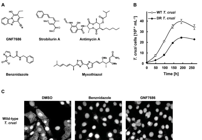
Fig 1. GNF7686 clearsT.cruziamastigotes from infected 3T3 cells.A) Structures of GNF7686, benznidazole and prototypic cytochromebinhibitors used in this study. B) Growth curves of wild-type and GNF7686-resistantT.cruziepimastigotes. C) Microscopy images of NIH/3T3 cells infected withT.cruzi
after 48 hour treatment with 0.2% DMSO, 5μM benznidazole or 1μM GNF7686.
form trypomastigotes (EC50>25μM). Curiously, GNF7686 was active onT.b.brucei
procyc-lics (EC50= 0.59μM), the parasite form found in the tsetse fly vector that mediatesT.b.brucei
transmission [34]. GNF7686 did not inhibit growth of 3T3 cell line (CC50>20μM).
Identification and characterization of GNF7686-resistant
T
.
cruzi
To investigate the mechanism of action of GNF7686, we selected a population of drug-resistant T.cruziepimastigotes through a long-term parasite culture in the presence of this compound. As tolerance for GNF7686 gradually increased over time, we periodically escalated the selection
pressure by raising the inhibitor concentration. In the course of eleven months, the EC50of the
T.cruziculture shifted ~ 4-fold from 0.16 µM to 0.73μM (Table 1).
Populations of evolving microbes often comprise cells harboring alternative mutations that
are derived from different mutation events [35,36]. To simplify analysis of genomic changes
accumulated during the selection by characterizing homogenous culture populations, we iso-lated three clones from the GNF7686-resistant culture. All three clones exhibited the same
extent of GNF7686 resistance (EC50~ 1μM) as the parent culture. Importantly, the sensitivity
of clones to benznidazole remained at the same level as observed for the wild-typeT.cruzi
strain (Table 1), demonstrating that resistance to GNF7686 did not arise through a broadly
pleiotropic mechanism. When cultured in medium lacking GNF7686, all three clones grew at a rate similar to the wild-type strain (mutant epimastigote doubling time = 5355 hours vs 60 hours for the wild-type strain), but reached stationary phase at a lower cell density (~60% of
the wild-type strain,Fig 1B).
Whole genome sequencing identified the same set of five mutations in all three clones that
included L197F in cytochromeb, L283M in the ATPase subunit of HsIVU protease, R75C in
TCSYLVIO008926 hypothetical protein, and two mutations in non-coding regions. The obser-vation that the three clones were identical at the genome sequence level suggests that they were siblings derived from one founding cell that expanded in the passaged culture during the selec-tion. During whole genome sequencing, multiple sequence reads (up to 100 in total) from many independent DNA molecules are obtained for each nucleotide position in the genome.
We uncovered that the L197F mutation in the cytochromebgene and one of two mutations
mapped to non-coding regions fully replaced the corresponding wild-type alleles, while the other three mutations remained heterozygous during the selection. Interestingly, the two
muta-tions map both to the kinetoplast maxicircle DNA, which is present in 2050 copies per cell [37,
38]. This indicates that these two mutations, presumably appearing on one maxicircle copy at
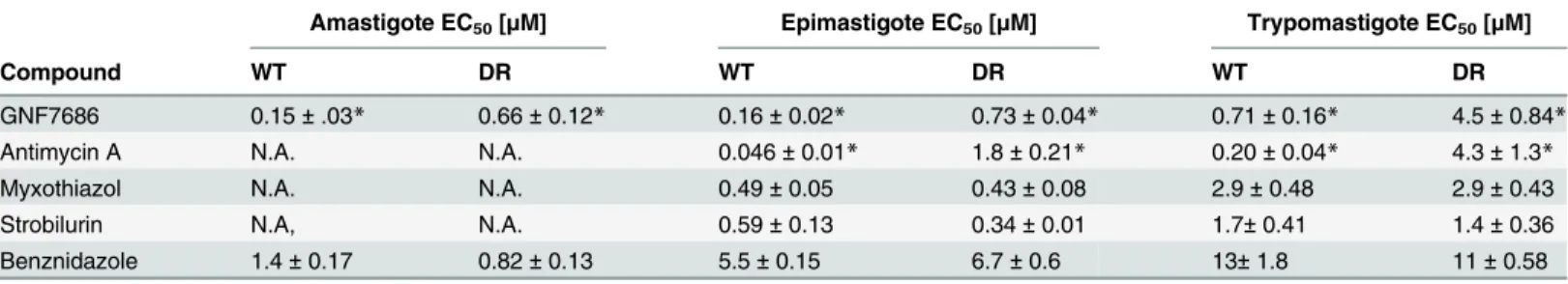
Table 1. Potency of GNF7686 and prototypic cytochromebInhibitors on wild-type (WT) and GNF7686-resistant (DR)T.cruzimorphological forms.
Amastigote EC50[μM] Epimastigote EC50[μM] Trypomastigote EC50[μM]
Compound WT DR WT DR WT DR
GNF7686 0.15±.03* 0.66±0.12* 0.16±0.02* 0.73±0.04* 0.71±0.16* 4.5±0.84*
Antimycin A N.A. N.A. 0.046±0.01* 1.8±0.21* 0.20±0.04* 4.3±1.3*
Myxothiazol N.A. N.A. 0.49±0.05 0.43±0.08 2.9±0.48 2.9±0.43
Strobilurin N.A, N.A. 0.59±0.13 0.34±0.01 1.7±0.41 1.4±0.36
Benznidazole 1.4±0.17 0.82±0.13 5.5±0.15 6.7±0.6 13±1.8 11±0.58
EC50values were calculated from three independent repeats (n = 3), each performed in duplicate. Standard error values are also shown. N.A. in the table
stands for‘not applicable’.
*P-value for wild-type versus drug-resistantT.cruziEC50values is<0.05.
first, replaced the corresponding wild-type alleles during the selection to the point of achieving homoplasmy.
Chemogenomic profiling of GNF7686 in yeast supports inhibition of the
respiratory chain at the level of complex III
In addition to selectionof T.cruzimutants resistant to GNF7686, we subjected the inhibitor
also to chemogenomic profiling inS.cerevisiae. The genome-wide deletion collections available
for this eukaryotic model system provide powerful genetic tools for investigation of bioactive
molecules [39,40], and the approach was successfully applied to mechanism of action studies
of various growth inhibitors in the past [41,42]. In the haploinsufficiency profiling assay
(HIP), complete collection of heterozygous yeast deletion strains, in which each strain has only one copy of a particular gene, is profiled for hypersensitivity to a compound. Gene deletions associated with increased compound sensitivity indicate pathways directly affected by the
com-pound [43].
We observed that growth ofS.cerevisiaeis inhibited when the yeast was cultured in media
containing glycerol but not glucose as the carbon source (see also below). We therefore con-ducted a HIP profiling experiment in medium containing glycerol. Testing of GNF7686 at its
EC30concentration in two independent HIP experiments resulted in a reproducible profile
(Fig 2A). Identified hypersensitive heterozygous strains included those with deletions in genes
involved in mitochondrial metabolism, such asCYT1(cytochromec1),HAP4(a transcription
factor involved in regulation of the respiratory chain includingCYT1),CBP1(a regulator of
cytochromebmRNA stability), andQCR6(a subunit of the cytochromecreductase complex)
[44]. As all these hits pointed at inhibition of mitochondrial respiration by GNF7686, we
per-formed additional HIP experiments with strobilurin, an inhibitor of cytochromebc1complex,
and venturicidin, an inhibitor of F-type ATPase [45,46]. In a control experiment, we also
col-lected the HIP profile for benomyl, a microtubule binding inhibitor, which does not interfere with ATP production during oxidative phosphorylation.
While both venturicidin and benomyl yielded HIP profiles distinctly different from that of
GNF7686 (Fig 2C and 2D), treatment of gene deletion strain collection with the cytochrome
bc1inhibitor strobilurin identified essentially the same set of sensitive, heterozygous mutants as
GNF7686 and includedCYT1,HAP4,CBP1andQCR6(Fig 2B). It is important to note that the
gene coding for cytochromeb, which is the proposed direct target of strobilurin [47], is
encoded by the mitochondrial genome inS.cerevisiaeand not amenable to standard gene
tar-geting protocols. Thus, the cytochromebgene deletion strain is not present in the yeast
hetero-zygous deletion pool and could not be directly identified by the HIP method.
The observed HIP compound profiles strongly suggested that GNF7686 directly interferes
with function of theS.cerevisiaerespiratory chain, possibly at the level of complex III.
Prototypical cytochrome
b
inhibitors block
in vitro
proliferation of
T
.
cruzi
Genomic analyses of GNF7686 resistance/sensitivity pointed to involvement of theT.cruzi
cytochromebin resistance to growth inhibition by GNF7686. Cytochromebis a component of
the multisubunit cytochromebc1complex, an asymmetric homodimer with two spatially
sepa-rated catalytic sites QNand QP(Fig 3A). In concert, QNand QPcatalyze oxidation of ubiquinol
formed by preceding steps of the respiratory chain [48,49].
Inhibitors of cytochromebare already of interest as anti-parasitic drugs. Atovaquone, a
hydroxy-naphthoquinone inhibitor of the QPsite, is used in the treatment of malaria and
fun-gal pneumonia [50]. Another hydroxy-naphthoquinone, buparvaquone, is used to treat cattle
cytochromebinhibitors for treatment of Chagas disease has not been explored, even though
cytochromebinhibitors including antimycin A were shown to affectT.cruzimitochondrial
respiration, bioenergetics, and calcium homeostasis [52–55]. To assess the effect of this class of
compounds onT.cruzigrowth and survival, we tested prototypical QNand QPsite inhibitors
on intracellular amastigotes, trypomastigotes, and epimastigotes (inhibitor structures shown in
Fig 1A) [46,49].
The QNsite inhibitor antimycin A potently inhibited the growth of epimastigotes and
rap-idly reduced viability of trypomastigotes. We also observedT.cruziinhibition by compounds
targeting the cytochromebQPsite. Two well-characterized QPsite inhibitors, myxothiazol and
Fig 2. Chemogenomic profiling inS.cerevisiaesuggests cytochromebas the target of GNF7686 in yeast.A) HIP profile of GNF7686. B-D) Alignment of the HIP profile of GNF7686 with profile of cytochromebinhibitor strobilurin (B), F-type ATPase inhibitor venturicidin (C), and microtubule-binding fungicide benomyl (D).
strobilurin, blocked growth of epimastigotes, and reduced viability of trypomastigotes (Table 1). The effect of antimycin A, myxothiazol, and strobilurin on intracellular amastigotes could not be accurately determined because of the inhibitor toxicity on the host 3T3 cells.
T.b.bruceiis a kinetoplastid parasite closely related toT.cruziat the genomic level [56].
While the parasite bloodstream (mammalian) form ofT.b.bruceirelies primarily on the
glyco-lytic pathway for ATP production, growth of the procyclic (insect) form requires activity of the
conventional respiratory pathway, including cytochromeb[48]. In line with the hypothesis
that GNF7686 is a cytochromebinhibitor, GNF7686 inhibited the growth of the procyclic but
not bloodstreamT.b.bruceiparasites (EC50= 0.59μM vs>25μM). We also observed similar
differential activity on the twoT.bruceiforms with the other tested cytochromebinhibitors
such as antimycin A (EC50= 0.03μM vs>25μM;Table 2).
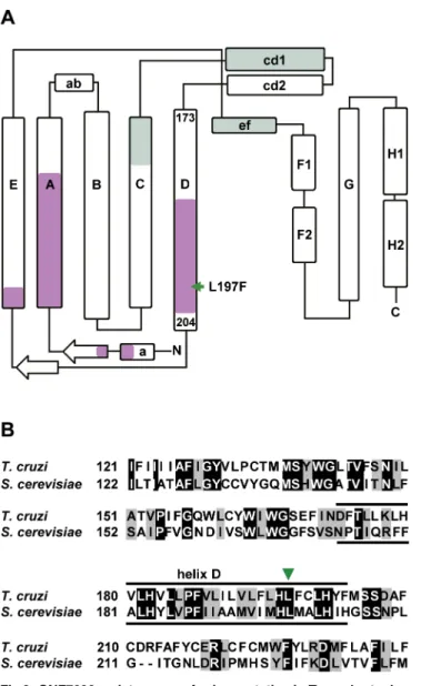
Fig 3. GNF7686 resistance-conferring mutation inT.cruzicytochromebstructure.A) Secondary structure of yeast cytochromeb(adapted from Dinget al. 2006 and Fisheret al. 2008, [50,57]) with amino acid sequence stretches that form QPand QNubiquinol-binding sites highlighted (green and violet,
respectively). The amino acid indicated by a green arrow corresponds to the L197F mutation (equivalent to L198F inS.cerevisiae) present in the GNF7686-resistantT.cruzistrain. B) Alignment ofS cerevisiaeandT.
cruzicytochromebamino acid sequence around L197F mutation. Identical amino acids are highlighted in black and conserved substitutions are highlighted in grey.
The inhibition of various morphological forms ofT.cruziby prototypical cytochromeb
inhibitors is consistent with the hypothesis that the cytochromebfulfills an essential function
in parasite physiology and that GNF7686 inhibits its function. However, with the exception of antimycin A, the anti-parasitic potency of the other tested inhibitors is too weak to be of thera-peutic significance.
GNF7686 is an inhibitor of cytochrome
b
Q
Nsite
Through a literature search, we identified a previously published report that described L198F
mutation inS.cerevisiaecytochromeb, which is equivalent to the L197F mutation inT.cruzi
cytochromeb(Fig 3B). The yeast L198F mutation confers resistance to ilicicolin H, a
cyto-chromebinhibitor with potent anti-fungal activity [46,57]. Inspection of high resolution
crys-tal structure of the yeast cytochromebc1complex further revealed that the Leu198 side chain is
positioned in close proximity (<5 Å) to ubiquinol bound inside the QNpocket and next to
His197, which coordinates the iron atom in the bHheme. In accordance with the structure,
L198F mutation also conferred resistance in yeast to other tested QNsite inhibitors such as
funiculosin and antimycin A [57,58].
Additional characterization of the GNF7686-resistantT.cruzimutants revealed that they
were selectively resistant to antimycin A, a QNsite inhibitor [46]. While antimycin A displayed
very potent activity on wild type epimastigotes, a sharp decrease in potency (40-fold) was
observed with GNF7686-resistant epimastigotes (EC50= 1.8μM,Table 1). Similarly, a steep
shift in potency (20-fold) was observed between the wild-type and mutant trypomastigotes (Table 1). In contrast, QPinhibitors myxothiazol and strobilurin showed comparable activity
on both wild-type and resistant strains (Table 1). In summary, the L197F mutation in theT.
cruzicytochromebis likely located within the QNsite and can interfere with binding of QNsite
inhibitors in a similar way as was previously described for the L198F mutation in theS.
cerevi-siaecytochromeb.
GNF7686 inhibits cytochrome
b
and respiration of
T
.
cruzi
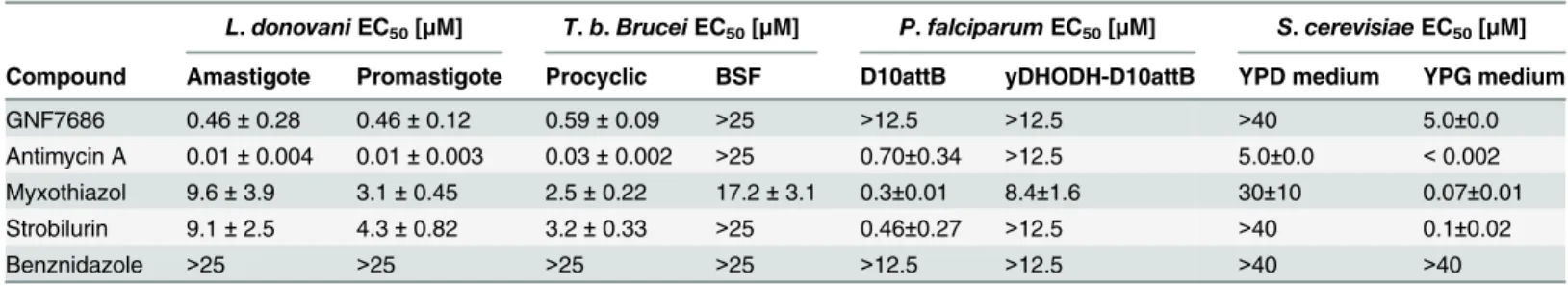
During aerobic respiration, the electron transport chain (ETC) conducts electrons derived from reduced carbon substrates through a series of redox reactions to the terminal electron Table 2. Potency of GNF7686 and prototypic cytochromebinhibitors inL.donovani,T.brucei,P.falciparumandS.cerevisiaeproliferation assays.
L.donovaniEC50[μM] T.b.BruceiEC50[μM] P.falciparumEC50[μM] S.cerevisiaeEC50[μM]
Compound Amastigote Promastigote Procyclic BSF D10attB yDHODH-D10attB YPD medium YPG medium
GNF7686 0.46±0.28 0.46±0.12 0.59±0.09 >25 >12.5 >12.5 >40 5.0±0.0 Antimycin A 0.01±0.004 0.01±0.003 0.03±0.002 >25 0.70±0.34 >12.5 5.0±0.0 <0.002 Myxothiazol 9.6±3.9 3.1±0.45 2.5±0.22 17.2±3.1 0.3±0.01 8.4±1.6 30±10 0.07±0.01 Strobilurin 9.1±2.5 4.3±0.82 3.2±0.33 >25 0.46±0.27 >12.5 >40 0.1±0.02
Benznidazole >25 >25 >25 >25 >12.5 >12.5 >40 >40
GNF7686 and cytochromebinhibitors were tested for cytotoxicity in various parasites using the CellTiter-Glo Luminescent Cell Viability assay reagent system (seeTable 1), SYBR Green Fluorescent dye (P.falciparumonly), or MIC visual growth inhibition screen (S.cerevisiaeonly). Luminescence or
fluorescence was monitored using the EnVision Multilabel Plate reader and EC50values (with respective standard errors) were determined based on three
(n = 3;L.donovaniandT.b.brucei) or two (n = 2;P.falciparumandS.cerevisiae) biological repeats with duplicate technical repeats. Abbreviations include: BSF (bloodstream form), DHODH (malarial dihydroorotate dehydrogenase), yDHODH-D10attB (fumarate-utilizingS.cerevisiaeDHODH), YPD (yeast extract peptone with dextrose), and YPG (yeast extract peptone with glycerol).
acceptor, molecular oxygen, which is then reduced to water [33,48]. Inhibition of electron flow
through the ETC at any step, including cytochromeb, results in a block of oxygen
consumption.
To evaluate whether GNF7686 disrupts the function of theT.cruziETC, oxygen
consump-tion by intactT.cruziepimastigotes was monitored in the presence of GNF7686 and prototypic
cytochromebinhibitors (Fig 4A and 4B). Antimycin A potently blocked respiration by
wild-type epimastigotes (IC50= 0.04μM), but was ~7-fold less potent on GNF7686-resistant
para-sites (IC50= 0.27μM). Two QPsite inhibitors employed in this report, myxothiazol and
strobi-lurin, both inhibited epimastigote respiration, but, in contrast to antimycin A, the respiratory
IC50values of the QPinhibitors were comparable between wild-type and GNF7686-resistantT.
cruzi.
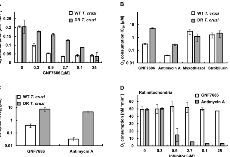
Fig 4. GNF7686 inhibits cellular respiration and cytochromebfunction inT.cruzi.A)T.cruziepimastigote respiration was monitored using the MitoXpress-Xtra HS phosphorescent probe (seeMethods) in the presence of 0.2% DMSO (control) and various concentrations of GNF7686. Oxygen consumption rates per 106T.cruziepimastigotes cells are shown. Plotted values were derived from three biological repeats (n = 3) with duplicate technical
repeats in either wild-type or evolved GNF7686-resistant (DR) parasites. B) Oxygen consumption IC50values for prototypic cytochromebinhibitorsin T.cruzi
epimastigote respiration assay on wild-type and GNF7686-resistant (DR) parasites. Shown GNF7686 IC50values were derived from the experiment shown
in the (A) panel. C) Mitochondrial complex III activity was monitored in digitonin-solubilizedT.cruziepimastigotes (both wild-type and evolved GNF7686-resistant (DR) parasite strains) utilizing a coupled decylubiquinol oxidation / cytochromecreduction reaction in the presence of a compound (GNF7686 or antimycin A). IC50values were determined based on three biological repeats (n = 3) with triplicate technical repeats in either wild-type or
evolved drug-resistant (DR) parasites relative to 0.2% DMSO conditions. D) High selectivity of GNF7686 forT.cruzicytochromebis reflected in the absence of inhibition of rat mitochondrial respiration by this compound up to 25μM concentration. For comparison, antimycin A potently inhibits mammalian
mitochondrial respiration. Oxygen consumption rates per 1 mg of total mitochondrial protein are shown.
GNF7686 inhibited respiration by wild-type parasites with an IC50= 0.21μM. A significant
drop in inhibitor potency was observed with the GNF7686-resistantT.cruziepimastigotes
(oxygen consumption IC50= 5.2μM). As seen for growth inhibition, the results on respiration
inhibition distinguish GNF7686 and antimycin A from the QPsite inhibitors (Fig 4B).
We then asked whether GNF7686 inhibitsT.cruzicytochromebc1(complex III) directly
(Fig 4C). Epimastigotes were permeabilized with digitonin, and KCN (complex IV inhibitor) was added to the permeabilized cells to block electron conductance by the parasite ETC. The reaction was then initiated by adding stoichiometric quantities of decylubiquinol (an electron
donor for complex III) and oxidized yeast cytochromec(an electron acceptor), and the
cata-lytic activity of complex III was monitored through accumulation of reduced yeast cytochrome c. A control reaction with antimycin A confirmed earlier observations with intact epimastigotes (Table 1). Antimycin A potently blocked reduction of cytochromecin the reaction with wild-type permeabilized epimastigotes, but a dramatic loss of inhibitor potency was observed
(~100-fold) when GNF7686-resistant permeabilized parasites were used (Fig 4C). In a similar
fashion, GNF7686 inhibited wild-type complex III activity with an IC50of 0.40μM, but a
20-fold loss of potency was observed with the mutant complex III (Fig 4C). These observations
validate GNF7686 as a complex III inhibitor that likely targets the QNsite of theT.cruzi
cyto-chromeb.
GNF7686 is highly selective for
T
.
cruzi
and does not inhibit mammalian
respiration
The inhibitory effect of GNF7686 on mammalian cytochromebfunction was assessed through
monitoring oxygen consumption by mitochondria isolated from rat skeletal muscle cells. In
the control reaction, antimycin A showed a potent inhibition (IC50= 0.81μM) of
mitochon-drial respiration (Fig 4D). In a parallel experiment, GNF7686 did not show any effect on
oxy-gen consumption up to 25μM concentration (Fig 4D). This observation validates GNF7686 as
a highly selective inhibitor of theT.cruzicytochromeband a promising starting point for
Cha-gas disease drug discovery.
We also examined effect of GNF7686 on cytochromebin the malaria parasite,P.
falcipa-rum, and in yeastS.cerevisiae, the latter being used as a surrogate for pathogenicPneumocystis jirovecii, a causative agent of a pneumocystis pneumonia [59]. Cytochromebis a validated
drug target in both organisms and atovaquone, an inhibitor of cytochromeb, is a clinical
treat-ment for these diseases.
For theP.falciparumstudies, all inhibitors were tested on two parasite lines—D10attB and
yDHODH-D10attB (Table 2). In the wild-type D10attB line, the parasitede novopyrimidine
biosynthesis is dependent on a type 2 dihydroorotate dehydrogenase (PfDHODH) and requires
a functionalP.falciparumETC, including cytochromeb, downstream fromPfDHODH [26,
27]. In contrast, the yDHODH-D10attB cell line is modified with a type 1A dihydroorotate
dehydrogenase fromS.cerevisiae(yDHODH), which is cytosolic and utilizes fumarate as the
terminal electron acceptor [25–27]. The assay was validated with antimycin A, which blocked
growth of the D10attB line (EC50= 0.7μM), but was inactive on the yDHODH-D10attB
para-site line (EC50>12.5μM). Myxothiazol and strobilurin also showed a similar preferential
activity on the D10attB line, whereas GNF7686 did not inhibit growth of eitherP.falciparum
cell line. This result suggests that GNF7686 is not active onP.falciparumcytochromeb.
A similar, growth inhibition-based assessment of GNF7686 effect on the ETC function was
also performed onS.cerevisiae(Table 2). Growth inhibition of a wild-typeS.cerevisiaestrain
by compounds in two different media was monitored. The first medium contained glucose as
the second medium, glucose was replaced with glycerol, a non-fermentable carbon source. Under the latter condition, yeast growth is dependent on cellular respiration and functional
cytochromeb[60]. All prototypic cytochromebinhibitors used in this report potently
inhib-ited yeast growth on medium with glycerol, but were inactive when yeast grew in medium with glucose. Following a similar pattern, GNF7686 weakly inhibited growth of yeast in glycerol
medium (EC50= 5.0μM), but did not affect yeast growing in the medium with glucose.
In summary, GNF7686 selectively inhibitsT.cruzicytochromeband does not affect
respira-tion of mammalian mitochondria nor does it significantly inhibit respirarespira-tion-dependent
growth ofP.falciparumandS.cerevisiae.
Discussion
We have shown that cytochromebis a possible target for new drug discovery efforts aimed at
treating kinetoplastid diseases. The importance of this finding is underlined by the paucity of
drug targets for these diseases. For Chagas disease, only sterol 14α-demethylase (CYP51) and
cruzain have been explored in depth as possible targets. However, low parasitological cure rates
that were observed (20–30%) during clinical testing of anti-fungal drugs targeting sterol 14α
-demethylase (posaconazole and ravuconazole prodrug E1224) in Chagas disease patients have
lessened enthusiasm for further work on repurposed CYP51 inhibitors as single agents [61,
62]. Additional clinical evaluation of this class of drugs partnered with benznidazole for
combi-nation treatments is still planned. It is also important to note that the failure of posaconazole and E1224 in phase 2 trials have been attributed to insufficient drug exposure or dosing
dura-tion [14], and work onT.cruzi-specific CYP51 inhibitors that could enter clinical development
in the future is also ongoing [63,64]. Finally, the anti-cancer drug BEZ235 has activity across
kinetoplastid parasites, but it requires additional optimization (improvement of therapeutic
index) before becoming a preclinical candidate [65]. Given this sparse landscape, new chemical
starts and new drug targets are urgently needed to anchor drug discovery efforts for the kineto-plastid diseases.
Many groups have resorted to a‘pre-genomic’approach to drug discovery, in which
com-pounds are screened to identify inhibitors of pathogen growth, without regard to mechanism of action. While this approach typically provides large number of chemical starting points and broad hit diversity, a lack of information on mechanism of action creates additional risk in chemical optimization, and in predicting possible toxicity liabilities. Next generation sequenc-ing of evolved resistant pathogens has been used successfully to identify resistance mechanisms and, in many cases, target mechanisms, for several pathogens. In our own program, we have
identified targets for malaria and tuberculosis [66,67]. However, the approach has not been
reduced to practice in kinetoplastid drug discovery until this study.
Several features of the results in this study bode well for future application of the approach. First, the number of mutations associated with the emergence of drug resistance was relatively low (five point mutations), which simplified subsequent target prediction and validation.
Sec-ond, we were able to generate resistance inT.cruziepimastigotes despite a relatively long
dou-bling time and stable genome. Selection process required almost a year of drug pressure in this
study but was ultimately successful. Finally, we found a mutation in a gene that is a‘plausible’
drug target, where plausibility is supported by essentiality of the mutated gene in other cellular systems, or precedence from drugs targeting homologous targets in other organisms.
Drugs targeting cytochromebare in clinical use for treatment of malaria and fungal
pneu-monia, and cytochromebwas also reported as a promising target for treatment of tuberculosis.
The current study extends utility of cytochromebas a drug target also to Chagas disease, and
of growth and biochemical inhibition that consistently confirmed that functional cytochromeb
is essential forT.cruzipropagation. Based on the presented validation data,T.cruzigrowth
can be inhibited through targeting either QNor QPcytochromebsite. GNF7686 represents a
new cytochromebinhibitor, likely targeting the QNsite,which has high selectivity forT.cruzi
and does not show any effect on respiration of mammalian mitochondria. Crystal structures of
cytochromebfrom several sources (bovine, chicken, yeast,Rhodobacter,Paracoccus) were
pre-viously published [68,69]. This opens opportunities for rational drug design based on
homol-ogy modeling ofT.cruzicytochromebstructure. Finally, a counter-screen assay to measure
inhibitory activity on the cognate human enzyme (as described here) can be used to guide chemical optimization away from host toxicity. While GNF7686 appears to be a promising
starting point for kinetoplastid drug discovery, it does not inhibit the growth ofP.falciparum
orS.cerevisiae, and thus may not be a suitable starting point for anti-malaria or anti-fungal
drug discovery. Furtherin vivocharacterization of GNF7686 revealed that it has poor
pharma-cokinetic properties, including low oral bioavailability (F = 6%) and highin vivomouse
clear-ance (53 mLmin-1kg-1); thus, extension of the current studies to anin vivomodel of
Chagas disease will require identification of a GNF7686 analogue that has improved pharma-cokinetic profile.
In summary, we have established an approach for identification of molecular targets ofT.
cruzigrowth inhibitors that enables transition to target-based drug discovery for compounds with previously unknown mechanism of action. The first application of this approach resulted
in identification of a highly selective inhibitor ofT.cruzicytochromeb, GNF7686, which can
serve as an excellent starting point for discovery of new drugs for Chagas disease and leishmaniasis.
Acknowledgments
We thank Rick Tarleton (University of Georgia) for providing theT.cruziCL strain. We are
grateful to Jason Matzen, Annie Mak and Steven Chen for providing technical support. Addi-tional acknowledgments go to: Xianzhong (Leo) Liu and Randy Soriano for profiling com-pounds on NIH/3T3 cells; Carolina Turk and Leonardo Vargas for preparations of rat skeletal muscle mitochondria used in respiration studies; Maureen Ibanez, Kerstin Gagaring, and
Shu-lin Han for screening compounds inPlasmodiumstrains; AnneMarie Nguyen and Glenn
Fed-ere for processing samples for whole genome sequencing; Jia Zhang for assistance with cloning for sequence confirmation analysis; and Jair L. Siqueira-Neto for critical reading of the manuscript.
Author Contributions
Conceived and designed the experiments: SK SLR SWB DH JRW AKC RJN MRA CWM YF VM VY JHM RJG FS. Performed the experiments: SK SLR SWB DH JB YL YF. Analyzed the data: SK SWB DH JRW AKC RJN MRA CWM JB YL YF RJG FS. Contributed reagents/materi-als/analysis tools: SLR JRW RJN CWM. Wrote the paper: SK DH MRA CWM VM VY JHM RJG FS.
References
1. Anis Rassi Jr AR, Jose Antonio Marin-Neto (2010) Chagas disease. Lancet 375: 1388–1402. doi:10. 1016/S0140-6736(10)60061-XPMID:20399979
3. Rodolfo Viotti CV, Bruno Lococo, Maria Gabriela Alvarez, Marcos Petti, Graciela Bertocchi and Alejan-dro Armenti (2009) Side effects of benznidazole as treatment in chronic Chagas disease: fears and realities. Expert Rev Anti Infect Ther 7: 157–163. doi:10.1586/14787210.7.2.157PMID:19254164
4. Bern C, Kjos S, Yabsley MJ, Montgomery SP (2011) Trypanosoma cruzi and Chagas' Disease in the United States. Clin Microbiol Rev 24: 655–681. doi:10.1128/CMR.00005-11PMID:21976603
5. Hurwitz I, Fieck A, Klein N, Jose C, Kang A, et al. (2012) A Paratransgenic Strategy for the Control of Chagas Disease. Psyche: A Journal of Entomology 2012: 1–10.
6. Buckner FS, Navabi N (2010) Advances in Chagas disease drug development: 2009–2010. Curr Opin Infect Dis 23: 609–616. doi:10.1097/QCO.0b013e3283402956PMID:20885320
7. Choi JY, Calvet CM, Gunatilleke SS, Ruiz C, Cameron MD, et al. (2013) Rational development of 4-aminopyridyl-based inhibitors targeting Trypanosoma cruzi CYP51 as anti-chagas agents. J Med Chem 56: 7651–7668. doi:10.1021/jm401067sPMID:24079662
8. Clayton J (2010) Chagas disease: pushing through the pipeline. Nature 465: S12–15. doi:10.1038/ nature09224PMID:20571548
9. Friggeri L, Hargrove TY, Rachakonda G, Williams AD, Wawrzak Z, et al. (2014) Structural Basis for Rational Design of Inhibitors Targeting Trypanosoma cruzi Sterol 14alpha-Demethylase: Two Regions of the Enzyme Molecule Potentiate its Inhibition. J Med Chem.
10. Bustamante JM, Craft JM, Crowe BD, Ketchie SA, Tarleton RL (2014) New, combined, and reduced dosing treatment protocols cure Trypanosoma cruzi infection in mice. J Infect Dis 209: 150–162. doi: 10.1093/infdis/jit420PMID:23945371
11. Pinazo MJ, Espinosa G, Gallego M, Lopez-Chejade PL, Urbina JA, et al. (2010) Successful treatment with posaconazole of a patient with chronic Chagas disease and systemic lupus erythematosus. Am J Trop Med Hyg 82: 583–587. doi:10.4269/ajtmh.2010.09-0620PMID:20348503
12. DNDi Press Release. Drug Trial for Leading Parasitic Killer of the Americas Shows Mixed Results but Provides New Evidence for Improved Therapy. Drugs for Neglected Diseases Initiative. 14 November 2013.http://www.dndi.org/media-centre/press-releases/1700-e1224.html. Accessed 01 December 2014.
13. VandeBerg JL (2014) Treatment Trials and Efficacy Determination in Non-human Primates with Chronic T. cruzi Infections. Southwest National Primate Research Center at the Texas Biomedical Research Institute, San Antonio, Texas, USA. pp. 23.
14. Urbina JA (2015) Recent clinical trials for the etiological treatment of chronic chagas disease: advances, challenges and perspectives. J Eukaryot Microbiol 62: 149–156. doi:10.1111/jeu.12184 PMID:25284065
15. McKerrow JH, Doyle PS, Engel JC, Podust LM, Robertson SA, et al. (2009) Two approaches to discov-ering and developing new drugs for Chagas disease. Mem Inst Oswaldo Cruz 104 Suppl 1: 263–269. PMID:19753483
16. Choy JW, Bryant C, Calvet CM, Doyle PS, Gunatilleke SS, et al. (2013) Chemical-biological characteri-zation of a cruzain inhibitor reveals a second target and a mammalian off-target. Beilstein J Org Chem 9: 15–25. doi:10.3762/bjoc.9.3PMID:23400640
17. Engel JC, Doyle PS, Hsieh I, McKerrow JH (1998) Cysteine protease inhibitors cure an experimental Trypanosoma cruzi infection. J Exp Med 188: 725–734. PMID:9705954
18. Doyle PS, Zhou YM, Engel JC, McKerrow JH (2007) A cysteine protease inhibitor cures Chagas' dis-ease in an immunodeficient-mouse model of infection. Antimicrob Agents Chemother 51: 3932–3939. PMID:17698625
19. Carneiro G, Aguiar MG, Fernandes AP, Ferreira LA (2012) Drug delivery systems for the topical treat-ment of cutaneous leishmaniasis. Expert Opin Drug Deliv 9: 1083–1097. doi:10.1517/17425247.2012. 701204PMID:22724539
20. Gilbert IH, Leroy D, Frearson JA (2011) Finding new hits in neglected disease projects: target or pheno-typic based screening? Curr Top Med Chem 11: 1284–1291. PMID:21401505
21. Bourguignon SC, de Souza W, Souto-Padron T (1998) Localization of lectin-binding sites on the sur-face of Trypanosoma cruzi grown in chemically defined conditions. Histochem Cell Biol 110: 527–534. PMID:9826132
22. Garcia-Silva MR, Frugier M, Tosar JP, Correa-Dominguez A, Ronalte-Alves L, et al. (2010) A popula-tion of tRNA-derived small RNAs is actively produced in Trypanosoma cruzi and recruited to specific cytoplasmic granules. Mol Biochem Parasitol 171: 64–73. doi:10.1016/j.molbiopara.2010.02.003 PMID:20156490
24. Engel JC, Ang KK, Chen S, Arkin MR, McKerrow JH, et al. (2010) Image-based high-throughput drug screening targeting the intracellular stage of Trypanosoma cruzi, the agent of Chagas' disease. Antimi-crob Agents Chemother 54: 3326–3334. doi:10.1128/AAC.01777-09PMID:20547819
25. Ke H, Morrisey JM, Ganesan SM, Painter HJ, Mather MW, et al. (2011) Variation among Plasmodium falciparum strains in their reliance on mitochondrial electron transport chain function. Eukaryot Cell 10: 1053–1061. doi:10.1128/EC.05049-11PMID:21685321
26. Nam TG, McNamara CW, Bopp S, Dharia NV, Meister S, et al. (2011) A chemical genomic analysis of decoquinate, a Plasmodium falciparum cytochrome b inhibitor. ACS Chem Biol 6: 1214–1222. doi:10. 1021/cb200105dPMID:21866942
27. Painter HJ, Morrisey JM, Mather MW, Vaidya AB (2007) Specific role of mitochondrial electron trans-port in blood-stage Plasmodium falciparum. Nature 446: 88–91. PMID:17330044
28. Plouffe D, Brinker A, McNamara C, Henson K, Kato N, et al. (2008) In silico activity profiling reveals the mechanism of action of antimalarials discovered in a high-throughput screen. Proc Natl Acad Sci U S A 105: 9059–9064. doi:10.1073/pnas.0802982105PMID:18579783
29. Ghannoum MA, Ibrahim AS, Fu Y, Shafiq MC, Edwards JE Jr., et al. (1992) Susceptibility testing of Cryptococcus neoformans: a microdilution technique. J Clin Microbiol 30: 2881–2886. PMID:1452658
30. Robinson JT, Thorvaldsdottir H, Winckler W, Guttman M, Lander ES, et al. (2011) Integrative genomics viewer. Nat Biotechnol 29: 24–26. doi:10.1038/nbt.1754PMID:21221095
31. Thorvaldsdottir H, Robinson JT, Mesirov JP (2013) Integrative Genomics Viewer (IGV): high-perfor-mance genomics data visualization and exploration. Brief Bioinform 14: 178–192. doi:10.1093/bib/ bbs017PMID:22517427
32. Telford JE, Kilbride SM, Davey GP (2010) Decylubiquinone increases mitochondrial function in synap-tosomes. J Biol Chem 285: 8639–8645. doi:10.1074/jbc.M109.079780PMID:20080966
33. Spinazzi M, Casarin A, Pertegato V, Salviati L, Angelini C (2012) Assessment of mitochondrial respira-tory chain enzymatic activities on tissues and cultured cells. Nat Protoc 7: 1235–1246. doi:10.1038/ nprot.2012.058PMID:22653162
34. International Glossina Genome I (2014) Genome sequence of the tsetse fly (Glossina morsitans): vec-tor of African trypanosomiasis. Science 344: 380–386. doi:10.1126/science.1249656PMID: 24763584
35. Holmes R K, Jobling M.G. (1996) Medical Microbiology. 4th Edition. Galveston, TX: University of Texas Medical Branch at Galveston,.
36. Lang GI, Rice DP, Hickman MJ, Sodergren E, Weinstock GM, et al. (2013) Pervasive genetic hitchhik-ing and clonal interference in forty evolvhitchhik-ing yeast populations. Nature 500: 571–574. doi:10.1038/ nature12344PMID:23873039
37. Lukes J, Guilbride DL, Votypka J, Zikova A, Benne R, et al. (2002) Kinetoplast DNA network: evolution of an improbable structure. Eukaryot Cell 1: 495–502. PMID:12455998
38. Messenger LA, Llewellyn MS, Bhattacharyya T, Franzen O, Lewis MD, et al. (2012) Multiple mitochon-drial introgression events and heteroplasmy in trypanosoma cruzi revealed by maxicircle MLST and next generation sequencing. PLoS Negl Trop Dis 6: e1584. doi:10.1371/journal.pntd.0001584PMID: 22506081
39. Giaever G, Shoemaker DD, Jones TW, Liang H, Winzeler EA, et al. (1999) Genomic profiling of drug sensitivities via induced haploinsufficiency. Nat Genet 21: 278–283. PMID:10080179
40. Smith AM, Ammar R, Nislow C, Giaever G (2010) A survey of yeast genomic assays for drug and target discovery. Pharmacol Ther 127: 156–164. doi:10.1016/j.pharmthera.2010.04.012PMID:20546776
41. Nyfeler B, Hoepfner D, Palestrant D, Kirby CA, Whitehead L, et al. (2012) Identification of elongation factor G as the conserved cellular target of argyrin B. PLoS One 7: e42657. doi:10.1371/journal.pone. 0042657PMID:22970117
42. Richie DL, Thompson KV, Studer C, Prindle VC, Aust T, et al. (2013) Identification and evaluation of novel acetolactate synthase inhibitors as antifungal agents. Antimicrob Agents Chemother 57: 2272–
2280. doi:10.1128/AAC.01809-12PMID:23478965
43. Giaever G, Flaherty P, Kumm J, Proctor M, Nislow C, et al. (2004) Chemogenomic profiling: identifying the functional interactions of small molecules in yeast. Proc Natl Acad Sci U S A 101: 793–798. PMID: 14718668
44. Cherry JM HE, Amundsen C, Balakrishnan R, Binkley G, Chan ET, Christie KR, Costanzo MC, Dwight SS, Engel SR, Fisk DG, Hirschman JE, Hitz BC, Karra K, Krieger CJ, Miyasato SR, Nash RS, Park J, Skrzypek MS, Simison M, Weng S, Wong ED (2012) Saccharomyces Genome Database: the geno-mics resource of budding yeast. Nucleic Acids Res. pp. D700–705.
46. Gutierrez-Cirlos EB, Merbitz-Zahradnik T, Trumpower BL (2004) Inhibition of the yeast cytochrome bc1 complex by ilicicolin H, a novel inhibitor that acts at the Qn site of the bc1 complex. J Biol Chem 279: 8708–8714. PMID:14670947
47. di Rago JP, Coppee JY, Colson AM (1989) Molecular basis for resistance to myxothiazol, mucidin (stro-bilurin A), and stigmatellin. Cytochrome b inhibitors acting at the center o of the mitochondrial ubiquinol-cytochrome c reductase in Saccharomyces cerevisiae. J Biol Chem 264: 14543–14548. PMID: 2547800
48. Trumpower BL (1990) Cytochrome bc1 Complexes of Microorganisms. MICROBIOLOGICAL REVIEWS 54: 101–129. PMID:2163487
49. Huang LS, Cobessi D, Tung EY, Berry EA (2005) Binding of the respiratory chain inhibitor antimycin to the mitochondrial bc1 complex: a new crystal structure reveals an altered intramolecular hydrogen-bonding pattern. J Mol Biol 351: 573–597. PMID:16024040
50. Fisher N, Meunier B (2008) Molecular basis of resistance to cytochrome bc1 inhibitors. FEMS Yeast Res 8: 183–192. PMID:18093133
51. Soni MP, Shelkar N, Gaikwad RV, Vanage GR, Samad A, et al. (2014) Buparvaquone loaded solid lipid nanoparticles for targeted delivery in theleriosis. J Pharm Bioallied Sci 6: 22–30. doi: 10.4103/0975-7406.124309PMID:24459400
52. Peloso Ede F, Vitor SC, Ribeiro LH, Pineyro MD, Robello C, et al. (2011) Role of Trypanosoma cruzi peroxiredoxins in mitochondrial bioenergetics. J Bioenerg Biomembr 43: 419–424. doi:10.1007/ s10863-011-9365-4PMID:21732175
53. Silva TM, Peloso EF, Vitor SC, Ribeiro LH, Gadelha FR (2011) O2 consumption rates along the growth curve: new insights into Trypanosoma cruzi mitochondrial respiratory chain. J Bioenerg Biomembr 43: 409–417. doi:10.1007/s10863-011-9369-0PMID:21732174
54. Docampo R, Moreno SN, Vercesi AE (1993) Effect of thapsigargin on calcium homeostasis in Trypano-soma cruzi trypomastigotes and epimastigotes. Mol Biochem Parasitol 59: 305–313. PMID:8341327
55. Martins RM, Covarrubias C, Rojas RG, Silber AM, Yoshida N (2009) Use of L-proline and ATP produc-tion by Trypanosoma cruzi metacyclic forms as requirements for host cell invasion. Infect Immun 77: 3023–3032. doi:10.1128/IAI.00138-09PMID:19433547
56. El-Sayed NM, Myler PJ, Blandin G, Berriman M, Crabtree J, et al. (2005) Comparative genomics of try-panosomatid parasitic protozoa. Science 309: 404–409. PMID:16020724
57. Ding MG, di Rago JP, Trumpower BL (2006) Investigating the Qn site of the cytochrome bc1 complex in Saccharomyces cerevisiae with mutants resistant to ilicicolin H, a novel Qn site inhibitor. J Biol Chem 281: 36036–36043. PMID:16987808
58. di Rago J.-P. JP, and Colson A.-M. (1990) Isolation and RNA sequence analysis of cytochromeb
mutants resistant to funiculosin, a center i inhibitor of the mitochondrial ubiquinol-cytochromec reduc-tase inSaccharomyces cerevisiae. FEBS Letters 263: 93–98. PMID:2158909
59. Stringer JR, Beard CB, Miller RF, Wakefield AE (2002) A new name (Pneumocystis jiroveci) for Pneu-mocystis from humans. Emerg Infect Dis 8: 891–896. PMID:12194762
60. Perocchi F, Jensen LJ, Gagneur J, Ahting U, von Mering C, et al. (2006) Assessing systems properties of yeast mitochondria through an interaction map of the organelle. PLoS Genet 2: e170. PMID: 17054397
61. (DNDi) DfNDI (2013) Drug Trial for Leading Parasitic Killer of the Americas Shows Mixed Results but Provides New Evidence for Improved Therapy. Media Center/Press Releases.
62. Molina I, Gomez i Prat J, Salvador F, Trevino B, Sulleiro E, et al. (2014) Randomized trial of posacona-zole and benznidaposacona-zole for chronic Chagas' disease. N Engl J Med 370: 1899–1908. doi:10.1056/ NEJMoa1313122PMID:24827034
63. Andriani G, Amata E, Beatty J, Clements Z, Coffey BJ, et al. (2013) Antitrypanosomal lead discovery: identification of a ligand-efficient inhibitor of Trypanosoma cruzi CYP51 and parasite growth. J Med Chem 56: 2556–2567. doi:10.1021/jm400012ePMID:23448316
64. Calvet CM, Vieira DF, Choi JY, Kellar D, Cameron MD, et al. (2014) 4-Aminopyridyl-based CYP51 inhibitors as anti-Trypanosoma cruzi drug leads with improved pharmacokinetic profile and in vivo potency. J Med Chem 57: 6989–7005. doi:10.1021/jm500448uPMID:25101801
65. Diaz-Gonzalez R, Kuhlmann FM, Galan-Rodriguez C, Madeira da Silva L, Saldivia M, et al. (2011) The susceptibility of trypanosomatid pathogens to PI3/mTOR kinase inhibitors affords a new opportunity for drug repurposing. PLoS Negl Trop Dis 5: e1297. doi:10.1371/journal.pntd.0001297PMID:21886855
67. Rao SP, Lakshminarayana SB, Kondreddi RR, Herve M, Camacho LR, et al. (2013) Indolcarboxamide is a preclinical candidate for treating multidrug-resistant tuberculosis. Sci Transl Med 5: 214ra168. doi: 10.1126/scitranslmed.3007355PMID:24307692
68. Xia D, Yu CA, Kim H, Xia JZ, Kachurin AM, et al. (1997) Crystal structure of the cytochrome bc1 com-plex from bovine heart mitochondria. Science 277: 60–66. PMID:9204897