Universidade de Brasília Faculdade de Medicina
Laborat ório Mult idisciplinar de Pesquisa em Doença de Chagas
METILTIOADENOSINA FOSFORILASE DE
TRYPANOSOMA
CRUZI
, UM ALVO POTENCIAL PARA QUIMIOTERAPIA
DA DOENÇA DE CHAGAS, APRESENTA AMPLA
ESPECIFICIDADE A SUBSTRATOS E ELEVADA
ESTABILIDADE ESTRUTURAL
DAVID NEVES
Orientador: Prof. Dr. Jaime Martins de Santana
Co-orientador: Prof. Dra. Sônia Maria de Freitas
Tese apresentada ao
programa de Pós-graduação
em Patologia Molecular da
Universidade de Brasília
como requisito parcial à
obtenção do título de
Doutor em Patologia
Molecular
DEDICATÓRIA
Dedico esta tese aos meus pais,
Telma e Osvaldo, pela vida, amor e apoio
incondicionais em mais essa etapa.
AGRADECIMENTOS
Ao professor Jaime Santana, pela confiança depositada, dedicação diária e
incentivo em galgar novos horizonte. Obrigado pelo exemplo de
competência como pesquisador e orientador.
À professora Sônia Freitas pela oportunidade de enveredar pelo fascinante
mundo da biofísica, acessibilidade e paciência de repetir conceitos por
tantas vezes confundidos e sempre com a mesma energia.
Ao professor Antônio Teixeira, pela oportunidade de trabalho e exemplo de
amor a investigação científica.
À Izabela, pelo acolhimento no LMPDC, amizade, “broncas” sempre
construtivas e exemplo de persistência.
Às eternas amigas de trabalho, Danielle, Carol, Glória, Flávia, Mariana,
Meire, Carla, Nadjar, Lina, Perla, Adriana e especialmente a Teresa
Cristina.
À minoria do LMPDC, Thiago, Alessandro, Cléver e Rubens pela amizade.
À Ana de Cássia, Márcia, Miguel e Geraldo pelo apoio técnico,
fundamental na realização desse trabalho.
Aos professores do LNLS Javier Medrano e João Alexandre pela ajuda na
elaboração e realização dos trabalhos de biofísica e cristalização.
À minha ex-orientadora e amiga, Cláudia Renata, pelos primeiros passos na
pesquisa, ensinamentos de vida e incentivo.
Aos amigos da UnB, Carla Tatiana, Daniela, Tainá, Patrícia, Regina,
Claudiner e Eduardo.
Aos companheiros de longa data Carpes, Alves e Zakir pela amizade
incondicional e sempre presente.
Ao imenso apoio da minha avó Catarina, assim como da tia Vitória, tia
Leila e primos Gabriela e Lucas.
LISTA DE ABREVIATURAS
[θ] Elipcidade média por resíduo
°C Grau centígrado
Ado Adenosina Arg Arginina
CD Dicroísmo circular cDNA Acido desoxirribonucléico complementar CDP Citidia-difosfato cm centímetro
CTAB Hexadecil-trimetilamonia Da Dalton
dAdo Deoxi-adenosina
dGTP Deoxi-guanidina-trifosfato DNA Ácido desoxirribonucléico DTT Ditiotreitol
g Grama Gnd-HCl Hidrocloreto de guanidina
GTP Guanidina-trifosfato HEPES Ácido (2-hidroxietil)-piperazina etanosulfônico His Histidina
IgG Imuno-globulina G IPTG Isopropil-β-D-tiogalactopiranosídeo kb Kilobase
kcat Constante catalítica
kcat/Km Eficiência catalítica
Km Constante de Michaelis-Menten
Ku Constante de equilíbrio do processo de desnovelamento
L-6 Célula muscular murina M Molar
PBS Tampão fosfato 50 mM, NaCl 0,15 M pH 7,2 Pro Prolina
RNA Ácido ribonucléico RPMI Meio Instituto Roswell Park Memorial
SDS-PAGE Eletroforese em gel de poliacrilamida contando dodecil sulfato de sódio
Tris Tris- hidroximetil-etano Trp Triptofano
U.I. Unidade internacional
UV Ultravioleta v/v Razão volume/volume
Vmax Velocidade máxima
ÍNDICE
Apoio Financeiro. ...1
Dedicatória...2
Agradecimentos . ...3
Abreviaturas...4
Índice...6
Resumo ...8
INTRODUÇÃO...10
Doença de Chagas ...10
Desenvolvimento da doença ...11
Purina nucleosídeo fosforilase. ...13
Classificação ...14
Subfamília MTAF ...16
MTAF humana ...16
MTAF de parasitas ...17
Via metabólica do MTA ...18
Princípios teóricos da caracterização termodinâmica ...21
Dicroísmo Circular e desnaturação térmica...21
Espectroscopia de Fluorescência...23
OBJETIVOS...27
RESULTADOS...29
The methylthioadenosine phosphorylase of trypanosoma cruzi is mesophilic and displays broad substrate specificity...30
Summary...30
Introduction...31
Results and Discussion ...33
Material and Methods...50
References...54
The drug target methylthioadenosine phosphorylase of trypanosoma cruzi exhibits remarkable resistance to thermal and chemical denaturation ...59
Introduction ... 60
Results ... 61
Discussion... 71
Material and Methods... 73
References ... 75
CONCLUSÕES...80
PERSPECTIVAS...83
RESUMO
I
INTRODUÇÃO
Doença de Chagas
A doença de Chagas, ou Tripanossomíase Americana, foi descoberta por Carlos Chagas, o qual identificou seu vetor, agente etiológico e descreveu suas características clínicas (Chagas, 1909). O mal de Chagas é prevalente no continente americano, representando a terceira maior endemia parasitária, atrás somente da Malária e da Esquistossomose (WHO, 1993). A doença é causada pelo parasita Trypanosoma cruzi, que infecta cerca de 21 milhões de pessoas (WHO, 2002), além de manter mais de 120 milhões indivíduos (25% da população da América Latina) sob risco de adquirir a infecção (WHO, 2002) .
Do ponto de vista econômico, a perda anual para o continente Latino Americano foi calculada em cerca de 6,5 bilhões de dólares (WHO, 1997); o Brasil gasta aproximadamente 750 milhões de dólares por ano no tratamento de pacientes chagásicos (WHO, 2000). Calcula-se que o investimento anual dos governos dos países latino-americanos, visando o controle da doença de Chagas, é muito inferior à perda econômica causada por esta endemia.
Vias de Transmissão
Desenvolvimento da doença
As infecções causadas pelo T. cruzi podem ocorrer em três fases: aguda, latente ou indeterminada e crônica. A fase aguda é usualmente assintomática na maioria das pessoas infectadas e é caracterizada por febre, mialgia e mal-estar (Deghaide et alii, 1998). Em alguns pacientes, o sinal de Romaña (inchaço unilateral, bipalpebral) e o chagoma de inoculação são sinais indicativos da porta de entrada do parasita no hospedeiro humano(Brener et alii, 2000). Miocardites e meningoencefalites podem ocorrer ocasionalmente (Deghaide et alii, 1998). Estima-se que metade dos indivíduos infectados pelo T. cruzi entram na fase latente, (Macedo, 1980) na qual não apresentam as alterações patológicas típicas da doença. Após 20 anos de fase indeterminada, aproximadamente 30% dos indivíduos infectados desenvolvem a fase crônica (ElMunzer et alii, 2004). As manifestações clínicas mais freqüentes são as cardiopatias, disritmias, tromboembolismo e infarto do miocárdio, sendo que alterações no trato digestivo (megaesôfago e megacólon) e no sistema nervoso também podem ocorrer (Prata, 2001).
Ciclo de Vida do Trypanosoma cruzi
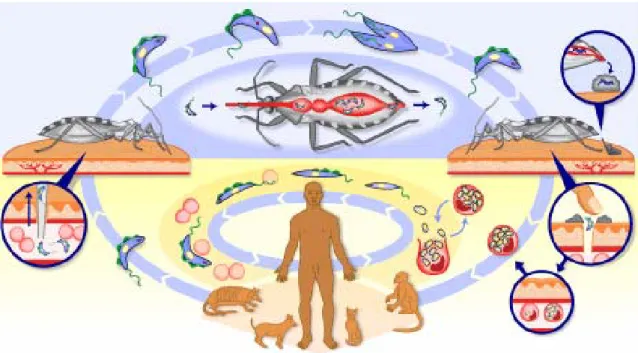
O ciclo evolutivo do T. cruzi no hospedeiro vertebrado inicia-se quando as formas tripomastigotas metacíclicas são eliminadas nas fezes e urina do inseto, durante seu repasto sangüíneo, penetrando na mucosa do hospedeiro. Nas células do hospedeiro, as tripomastigotas convertem-se em amastigotas replicativas que residem no citoplasma da célula do hospedeiro. Independente da via de infecção, amastigotas intracelulares, após sucessivas divisões binárias, transformam-se em tripomastigotas flageladas que, após romper a célula do hospedeiro, circulam na corrente sangüínea. As formas tripomastigotas podem invadir qualquer tipo celular, propagando a infecção pelo corpo (Brener et alii, 2000). Por fim, os tripomastigotas adquiridos durante a alimentação do triatomíneo com o sangue contaminado transformam-se em epimastigotas no trato digestivo do inseto antes de diferenciarem-se em formas tripomastigotas metacíclicas infectantes (Figura 1) completando o ciclo.
Purina nucleosídeo fosforilase.
As enzimas da família Purina nucleosídeo fosforilase, do inglês “purine nucleoside phosphorylase” (PNP, E.C. 2.4.2.1) catalisam a quebra da ligação glicosídica de ribo e deoxiribonucleosídeos gerando base purínica e ribose (deoxiribose)-1-fosfato, na presença de fosfato inorgânico (Pi), seu segundo substrato. Esta reação é direcionada, termodinamicamente, em favor da síntese de nucleosídeo (Friedkin, 1950). Contudo, nas células a fosforólise é altamente favorecida em detrimento da síntese, devido ao acoplamento com reações enzimáticas subsequentes. PNP é uma enzima ubíqua do metabolismo de purina, desempenhando papel na via de salvação dessa base, incluindo aquelas de parasitas protozoários, o que permite as células utilizarem as bases purínicas recicladas para síntese de nucleotídeos purínicos (Bzowska et alii, 2000).
A ubiqüidade das PNPs e sua distribuição em diversos tecidos e células foi documentado no passado por (Agarwal et alii, 1972; Stoeckler, 1984). Em humanos, a atividade mais intensa foi encontrada no rim, linfócitos periféricos e granulócitos. Contudo, levado em conta o volume celular, as células vermelhas mostram o mesmo nível de atividade que linfócitos periféricos. Logo, eritrócitos humanos, deficientes na síntese de novo de purinas e consequentemente dependente da reciclagem da mesma, são a maior fonte de PNP. Entretanto, eritrócitos de camundongos, ratos e outros animais contêm níveis de PNP consideravelmente menores, um fator importante na hora de extrapolar resultados de animais de laboratório para humanos (Stoeckler, 1984).
Além da fosforólise de nucleosídeos oriundos da degradação do DNA, as PNPs também atuam como enzima de salvação de substratos purínicos requeridos pela hipoxantina-guanina fosforibosiltransferase (HGFRT) para sintetizar os monofosfatos de inosina e guanosina, que serão por fim convertidos a deoxi-ribonucleotídeos pela enzima ribonucleotídeo redutase (Bzowska et alii, 2000).
As apresentações clínicas da deficiência homozigótica de PNPs em humanos incluem infecções recorrentes e linfopenia severa nos 2 primeiros anos de vida. Populações e funções de células T ficam reduzidas, enquanto as funções das células B são preservadas. Anemia hemolítica autoimune e neutropenia, lupus eritrematoso sistêmico, e linfomas de células B são observados nesses pacientes. Anormalidades neurológicas foram reportadas em mais de 50% das crianças afetadas pela deficiência de PNPs (Gilbertsen et alii, 1990; Hershfield et alii, 1995).
Acredita-se que as alterações em células T deficientes em PNP está relacionada com o acúmulo de dGTP que causa inibição da ribonucleotídeo redutase via retroalimentação negativa, bloqueando a redução da citidina di-fosfato (CDP) em deoxi-CDP (ddeoxi-CDP), necessária para a síntese de DNA, o que impede a maturação e diferenciação das células T. Contudo, em células B, não ocorre o acúmulo de dGTP pois estas apresentam atividade de diversas nucleotidases (Hershfield et alii, 1995). Contudo, mecanismos adicionais para o efeito do acúmulo de dGTP sobre as células T foram sugeridos (Duan et alii, 1990), onde a inibição da ribonucleotídeo redutase é um evento acessório, e outros mecanismos podem estar operando, como a inibição da síntese de RNA. Inclusive estudos com inibidores específicos para PNP apontam para a inibição de uma enzima que participa da síntese de DNA, mas que seja improvável ser a ribonucleotídeo redutase, pois o efeito dos inibidores não foi aumentado com a adição de dGTP.
Além disso, as PNPs têm a capacidade de realizar a reação oposta, síntese de nucleosídeos. Esta característica, apesar de menos estudada, é explorada para fins biotecnológicos, servindo de ferramenta para produção de análogos de nucleosídeos com potencial antiviral (Burns et alii, 1993) e anti-neoplásico (Chae et alii, 1998). A descrição dessa aplicação dessas enzimas foi documentado por Krenitsky et al (Krenitsky et alii, 1981). Outra aplicação relatada é utilização em sensores microeletrônicos, onde a partir da associação de enzimas foi possível detectar liberação de purina ou fosfato in vitro e in vivo (Haemmerli et alii, 1990; Llaudet et alii, 2003).
Classificação:
Uma delas baseia-se na especificidade a substratos e número de subunidades por oligômero, e foi dividida em duas categorias principais:
(1) Homotrímeros de baixa massa molecular, peso molecular (Mr) ~ 80 - 100
kDa, especificidade para 6-oxopurinas e seus nucleosídeos. Essa categoria também é chamada de “Ino-Guo fosforilases”, e listadas no banco de dados SWISS-PROT como “Fosforilases família 2 - PNP/5’-deoxi-5’-metiltioadenosine fosforilase (MTAF) (Pugmire et alii, 2002). Enzimas desse tipo foram isoladas de diversos tecidos de mamíferos (Agarwal et alii, 1972; Stoeckler, 1984) e também de microorganismos como Bacillus stearothermophilus (Hamamoto et alii, 1996), Cellulomonas sp. (Bzowska et alii, 1998).
(2) Homohexâmeros de alta massa molecular, Mr ~ 110 – 160 kDa, ampla
especificidade, aceitando tanto 6-oxo e/ou 6-aminopurinas e seus nucleosídeos. No banco de dados SWISS-PROT recebem a denominação de “Fosforilases família 1 – PNP/UDP”. Enzimas dessa categoria foram identificadas principalmente em microorganismos como Salmonella typhimurium (Jensen et alii, 1975), Klebsiella sp. (Takehara et alii, 1995) and Sulfolobus solfataricus (Cacciapuoti et alii, 1994).
Contudo essa é uma tentativa de classificação que se baseia nas características supracitadas e nas propriedades das quatro enzimas, de organismos diferentes, que tiveram suas estruturas tridimensionais resolvidas e depositadas no Protein Data Bank
(PDB). (Ealick et alii, 1990; Mao et alii, 1997; Bzowska et alii, 1998). Essas enzimas foram isoladas de eritrócitos humanos (PDB 1ULA), baço bovino (1VFN),
Cellulomonas sp (PDB 1QE5) e Escherichia coli (PDB 1ECP).
perfeitamente nas características gerais descritas ficam agrupadas em um terceiro grupo, onde as enzimas podem apresentar especificidade das PNPs de baixa massa mas não são homotrímeros ou apresentar especificidade das de alta massa molecular e não são homohexâmeros. Alguns exemplares desse grupo foram identificadas em bactérias (e.g.,
Proteus vulgaris), mamíferos (e.g., mitocôndria hepática bovina) e parasitas (e.g.,
Plasmodium falciparum) (Bzowska et alii, 2000).
Subfamília MTAF
Dentre os membros da família PNP, a enzima MTAF (E.C. 2.4.2.28) tem sido amplamente estudada, tanto a de origem humana quanto a de parasitas e microorganismos. Como esperado, esta enzima catalisa a clivagem nucleosídeos pelo intermédio de fosfato, contudo sua especificidade está voltada para o nucleosídeo modificado metiltioadenosina (MTA), porém com capacidade de metabolizar outros nucleosídeos purínicos.
MTAF humana
A MTAF humana, abundantemente encontrada em todas as células normais incluindo eritrócitos e células da medula óssea, é ausente em diversos tipos de cânceres como gliomas, câncer de pulmão de células não-pequenas, leucemia não-linfóide aguda, e melanoma (Della Ragione et alii, 1996; Schmid et alii, 1998; Behrmann et alii, 2003). O gene mtap está localizado no cromossomo 9p21 a uma distancia de 100 Kb dos genes supressores de tumor p16 e p14, igualmente ausentes em diversos tipos de tumor (Della Ragione et alii, 1996). Uma primeira hipótese para explicar a ausência de atividade da MTAF em tumores era a co-remoção homozigótica do gene mtaf com os genes supressores de tumor devido apenas a distância entre eles. Contudo, dados mais recentes sugerem que MTAF por si pode funcionar como um supressor de tumor. Por exemplo, demonstraram a ausência de MTAF não concomitante com a perda de p16 tanto em câncer de pulmão de células-não-pequenas como em gliomas (Schmid et alii, 1998; Brat
(Subhi et alii, 2003). Inclusive demonstrou-se que a sua super-expressão maligniza fibroblastos in vitro como também aumenta a freqüência de tumores de pele em camundongos transgênicos (Moshier et alii, 1993; O'Brien et alii, 1997). A partir da deficiência encontrada nesses diversos tipos de tumores vislumbrou-se a oportunidade de desenvolver uma terapia seletiva que pouparia as células normais. Esta parte do pressuposto que células deficientes em MTAF têm maior dependência da síntese de novo de nucleotídeos de adenina. A utilização bloqueadores da síntese de novo mataria as células MTAF-deficientes e as células normais seriam poupadas devido à reciclagem do MTA pela MTAF, suprindo adenina dessa forma (Batova et alii, 1999).
MTAF de parasitas
Esta enzima está presente em diversos parasitas como Trypanosoma cruzi
(Miller et alii, 1987), Trypanosoma brucei (Ghoda et alii, 1988), Schistosoma mansoni
(Savarese et alii, 1989), Leishmania donovani (Koszalka et alii, 1986) e é considerada um promissor alvo de drogas. Isto se deve principalmente porque a MTAF de parasitas, apesar de apresentar similaridades com a enzima de mamíferos, tem importantes diferenças. A enzima dos parasitas demonstra uma elevada especificidade em relação a outros nucleosídeos como adenosina e seus análogos (Ghoda et alii, 1988), o que não ocorre com a enzima de mamíferos. Tal fato auxilia o desenvolvimento de substratos subversivos seletivos apenas contra a enzima dos patógenos. Outra importante diferença, relacionada apenas aos tripanossomatídeos, é que estes não apresentam a síntese de novo de purinas. Fato que reforça esta via como alvo devido à grande dependência por fontes exógenas desses nucleosídeos, principalmente de adenina (Fish
Via metabólica do MTA
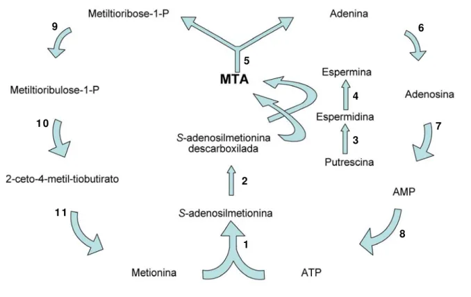
Poliaminas são cátions presentes em praticamente todas as células. As mais comuns são putrescina, espermidina e espermina. Pelo fato de serem cátions em pH fisiológico, por apresentarem conformação flexível e se ligarem reversivelmente a moléculas carregadas negativamente, as poliaminas desempenham uma variada gama de papéis bioquímicos, incluindo: cofator para síntese de macromoléculas, divisão e diferenciação celular e também como estabilizador conformacional de ácidos nucléicos (Cohen, 1998). Basicamente a produção delas se dá pela adição de grupos aminopropil transferidos da S-adenosilmetionina descarboxilada (dAdomet) para putrescina e espermidina, pelas enzimas espermidina e espermina sintase, respectivamente. Como subproduto dessa reação ocorre a formação do nucleosídeo metiltioadenosina (MTA). O MTA também é produzido na via da homoserina lactona, contudo sua contribuição para o total produzido não é conhecida, conferindo à via das poliamias a maior fonte desse metabólito (Williams-Ashman et alii, 1982; Walker et alii, 1997).
A existência do nucleosídeo metiltioadenosina (5’-deoxi-5’-metiltioadenosina), comummente abreviado a MTA, foi descoberta há quase um século (Williams-Ashman
et alii, 1982). Sua estrutura molecular foi descrita em 1924 e sua importância metabólica ficou aparente em 1952, em estudos sobre a relação entre metionina e 5’-tiometilribose. Devido à sua concomitância com a produção de poliaminas, o MTA está presente em todos os tipos de organismos, incluindo procariotos, protozoários, levedura, plantas e eucariontes (Williams-Ashman et alii, 1982).
O MTA está localizado numa encruzilhada do metabolismo celular, fazendo parte das vias de produção de poliaminas, salvação de purinas e de metionina. A Figura 2 representa um esquema das vias metabólicas às quais o nucleosídeo está integrado. O MTA é rapidamente clivado pela MTAF em adenina e metiltioribose-1-fosfato (MTR1P). A adenina é regenerada a adenosina e posteriormente a AMP em mamíferos ou no caso de vários parasitas diretamente a AMP pela enzima fosforibosiltransferase (el Kouni, 2003). Já o MTR1P é regenerado a metionina através de uma cascata enzimática.
Purinas são de vital importância para todos os organismos. Elas são essenciais para a síntese de ácidos nucléicos, proteínas e outros metabólicos, como também reações que requerem energia. Já a metionina, além do papel indispensável na formação proteínas, também é o metabólito precursor da Adomet, que está diretamente ligada à síntese de poliaminas, e ao processo de metilação de proteínas, lipídios e DNA.
Devido ao fato do MTA estar relacionado à via de salvação de dois precursores metabólicos de ávido consumo em células que se replicam constantemente, por exemplo
Figura 2. Via metabólica de síntese e metabolismo do nucleosídeo 5’- metiltioadenosina. O precursor do MTA, Adomet, é sintetizado pela enzima metionina adenosiltransferase (1). Em seguida, Adomet é descarboxilado pela Adomet descarboxilase (2). Adomet descarboxilada então é utilizada na síntese de poliaminas. Espermidina sintase (3) e espermina sintase (4) transferem o grupo propilamina da Adomet descarboxilada para putrescina e espermidina, respectivamente, em duas reações seqüenciais. A partir dessas duas reações ocorre a produção de MTA, substrato para a metiltioadenosina fosforilase (5). A última então catalisa a primeira reação de reciclagem de adenina e de metionina. Adenina é convertida a em ATP pela ação das enzimas Ade-fosforibosiltransferase (6) seguida da adenosina quinase (7) e finalmente fosforilado a ATP (8). A partir do outro produto da ação da MTAP, metiltioribose-1-P, o isômero metiltioribulose-1-P é gerado pela aldose-cetose isomerase (9). Este metabólito então sofre uma série de oxidações (10) resultando no 2-ceto-4-metil-tiobutirato, que é finalmente transaminado a metionina por uma aminotransferase (11).
6
2
3 4 5
1
8
7 9
Princípios teóricos da caracterização termodinâmica
Dicroísmo circular (CD do inglês circular dichroism) é a técnica espectroscópica relacionada à absorção diferenciada dos componentes circularmente polarizados para esquerda e direita de uma radiação plano-polarizada. Este efeito ocorre quando um cromóforo é quiral (opticamente ativo) pelo fato: (a) de ser intrínseco a sua estrutura, ou (b) estar covalentemente ligado a um centro quiral ou (c) estar situado em um ambiente assimétrico. Após os componentes da radiação eletromagnética passarem pela amostra e sendo um deles absorvido em maior amplitude, a radiação resultante estará elipticamente polarizada, isto é, a resultante terá uma trajetória em forma de elipse.
As propriedades biológicas de uma proteína estão intimamente relacionadas à sua estrutura tridimensional, as quais podem ser influenciadas por fatores químicos e físicos como, temperatura, pH e ligantes. Apesar do número elevado de estruturas atômicas de proteínas depositadas no bando de dados PDB pouco se sabe sobre o processo de dobramento destas moléculas. Decifrar o mecanismo responsável pelo dobramento correto e assim estabelecer a relação entre seqüência e estrutura tridimensional é de grande interesse biológico como também representaria grande avanço tecnológico e médico. A relevância clínica pode ser ilustrada pelo fato de que certas doenças são causadas pelo dobramento incorreto de proteínas, como a fibrose cística (Sifers, 1995; Thomas et alii, 1995). Informações sobre as etapas de dobramento protéico foram adquiridas principalmente a partir de polipeptídios pequenos, inclusive com a definição de características estruturais de fases intermediárias e da cinética de processos-chave, sendo que o DC desempenhou importante papel na obtenção desses dados.
Dicroísmo Circular e desnaturação térmica
cristalografia de raios-X (Provencher et alii, 1981). Outra utilidade decorrente é acompanhar as modificações da proteína durante processo de desdobramento por calor ou por agentes denaturantes. Essas modificações também podem ser acompanhadas por outros métodos espectroscópicos como emissão de fluorescência dos resíduos aromáticos da proteína, este tópico será posteriormente analisado.
Com a obtenção de espectros em temperatura crescente pode-se analisar o efeito desta sobre a molécula e determinar a região da transição entre estado nativo e desnaturado. A curva de desdobramento analisada matematicamente permite calcular os parâmetros que caracterizam a estabilidade de proteínas. Previamente, é importante estabelecer se a molécula em questão desnatura em um modelo de dois estados (isto é, nativo e desdobrado) ou outro mais complexo. Isto pode ser observado (a) pelo formato da curva de transição entre os estados e/ou (b) coincidência das curvas de transição monitoradas por diferentes métodos espectrofotométricos e/ou (c) equivalência do ΔH
Espectroscopia de Fluorescência
A espectroscopia de fluorescência, com suas múltiplas aplicações para análise de proteínas e para ciências da vida em geral, passou por rápido desenvolvimento na última década. Quando uma molécula absorve radiação visível e UV ela passa para o estado de excitação eletrônica ou eletrônica e vibracional simultaneamente. Uma parte da energia absorvida é transferida para outros níveis energéticos como os níveis vibracionais, fônons, energia térmica, entre outros. A energia remanescente é emitida como fótons com comprimento de onda maior do que o absorvido. Inclusive, algumas reações químicas têm a característica de emitir radiação enquanto estão ocorrendo. Em termos gerais, a radiação emitida nesses processos é chamada luminescência. Métodos de análise muito sensíveis utilizam fenômenos específicos da luminescência como a fluorescência, fosforescência e quimiluminescência.
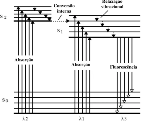
As transições no nível eletrônico e vibracional envolvidas no fenômeno da luminescência podem ser entendidas com a ajuda do diagrama de energia de Jablonski (Figura 2). Um estado eletrônico excitado (S1 ou S2) é observado quando um elétron,
localizado na orbital molecular ocupada de mais alta energia de um estado eletrônico fundamental (S0), passa para a próxima orbital de maior energia desocupada. A
excitação geralmente é acompanhada por mudanças no estado vibracional da molécula. Primeiramente ocorre a relaxação do estado excitado para um nível vibracional mais baixo, que ocorre por meio de uma transição não radioativa. Esta é seguida pela fluorescência, que é a relaxação para o estado eletrônico fundamental por meio da emissão de radiação eletromagnética.
Absorção
Absorção Fluorescência
Conversão interna
Relaxação vibracional
Figura 2. Diagrama de Jablonski, parcialmente representado, mostrando os processos que levam a luminescência.
A sensibilidade do triptofano à polaridade e mobilidade do ambiente faz de sua fluorescência uma importante ferramenta em estudos de dinâmica e estrutura de proteína. A fluorescência do triptofano sofre atenuação (quenching) pela água e que leva frequentemente a um decréscimo no rendimento quântico em situações de desnaturação da proteína, momento em que o triptofano se encontra exposto a um ambiente aquoso. Contudo existem exceções a essa regra, que causa a atenuação da emissão do triptofano mesmo com a proteína em seu estado nativo, por exemplo, ligações dissulfeto, os aminoácidos lisina e arginina quando estão carregados, histidina quando se encontra protonada, absorção por grupo heme, entre outros (Burstein, 1976; Burstein, 1977; Colucci et alii, 1990). A atenuação também pode ser causado por uma variedade de substâncias entre elas, oxigênio, iodeto, acrilamida, Cs2+, Cu2+. Além disso, a partir da atenuação causada por esses reagentes causam é possível mensurar o quão acessível está um resíduo de triptofano de uma proteína.
proteínas que não contém triptofanos em sua seqüência. Estudos que utilizam a fluorescência da fenilalanina como parâmetro são raros devido ao seu fraco rendimento quântico.
O
OBJETIVOS
OBJETIVO GERAL:
O objetivo geral do nosso trabalho é melhor entender a fisiologia do parasita T. cruzi e identificar diferenças entre as suas vias metabólicas e as do hospedeiro mamífero permitindo determinar novos alvos de drogas. Neste contexto, a caracterização molecular e biofísica da TcMTAP mostrou-se interessante devido à grande dependência do parasita pelas vias associadas a esta enzima. Acreditamos que essa fosforilase desempenha papel importante na manutenção da vida do T. cruzi e que apresenta diferenças moleculares suficientes para torna-la um promissor objeto de estudo vislumbrando o desenvolvimento de quimioterápicos mais específicos para o tratamento da doença de Chagas. Nossa pesquisa tem como objetivos específicos:
• Determinar a quantidade de cópias do gene mtaf presentes no genoma do
T. cruzi.
• Caracterizar as propriedades cinéticas da enzima MTAF recombinante.
• Determinar o padrão de expressão da MTAF nativa nas diferentes formas
do parasita.
• Caracterizar a estabilidade estrutural da MTAF recombinante.
R
Os resultados deste estudo estão apresentados na forma de manuscritos a serem submetidos à publicação em periódicos internacionais indexados. Os manuscritos são:
1. “The methylthioadenosine phosphorylase of Trypanosoma cruzi is
mesophilic and displays broad substrate specificity.”
2. “The drug target methylthioadenosine phosphorylase of Trypanosoma cruzi exhibits remarkable resistance to thermal and chemical
THE METHYLTHIOADENOSINE PHOSPHORYLASE of Trypanosoma cruzi IS MESOPHILIC AND DISPLAYS BROAD SUBSTRATE SPECIFICITY
David Neves, Izabela Dourado Bastos, Meire Maria de Lima, Gloria Restrepo-Cadavid, Antonio R. L. Teixeira, Francisco Javier Medrano, Philippe Grellier, Joseph Schrével, David Engman and Jaime Martins Santana.
Summary
Introduction
Chagas’ disease is an illness caused by the trypanosomatid protozoan
Trypanosoma cruzi and afflicts more than 18 million people in the American continent, from the south of Argentina to the south of the USA [1]. The disease is known over 90 years but a total curative drug is not available yet. The drugs currently used to treat T. cruzi infections often present low specificity and considerable toxicity, therefore newer compounds are being tested [2]. In lack of an efficient treatment for Chagas disease, there is an urge to define metabolic targets for the development of new drugs to treat and control consumptive chronic disease. The existing evolutionary differences between parasitic and human enzymes allow rational drug design, aiming at the development of new and specific compounds for the chemotherapy of “so called” incurable diseases [3,4]. For example, the pathogenic trypanosome´s purine transport system, which is absent in mammalian cells, allows the use of subversive analogs [5-7]. It has been shown that the purine and polyamine metabolism are essential to the trypanosomatids’ life and, thus, promising pathways for such approach [8]. Purines serve as precursor molecules for DNA and RNA, as carriers of high-energy phosphate bonds, as constituents of coenzymes and as modulators of certain enzymes. Methionine besides a constituent of proteins also has a key participation in protein methylation and polyamine biosynthesis. For instance, Trypanosoma brucei intensively uses methionine to methylate proteins and lipids and to form polyamines that play crucial roles in replication, differentiation and cellular growth. [9-11].
MTR1P is converted again to methionine in a five-step enzyme reaction [13]. The amount of recycled methionine plays an important role in polyamine production [4]. The purine salvage pathway, part of the purine metabolism, becomes even more important in trypanosomatids because they lack the molecular machinery to the de novo
synthesis of the purinic ring [14,15]. Therefore, uptake and salvage pathways are the main sources of purines for these protozoans [16,17]. Purine metabolism in pathogenic protozoans appears to offer several opportunities for chemotherapy: a) because no de novo synthesis of these compounds occurs, interdiction of the salvage pathway has far greater implications than interruption of similar pathways in mammals; b) some of the enzyme systems are capable of accepting purine analogs and metabolizing them to nucleotides; c) these analogs serve as metabolic inhibitors [18,19].
MTAPs (E.C. 2.4.2.28) belong to the purine nucleoside phosphorylase (PNP) family. It has been proposed that this family should be divided into low- and high-molecular mass categories based on primary sequence homology, quartenary structure and substrate specificity. Whereas PNP proteins do not usually show high sequence homology, structure comparisons show striking significant similarities [20,21]. The members of this family are ubiquitously distributed in nature, from Archaea to human, showing heterogeneous kinetics and substrate preferences [20].
MTAPs of protozoan parasites have been categorized as potential chemotherapeutic targets because of their relation to crucial physiological processes, purine and methionine salvage and the important differences between the host and the parasite metabolism in these pathways. [10,22,23]. The African trypanosome T. brucei
showing a molecular mass of 68-kDa, as determined by gel filtration chromatography [26].
In this study we report the identification and characterization of the gene encoding for TcMTAP (Tcmtap). The amino acid deduced sequence of TcMTAP shares significant identity with other MTAPs. The gene was cloned and expressed in heterologous system allowing substrate characterization, kinetic constants calculation and determination of optimal temperature and pH. Its biochemical and enzymatic properties lead us to consider TcMTAP a member of PNP/MTAP family 2. TcMTAP is differentially expressed by the three developmental forms of T. cruzi. Next, we showed that the enzyme recovers its structure and function after chemical denaturation and assembles into dimer in non-reducing environment.
RESULTS AND DISCUSSION
The TcMTAP belongs to the MTAP/PNP family 2
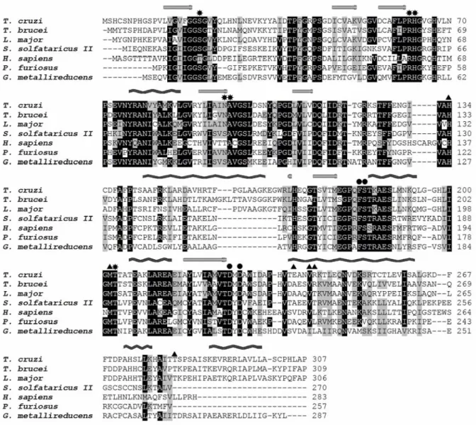
The Tcmtap gene was identified through screening of a T. cruzi cDNA library with a specific probe and its sequence contains an ORF of 921 bp that encodes a predicted 307 amino acid protein with a calculated molecular mass of 33,184 Da (Fig. 1). The functional identification of Tcmtap is corroborated by the presence, in the translated gene sequence, of the PNP/MTAP family 2 domain signature [LIVF] - x[7] - [GS] - x(2) - H - x - [LIVMFY] - x(4) - [LIVMF] - x[7] - [ATV] - x(1,2) - [LIVM] - x - [ATV] - x(4) - [GN] - x(3,4) - [LIVMF](2) - x(2) - [STN] - [SAGT] - x - G - [GS] - [LIVM]. Multiple amino acid sequence alignment shows that TcMTAP shares high identity with its kinetoplastid counterparts, 59% with T. brucei and 58% with
Leishmania major. The enzyme also exhibits considerable identity with
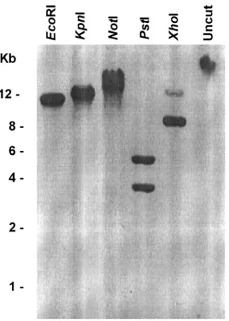
and Pyrococcus furiosus enzymes, respectively, while it is only 35% identical with human MTAP. Our sequence is almost identical, nine amino acids differing, to the sequences from the TIGR T. cruzi genome project (data not show; http://www.tigr.org/tdb/e2k1/tca1/; accession number XM_814834). The presented differences are probably because we sequenced the gene from Berenice stock whereas the genome sequences are from the hybrid CL. Brener, which is heterozygous at many loci [27]. The Tcmtap organization in the T. cruzi genome was verified by Southern blot analysis. A single band was revealed when the genomic DNA was digested with EcoRI,
KpnI or NotI and probed with the full-length Tcmtap (Fig. 2). Differently, when the DNA was digested with PstI and XhoI enzymes, the probe hybridized with two bands. This pattern correlates well with sites for PstI and XhoI at position 14 and 734 at the gene sequence, respectively. These results suggest that the Tcmtap gene is represented as a single copy per haploid genome of the parasite.
The active site of human MTAP (hMTAP) [12] is divided in three specialized parts: phosphate-, pentose-, and base-binding sites. The amino acid residues involved in these three parts of hMTAP active site are present in the sequences of other MTAPs (Fig. 1). Since the base-binding region is completely conserved among MTAPs, we speculate that these enzymes may share substrate specificity towards MTA. The phosphate- and methylthioribose-binding sites of hMTAP are also conserved among the MTAPs but few conservative substitutions occur. Then, we asked whether TcMTAP
also shares secondary structure with other MTAPs. The consensus prediction of α-helix
and β-sheet structures is shown in Fig. 1. Extended loops, characteristic to all PNPs, link the secondary structural elements and form the contacts between the monomers can
be observed. The β-sheets form the monomer core, known to be similar throughout the
Fig. 2.Tcmtap is a single-copy gene. Southern blot of T. cruzi genomic DNA digested with EcoRI, KpnI, NotI, PstI or XhoI was performed using a tcmtaf cDNA probe. The probe revealed two bands at PstI and XhoI lanes as expected by the analysis of Tcmtap
homology is low or nil, structural similarity is quite high among PNPs [20]. It has been postulated (Pugmire, 2002) that sequence divergence and structure similarity may be consequence of a divergent evolutionary event in ancient PNP fold. This common ancestor had probably accepted a wide range of nucleosides substrates, becoming more specialized over time, a specialization observed in eukaryotic organisms. In conjunction, these results indicate that TcMTAP is a member of the MTAP/PNP family 2.
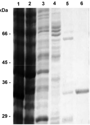
rTcMTAP monomer shows 33-kDa mass
monomer. Similar phenomenon was reported in the heterologous expression of MTAP from S. solfataricus [28]. A factor contributing to this issue is the inability of bacteria to attain an entire gamut of post-translational modifications a protein requires. An example and most important in our case, the formation of intra- or intermolecular disulfide bonds do not occur in the reducing cytoplasm of E. coli, where the recombinant proteins are stored in absence of a secretion signal [29]. Considering that during purification the rTcMTAP faces an oxidative environment that facilitates the formation of disulfide bonds, we added the reducing agent DTT to perform activity assays intending to avoid misplaced disulfide bonds influencing the kinetics measurements.
Native TcMTAP shows 30-kDa mass
The specificity of polyclonal antibodies raised against rTcMTAP was tested against E. coli total protein extract and identified a single band, corresponding to the enzyme expected mass (data not shown). Western blotting performed with T. cruzi
native TcMTAP that was estimated by gel filtration [26]. Regarding this issue, previous reports on PNPs have showed problems defining molecular mass and subunit composition based on electrophoresis, gel filtration and other low resolving methods [20]; for e.g. a dimer composition had been proposed to the human erythrocyte PNP based on gel filtration and sedimentation equilibrium assays [30], but it has been considered obsolete since the establishment of its trimeric structure in crystal (PDB 1ULA). A reasonable explanation for this incongruence is the existence of an equilibrium mixture of species with different subunit composition when the protein is in solution, previously reported fact in PNP family [20].
TcMTAP is differentially expressed through T. cruzi’s developmental stages
Enzymatic Assay
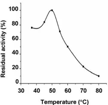
To test the recombinant TcMTAP activity and to calculate the kinetics constants, a spectrophotometric assay was employed to measure the conversion of MTA into adenine as previously reported [32]. The temperature dependence of rTcMTAP activity assayed in the range from 37 °C to 80 °C is reported in Fig. 6. The enzyme appears moderately thermophilic, showing an optimal temperature of 50 °C, keeping 35% activity at 70 °C. Thermostability is a common characteristic among PNPs from thermophilic and non-thermophilic organisms, e.g. E. coli PNP is stable for 10 min at 55 °C [33] and that of Klebsiella for 16 h at 60 °C [34]. Forterre [35] has postulated that the mesophilic prokaryotes existing today must have evolved through a gradual adaptation of thermophilic enzymes to lower temperature optima. The presence of thermophilic enzymes may represent an evolutionary holdover from a thermophilic ancestor. We also tested the pH effect on MTA phosphorolysis (Fig. 7). It was shown that the enzyme has a dependence on neutral pH but maintains its activity over a broad pH range. Although maximum activity was observed at pH 7.4, the measured enzymatic activities at pH 5.0 and 8.0 were 51 and 78 % of the optimal. These findings are in accordance with the results obtained for the native enzyme [26]. Furthermore, PNPs, with few exceptions, show broad pH activity [20].
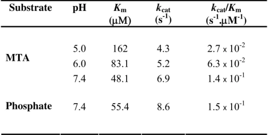
The kinetics parameters of rTcMTAP for phosphate and MTA substrates were determined at 37 °C that is closer to the natural conditions encountered at the mammal host. The Km, kcat and kcat/Km values for the substrates MTA and phosphate were
calculated and typical Michaelis-Menten kinetics were observed (Table 1). In previous reports the Km and substrate specificity from the partially purified native T. cruzi and T.
brucei MTAPs were calculated [22,26], exhibiting a Km of 3 and 2 μM for MTA
Fig. 6.rTcMTAP exhibited maximum activity at 50 °C. The effect of temperature on recombinant TcMTAP activity was determined, revealing a possible heritage from a thermophilic ancestor. Activity rapidly drops above 50 °C.
for MTA of the recombinant enzyme. This affinity difference between the native and recombinant enzymes may be due to the fact that the native proteins were partially purified from the parasite extract, allowing a masked promiscuous nucleoside phosphorylase to alter the rates. The substrate specificity results were very similar between the native enzymes and rTcMTAP, being MTA the preferred substrate in all cases.
The enzyme shows an increasing affinity for MTA when the pH moves towards 7.4 (Km 48.1 µM) as compared to pH 5.0 (Km 162 µM) and 6.0 (Km 83.2 µM). The
increasing affinity is expected since pH 7.4 is the optimal and, most likely, at this condition intra chain interactions are at their best configuration and the protein is more stabilized. The Km for the substrate phosphate was almost identical to that of MTA,
indicating no preference in the order of substrate binding.
Table 1. Kinetic parameters of rTcMTAP. Kinetics parameters against MTA and
phosphate were determined at 37 °C at different pH.
Km kcat kcat/Km Substrate pH
(s-1)
(μΜ) (s-1.μM-1)
5.0 162 4.3 2.7 x 10⁻²
6.0 83.1 5.2 6.3 x 10⁻² MTA
7.4 48.1 6.9 1.4 x 10⁻¹
7.4 55.4 8.6 1.5 x 10⁻¹
Phosphate
Table 2. Substrate specificity of rTcMTAP against nucleosides substrates.N.d. non
detected. Rates were normalized by setting the rate of MTA cleavage at 100%. The
actual rate was 2.2 μmoles/ min.mg protein.
Substrate Relative rate of cleavage (%)
MTA 100 Adenosine 83 Deoxy-adenosine 78
N.d.
Inosine
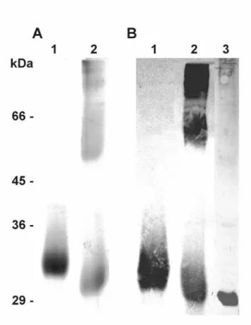
rTcMTAP reassumes its native structure after denaturation.
To analyze the rTcMTAP capability of renaturation, we first incubated the enzyme in the presence of 6 M guanidine-HCl during 24 h at 25 °C. The denaturation was evaluated by monitoring the shift in fluorescence maximum wavelength upon excitation at 295 nm. In the folded state the protein exhibited relative fluorescence emission maximum at 333 nm, which is considered a characteristic of tryptophan that is in a low-polar hydrophobic environment (Fig. 8, slope A) [37]. After denaturation, the fluorescence showed a large red shift due to the increased exposure of the tryptophanyl residues to the more polar aqueous environment (Fig. 8, slope C). The refolding process was started by 20-fold dilution of the sample. Extensive dialyses were performed until complete removal of the denaturant. The refolding process was monitored by fluorescence measurements and activity assays. After refolding, rTcMTAP exhibited a fluorescence spectrum with almost the same profile as the native protein, i.e. a fluorescence maximum emission at 337 nm (Fig. 8, slope B). The refolded enzyme activity was 51% of the control protein. On the basis of the reported data, we conclude that rTcMTAP can recover its fluorescence spectrum to that prior to the unfolding process, which can be also associated with recovered tertiary structure. The MTAP of P. furiosus subjected to a similar experiment also exhibited a fluorescence spectrum with the same features as the native enzyme after the refolding process [38]. Nevertheless, the P. furiosus protein only exhibited the correct spectrum when the denaturation process was carried out in the presence of reducing agents, which led us to assume that intact disulfide bonds in rTcMTAP do not interfere with the refolding process.
good targets for drugs [23]. Since a biosynthetic pathway is lacking, T. cruzi must acquire purine ring through host and endogenous metabolite recycling. Then, inactivation of TcMTAP by specific inhibitors or through gene disruption would reveal its function in both T. cruzi biology and pathogenesis of Chagas disease. The molecular and biochemical features of TcMTAP presented here and further investigations would contribute to rational development of leads aiming at chemotherapy of T. cruzi
Fig. 8. Recombinant TcMTAP recovers native folding after chemical denaturation.
MATERIALS AND METHODS
Parasites
T. cruzi epimastigote forms from Berenice stock were grown in liver infusion tryptose medium supplemented with 100 units/ml penicillin, 100 µg/ml streptomycin and 10 % (v/v) fetal calf serum at 28 °C with continuous agitation. Trypomastigote and amastigote forms of the parasite were obtained by monolayer culture of murine muscle L-6 cells grown in RPMI medium containing 10 % fetal calf serum at 37 °C in 5 % CO2 and then purified as described previously [39,40].
Cloning and expression of T. cruzi MTAP
The Tcmtap gene was identified upon screening a cDNA library with serum from T. cruzi-infected rabbit as described (Caetano MAI, Garcia M MBP) and completely sequenced on both sides. Specific primers MTAP1 (forward,
5´-cgcgagCATATGTCCCACTGCAGCATTCCAC-3´; lowercase, random bases; underlined, NdeI site; bold, initiation codon) and MTAP2 (reverse,
5´-cacagaCTCGAGCAATCATGGGGCGAGATGCGGG-3´ lowercase, random bases; underlined, XhoI site; bold, stop codon) were synthesized. T. cruzi cDNA was synthesized using total RNA primed with oligo(dT) and mini-exon primers to assure amplification of complete and spliced mRNAs (Thermoscript RT-PCR system, Invitrogen). Then, the cDNA product was used to amplify the complete ORF with primers MTAP1 and MTAP2 using Platinum Taq DNA polymerase high fidelity
confirmed by sequencing. Secondary structure of the protein was predicted by submitting its primary sequence to the Jpred server (http://www.compbio.dundee.ac.uk/~www-jpred/). The N-terminal His tagged
TcMTAF was expressed in E. coli BL21(DE3) strain at 37 °C upon induction with 1
mM isopropyl-β-D-thiogalactoside (IPTG) during 5 h. Cells were harvested by centrifugation, lysed with BugbusterTM (Novagen) and cell debris were removed by centrifugation at 20,000 g for 20 min at 4 °C.
The recombinant protein was purified with the His-Bind resin and buffer kit (Novagen). The bacterial protein extract supernatant was loaded on a nickel charged column previously equilibrated with binding buffer. After the non-ligated proteins were eluted, the contaminant proteins were washed away from the column with 10 volumes of binding buffer, then with 3 volumes of washing buffer, and a final wash containing 100 mM imidazole. Finally, the His10 N-terminal tagged MTAP was eluted with 6 volumes
of elution buffer, containing 400 mM imidazole. After dialysis against 10 mM Tris-HCl pH 7.4 overnight at 4 °C, the purified protein was concentrated with a Centricon 10 (Millipore). The protein concentration was determined by measuring A280 using the
predicted extinction coefficient of 21980 M-1 cm-1, calculated with ProtParam tool
(http://www.expasy.org/tools/protparam.html). The cDNA sequence of TcMTAP is available in GenBank® under the accession number AY144609.
Enzymatic assay
The activity of TcMTAP was spectrophotometrically determined by measuring the
conversion of MTA into adenine as a decrease in absorbance at 275 nm to give Δε
mM DTT (reaction buffer) and 250 μM MTA at 37 °C. The optimal pH of TcMTAP activity was assayed as above in the following 100 mM buffers: Na-acetate pH 4.5 and 5.0, Na-citrate pH 5.6, Bis-Tris pH 6.0 and 6.8, K-phosphate pH 7.4 (no phosphate added), HEPES pH 7.7 and 7.9, Bicine pH 8.6, and Tris-HCl pH 9.0. To determine rTcMTAP optimal temperature activity, the reactions were performed at 37, 45, 50, 55, 60, 70, or 80 °C. The reaction mixture to calculate the Km and Vmax for MTA (20 – 333
μM) and phosphate substrates (50 – 2,200 μM) were reaction buffer and 100 mM Hepes
pH 7.4, 2 mM DTT and 500 μM MTA, respectively. The kinetics parameters were
calculated using a nonlinear regression of Michaelis-Menten equation in a Beckman Coulter spectrophotometer DU640 equipped with a temperature control system. All constants have a p <0.05.
Activity against 1 mM adenosine (Ado), 1 mM inosine (Ino), 1 mM 2’-deoxyadenosine (dAdo) or 0.04 mM guanosine (Guo) substrates was measured in a xanthine oxidase-coupled spectrophotometric assay [41,42]. This assay was carried out as described above in reaction buffer containing 0.1 UI xanthine oxidase /mL. The extinction coefficient for Ado and dAdo is 15.5, 12.0 for Ino and -4.2 mM-1 cm-1 for Guo, monitored at 305, 293 and 255 nm, respectively [41]. The reactions were initiated by addition of purified recombinant TcMTAP.
Anti-TcMTAP antiserum production
Polyclonal antibodies against T. cruzi MTAP was generated by subcutaneous inoculation of the purified recombinant protein into New Zealand rabbit. Pre-immune blood was drawn prior to the initial immunization, and the serum was used as a negative
enzyme in incomplete adjuvant, followed by a final booster with the enzyme alone. Sera were stored at - 20 °C in the presence of 50% glycerol.
Immunobloting
Soluble protein extracts (20 µg) from IPTG-induced BL21 bacteria either carrying pTcMTAP or empty vector, purified rTcMTAP or total proteins from amastigotes, epimastigotes or trypomastigotes of T. cruzi, corresponding to 1 × 10⁷ cells/well, were subjected to SDS-PAGE (10 % polyacrylamide) under reducing conditions. Parasites were solubilized directly in the electrophoretic sample buffer. Proteins were transferred on to a nitrocellulose membrane and blocked by incubation in 5 % (w/v) non-fat milk/PBS overnight at 4 °C. Blots were incubated for 2 h with control or anti-rTcMTAP serum diluted in 1 % non-fat milk/PBS. After three washes of 5 min each with PBS, membranes were incubated for 2 h with alkaline phosphatase-conjugated goat anti-rabbit IgG (Zymed) diluted to 1:2000, washed as above, and the immunocomplexes were revealed with the alkaline-phosphatase substrate 5-bromo-4-chloro-3-indolyl-1-phosphate /Nitro Blue Tetrazolium (Promega).
Refolding
in the absence or presence of 6 M Gnd-HCl in 20 mM K-phosphate pH 7.4. The unfolding process was followed by changes in fluorescence emission. After 24 h, the emission spectra were recorded; refolding was started by 20-fold dilution of the samples in 20 mM K-phosphate pH 7.4 at 25 °C. The final concentration of Gnd-HCl in the renaturation mixture was 0.3 M, whereas the protein concentration was 90 μg/mL. The
samples were extensively dialyzed against 20 mM K-phosphate pH 7.4 until complete removal of Gnd-HCl, and then activity measurements under standard conditions and fluorescence emission spectra were recorded. Experiments were normalized in accordance with protein concentration before and after renaturation and buffer fluorescence emission were subtracted [38].
REFERENCES
[1] Prata A (2001). Clinical and epidemiological aspects of Chagas disease. Lancet Infect Dis1, 92-100.
[2] Urbina JA (2001). Specific treatment of Chagas disease: current status and new developments. Curr Opin Infect Dis14, 733-741.
[3] Bacchi CJ, Goldberg B, Rattendi D, Gorrell TE, Spiess AJ & Sufrin JR (1999). Metabolic effects of a methylthioadenosine phosphorylase substrate analog on African trypanosomes. Biochem Pharmacol57, 89-96.
[4] Bacchi CJ & Yarlett N (2002). Polyamine metabolism as chemotherapeutic target in protozoan parasites. Mini Rev Med Chem2, 553-563.
[5] Marasco CJ, Jr., Kramer DL, Miller J, Porter CW, Bacchi CJ, Rattendi D, Kucera L, Iyer N, Bernacki R, Pera P & Sufrin JR (2002). Synthesis and evaluation of analogues of 5'-([(Z)-4-amino-2-butenyl]methylamino)-5'-deoxyadenosine as inhibitors of tumor cell growth, trypanosomal growth, and HIV-1 infectivity. J Med Chem45, 5112-5122.
[6] Byers TL, Casara P & Bitonti AJ (1992). Uptake of the antitrypanosomal drug 5'-([(Z)-4-amino-2-butenyl]methylamino)-5'-deoxyadenosine (MDL 73811) by the purine transport system of Trypanosoma brucei brucei. Biochem J 283 ( Pt
[7] Tolbert WD, Ekstrom JL, Mathews, II, Secrist JA, 3rd, Kapoor P, Pegg AE & Ealick SE (2001). The structural basis for substrate specificity and inhibition of human S-adenosylmethionine decarboxylase. Biochemistry40, 9484-9494. [8] Berger BJ, Carter NS & Fairlamb AH (1993). Polyamine and pentamidine
metabolism in African trypanosomes. Acta Trop54, 215-224.
[9] Goldberg B, Yarlett N, Rattendi D, Lloyd D & Bacchi CJ (1997). Rapid methylation of cell proteins and lipids in Trypanosoma brucei. J Eukaryot Microbiol44, 345-351.
[10] Sufrin JR, Meshnick SR, Spiess AJ, Garofalo-Hannan J, Pan XQ & Bacchi CJ (1995). Methionine recycling pathways and antimalarial drug design.
Antimicrob Agents Chemother39, 2511-2515.
[11] Goldberg B, Rattendi D, Yarlett N, Lloyd D & Bacchi CJ (1997). Effects of carboxylmethylation and polyamine synthesis inhibitors on methylation of Trypanosoma brucei cellular proteins and lipids. J Eukaryot Microbiol44, 352-358.
[12] Appleby TC, Erion MD & Ealick SE (1999). The structure of human 5'-deoxy-5'-methylthioadenosine phosphorylase at 1.7 A resolution provides insights into substrate binding and catalysis. Structure7, 629-641.
[13] Williams-Ashman HG, Seidenfeld J & Galletti P (1982). Trends in the biochemical pharmacology of 5'-deoxy-5'-methylthioadenosine. Biochem Pharmacol31, 277-288.
[14] Ceron CR, Caldas RD, Felix CR, Mundim MH & Roitman I (1979). Purine metabolism in trypanosomatids. J Protozool26, 479-483.
[15] Berens RL KE, Marr JJ (1995) in: Biochemistry and molecular biology of parasites., pp. 89-117 (M, M.J.a.M., Ed.) Academic Press, London.
[16] Carter NS & Fairlamb AH (1993). Arsenical-resistant trypanosomes lack an unusual adenosine transporter. Nature361, 173-176.
[17] Goldberg B, Rattendi D, Lloyd D, Sufrin JR & Bacchi CJ (2001). In situ kinetic characterization of methylthioadenosine transport by the adenosine transporter (P2) of the African Trypanosoma brucei brucei and Trypanosoma brucei rhodesiense. Biochem Pharmacol61, 449-457.
[19] Marr JJ (1991). Purine analogs as chemotherapeutic agents in leishmaniasis and American trypanosomiasis. J Lab Clin Med118, 111-119.
[20] Bzowska A, Kulikowska E & Shugar D (2000). Purine nucleoside phosphorylases: properties, functions, and clinical aspects. Pharmacol Ther 88, 349-425.
[21] Pugmire MJ & Ealick SE (2002). Structural analyses reveal two distinct families of nucleoside phosphorylases. Biochem J361, 1-25.
[22] Ghoda LY, Savarese TM, Northup CH, Parks RE, Jr., Garofalo J, Katz L, Ellenbogen BB & Bacchi CJ (1988). Substrate specificities of 5'-deoxy-5'-methylthioadenosine phosphorylase from Trypanosoma brucei brucei and mammalian cells. Mol Biochem Parasitol27, 109-118.
[23] el Kouni MH (2003). Potential chemotherapeutic targets in the purine metabolism of parasites. Pharmacol Ther99, 283-309.
[24] Bacchi CJ, Sufrin JR, Nathan HC, Spiess AJ, Hannan T, Garofalo J, Alecia K, Katz L & Yarlett N (1991). Alkyl-substituted analogs of 5'-methylthioadenosine as trypanocides. Antimicrob Agents Chemother 35, 1315-1320.
[25] Bacchi CJ, Sanabria K, Spiess AJ, Vargas M, Marasco CJ, Jr., Jimenez LM, Goldberg B & Sufrin JR (1997). In vivo efficacies of 5'-methylthioadenosine analogs as trypanocides. Antimicrob Agents Chemother41, 2108-2112.
[26] Miller RL, Sabourin CL & Krenitsky TA (1987). Trypanosoma cruzi adenine nucleoside phosphorylase. Purification and substrate specificity. Biochem Pharmacol36, 553-560.
[27] Sturm NR, Vargas NS, Westenberger SJ, Zingales B & Campbell DA (2003). Evidence for multiple hybrid groups in Trypanosoma cruzi. Int J Parasitol 33, 269-279.
[28] Cacciapuoti G, Fusco S, Caiazzo N, Zappia V & Porcelli M (1999). Heterologous expression of 5'-methylthioadenosine phosphorylase from the archaeon Sulfolobus solfataricus: characterization of the recombinant protein and involvement of disulfide bonds in thermophilicity and thermostability.
Protein Expr Purif16, 125-135.
[30] Lewis AS & Lowy BA (1979). Human erythrocyte purine nucleoside phosphorylase: molecular weight and physical properties. A Theorell-Chance catalytic mechanism. J Biol Chem254, 9927-9932.
[31] Hirschhorn R (1985). Complete and partial adenosine deaminase deficiency. Relationship of immune function to metabolite concentrations, enzyme activity, and effects of therapy. Ann N Y Acad Sci451, 20-25.
[32] Singh V, Shi W, Evans GB, Tyler PC, Furneaux RH, Almo SC & Schramm VL (2004). Picomolar transition state analogue inhibitors of human 5'-methylthioadenosine phosphorylase and X-ray structure with MT-immucillin-A.
Biochemistry43, 9-18.
[33] Krenitsky TA, Koszalka GW & Tuttle JV (1981). Purine nucleoside synthesis, an efficient method employing nucleoside phosphorylases. Biochemistry 20, 3615-3621.
[34] Ling F, Inuoe Y & Kimura A (1994). Induction, purification and utilization of purine nucleoside phosphorylase and uridine phosphorylase from Klebsiella sp.
Process biochemistry29, 355-361.
[35] Forterre P (1995). Looking for the most "primitive" organism(s) on Earth today: the state of the art. Planet Space Sci43, 167-177.
[36] Toorchen D & Miller RL (1991). Purification and characterization of 5'-deoxy-5'-methylthioadenosine (MTA) phosphorylase from human liver. Biochem Pharmacol41, 2023-2030.
[37] Volotovskii ID & Konev SV (1967). [Relation between the conformation and UV luminescence of proteins]. Biofizika12, 200-205.
[38] Cacciapuoti G, Moretti MA, Forte S, Brio A, Camardella L, Zappia V & Porcelli M (2004). Methylthioadenosine phosphorylase from the archaeon Pyrococcus furiosus. Mechanism of the reaction and assignment of disulfide bonds. Eur J Biochem271, 4834-4844.
[39] Ley V, Robbins ES, Nussenzweig V & Andrews NW (1990). The exit of Trypanosoma cruzi from the phagosome is inhibited by raising the pH of acidic compartments. J Exp Med171, 401-413.
[41] Miller RL & Lindstead D (1983). Purine and pyrimidine metabolizing activities in Trichomonas vaginalis extracts. Mol Biochem Parasitol7, 41-51.
THE DRUG TARGET METHYLTHIOADENOSINE PHOSPHORYLASE of
Trypanosoma cruzi EXHIBITS REMARKABLE RESISTANCE TO THERMAL AND
CHEMICAL DENATURATION.
Summary
Methylthioadenosine phosphorylase (MTAP) belongs to the family 2 of purine nucleoside phosphorylase/MTAP, whose members assembles into different oligomeric constitutions. These widely distributed enzymes catalyze the phosphorolysis of MTA, a byproduct of polyamine biosynthesis and are the starting point of purine and methionine salvage pathways. Herein, we report the physicochemical characterization of the MTAP of the trypanosomatid Trypanosoma cruzi (TcMTAP) that is considered a potential target for chemotherapy of Chagas disease, an incurable sickness responsible for thousands of deaths in Latin America. Thermal denaturation followed by circular dichroism performed at broad range of pH showed reduced dependence between enzyme stability and environment ionization condition. TcMTAP exhibited striking resistance to thermal denaturation, unfolding only above 79 °C. Its secondary structure content is significantly composed of α-helices, as predicted from circular dichroism data. However, even after thermal denaturation, the content of α-helices showed little variation, markedly at pH 7.4 and 9.0. Chemical denaturation of TcMTAP, induced by guanidine-hydrochloride or urea, was monitored by far-UV circular dichroism and fluorescence spectroscopy. The enzyme kept folded in concentration as high as 3.6 M of guanidine-hydrochloride and 8 M of urea. The thermodynamic parameters calculated from thermal and chemical denaturation assays were in good agreement, ΔG(H2O) =
INTRODUCTION
Methylthioadenosine phosphorylase (MTAP; E.C. 2.4.2.28) belongs to the purine nucleoside family 2 (PNP, E.C. 2.4.2.1) and is ubiquitously distributed in nature. It phosphorolytically cleaves the glycosidic bond of the sulfur-containing nucleoside methylthioadenosine (MTA), which is a metabolic product of S-adenosylmethionine (AdoMet) in polyamine biosynthesis. MTA is also the starting point of two major salvage pathways, the purine and methionine ones [1]. MTAP readily metabolizes MTA to adenine and methylthio-ribose-1-phosphate (MTR1P). Adenine will ultimately replenish the AMP and ATP pool whereas MTR1P will be converted to methionine. The MTAP and the intermediates of the pathways associated with MTA recycling have been considered chemotherapy targets against human cancers related with alterations at the chromosomal locus 9p21 and against protozoan diseases such as trypanosomiasis, leishmaniasis and malaria. This is because those cancer cells and protozoa parasites lack the machinery to synthesize the purine ring de novo and, thus, depend on the salvage pathway [2,3]. Differently from human MTAP, that show high specificity for MTA, MTAPs of parasitic protozoa also displays activity on other nucleosides and its analogues [4]. For instance, it has been shown that Trypanosoma brucei and
Trypanosoma rhodesiense are susceptible to the MTA analog, 5’-deoxy-5’-(hydroxyethylthio)adenosine (HETA), which was able to cure trypanosoma-infected mice without damaging mammalian cells [5,6].
Thermodynamic characterization may contribute to explain enzyme’s biochemical properties, for example a broad pH range activity, thermostability, and substrate preference [7]. In addition, the biophysical data can also provide insights into the molecular mechanisms related with protein stability, overall structure and biotechnological applicability [8], e. g. protein-ligand affinities, presence of intermediate in the folding/unfolding process and rational drug design [9,10].
likewise other kinetoplastids, is unable to synthesize the purine ring de novo [4,12,13]. We have biochemically characterized the recombinant purine-salvage-pathway participating enzyme MTAP of T. cruzi (rTcMTAP) [14]. The enzyme is active over a broad pH range, displays considerable thermostability and is capable to renature after chemical unfolding. rTcMTAP assembles into oligomers in solution, which apparently is composed by a mixture of association species [14]. Similar results were found by, Wielgus-Kutrowska [15], on Cellulomonas sp. PNP.
In this study we calculated the thermodynamic parameters and characterized the structural stability of the recombinant MTAP from T. cruzi. rTcMTAP exhibited striking resistance to thermal and chemical denaturation, followed by circular dichroism (CD) and fluorescence spectroscopy. The enzyme unfolds as a two-state model and increases its thermal stability at acid to neutral pH. In addition, the secondary structure content suffers little variation during the thermal unfolding process. We also tested the presence of stabilizers hexadecyltrimethylammonium bromide (CTAB) and sorbitol. which altered the enzyme stability and its denaturation pattern. Until present date, only the Cellulomonas sp. PNP [15] had its biophysical parameters fully determined, but no member of MTAP sub-family.
RESULTS
Thermal stability
Far-UV circular dichroism spectra of rTcMTAP at pH ranging from 4.0 to 9.0, recorded during thermal denaturation, show maximum peak at 192 nm and dichroic bands at 208 and 222 nm (Fig. 1). In addition, the protein spectra pattern, even after the unfolding transition, kept similar to the native, markedly at pH 7.4 and 9.0. To confirm this hypothesis, the secondary structure content was predicted [16] at 20 and 95 °C for all pH (Table 1) using the CDPro software package. The α-helix content showed modest decrease after denaturation, more strikingly at pH 7.4 and 9.0. These data indicate the secondary structure content of rTcMTAP is formed predominantly of α -helix and that thermal denaturation has diminute effect over its content.
process (Fig. 2). By these results, we assumed that rTcMTAP unfolding process follows a two-state model since the shape of the transition curve is characteristic of a highly cooperative process. The sharp transition curves resulted in melting temperatures (Tm)
equal or slightly greater than 79.8 °C (Fig. 2 and Table 2), which were extrapolated from the fitted transition curves. The highest stability at 25 °C (ΔG25) in the absence of additives was at pH 7.4, however its value was very similar to the others pH conditions (Table 2). We also performed thermal denaturation at pH 7.4 in the presence of phosphate, a MTAP substrate, to evaluate its effect on enzyme stability. The ΔG25 value almost doubled, from 36 to 61.2 kcal/ mol, when it was added. At pH 4.0, the enzyme showed lower ΔG25 when compared to 5.2, 6.0 and 7.4. Although significant differences were observed in ΔG25, Tm values were similar, ranging from 79.8 °C at pH 4.0 to 81.2
°C at pH 7.4.
We also investigated the effect of higher pH over rTcMTAP stability. Although the CD spectra indicated that at pH 9.0 the environment had modest effect over enzyme secondary structure (Fig. 1E, Table 1), the thermal denaturation at pH 8.0, 8.6 and 9.0 exhibited an altered transition slope (Fig. 3). Since the shape of the denaturation slopes were not characteristic of a two-state model transition the thermodynamic parameters could not be calculated for these conditions.
To determine the effects of known protein stabilizers on rTcMTAP, we incubated the enzyme with CTAB or sorbitol at pH 6.0 and 7.4. The thermodynamic parameters were determined (Table 2) and the denaturation curves in the presence (0.1, 0.5 and 1.0 mM) of detergent are shown in Fig. 2 (C and D, respectively). At pH 6.0, CTAB strikingly increased rTcMTAP stability. For instance, in the presence of 0.5 mM CTAB the value of ΔG25 triplicate and Tm changed to 97.6 °C. At pH 7.4, CTAB
D C
A
B
E