Cyclomodulin and Genotoxin in Colon Cancer
Emmanuel Buc1,2,4., Damien Dubois1,2,3., Pierre Sauvanet1,2,4
, Jennifer Raisch1,2, Julien Delmas1,2,3, Arlette Darfeuille-Michaud1,2,3., Denis Pezet1,2,4., Richard Bonnet1,2,3
*.
1UMR 1071 Inserm/Universite´ d’Auvergne, Clermont Universite´, Clermont-Ferrand, France,2USC 2018, INRA, Clermont-Ferrand, France,3Service de Bacte´riologie, CHU Clermont-Ferrand, Clermont-Ferrand, France,4Service de Chirurgie digestive, CHU Clermont-Ferrand, Clermont-Ferrand, France
Abstract
SomeEscherichia colistrains produce toxins designated cyclomodulins (CMs) which interfere with the eukaryotic cell cycle of host cells, suggesting a possible link between these bacteria and cancers. There are relatively few data available concerning the colonization of colon tumors by cyclomodulin- and genotoxic-producingE. coli. We did a qualitative and phylogenetic analysis of mucosa-associatedE. coliharboring cyclomodulin-encoding genes from 38 patients with colorectal cancer (CRC) and 31 with diverticulosis. The functionality of these genes was investigated on cell cultures and the genotoxic activity of strains devoid of known CM-encoding gene was investigated. Results showed a higher prevalence of B2 phylogroup E. coli harboring the colibatin-producing genes in biopsies of patients with CRC (55.3%) than in those of patients with diverticulosis (19.3%), (p,0.01). Likewise, a higher prevalence of B2E. coliharboring the CNF1-encoding genes in biopsies of patients with CRC (39.5%) than in those of patients with diverticulosis (12.9%), (p = 0.01). Functional analysis revealed that the majority of these genes were functional. Analysis of the ability ofE. colito adhere to intestinal epithelial cells Int-407 indicated that highly adherentE. colistrains mostly belonged to A and D phylogroups, whatever the origin of the strains (CRC or diverticulosis), and that mostE. colistrains belonging to B2 phylogroup displayed very low levels of adhesion. In addition, 27.6% (n = 21/76)E. colistrains devoid of known cyclomodulin-encoding genes induced DNA damage in vitro, as assessed by the comet assay. In contrast to cyclomodulin-producingE. coli, these strains mainly belonged to A or DE. coliphylogroups, and exhibited a non significant difference in the distribution of CRC and diverticulosis specimens (22% versus32.5%, p = 0.91). In conclusion, cyclomodulin-producing E. coli belonging mostly to B2 phylogroup colonize the colonic mucosa of patients with CRC.
Citation:Buc E, Dubois D, Sauvanet P, Raisch J, Delmas J, et al. (2013) High Prevalence of Mucosa-AssociatedE. coliProducing Cyclomodulin and Genotoxin in Colon Cancer. PLoS ONE 8(2): e56964. doi:10.1371/journal.pone.0056964
Editor:John R. Battista, Louisiana State University and A & M College, United States of America
ReceivedMay 12, 2012;AcceptedJanuary 18, 2013;PublishedFebruary 14, 2013
Copyright:ß2013 Buc et al. This is an open-access article distributed under the terms of the Creative Commons Attribution License, which permits unrestricted use, distribution, and reproduction in any medium, provided the original author and source are credited.
Funding:This work was supported by the Ministe`re Franc¸ais de l’Education Nationale, de la Recherche et de la Technologie, l’Institut national de la sante´ et de la recherche me´dicale (Inserm U1071), l’Institut National de la Recherche Agronomique (USC-2018) and la Ligue contre le cancer. The funders had no role in study design, data collection and analysis, decision to publish, or preparation of the manuscript.
Competing Interests:The authors have declared that no competing interests exist.
* E-mail: rbonnet@chu-clermontferrand.fr
.These authors contributed equally to this work.
Introduction
Colorectal cancer (CRC) is currently the third most common cancer in men and women, and the fourth leading cause of cancer death worldwide, with 1.2 million estimated cases and 609,000 estimated deaths per year [1]. Sporadic CRC accounts for about 95% of colorectal cases [2]. Genetic factors are usually thought to account for about 20% of cancer causation with environment contributing the remaining 80% risk [3]. CRC is therefore strongly associated with environmental exposures including bacteria that contribute significantly to the colonic environment. Evidence has accumulated showing that the composition of human intestinal microbiota influences host health status. Micro-bial dysbiosis is observed in CRC patients and evidence of bacterial interactions in CRC has been reported for Streptococcus bovis, Enterococcus spp., Helicobacter pylori, Bacteroides fragilis and
Escherichia coli (for review see [4]). Various studies clearly demonstrate a link between mucosally adherent E. coli and CRC. Studies of cancer patients in the UK and Germany revealed that mucosa-associated E. coli are more frequently identified in
colon tissue from patients with adenocarcinomas than in that of controls [5,6]. Swidsinski et al. reported that only 3% of colon mucosa biopsies from asymptomatic controls tested were positive forE.coli with a bacterial universal PCR [5]. In contrast, biopsies from 92% of patients with colonic adenomas or carcinomas harbored bacteria, with E. coli being predominant in 70% of patients [5]. Similarly, Martin et al. found that 70% of CRC patients had mucosa-associated bacteria, and that a significant proportion of bacteria belonged to theE. colispecies. Interestingly, Bronowskiet al.showed that some E. coli strains carry virulence genes previously categorized as specific to uropathogenicE. coli
(UPEC) [7]. SuchE. colistrains may trigger cellular proliferation in intestinal tract [8], as seen with other bacteria [9,10].
enable them to cause intestinal (InPEC, Intestinal PathogenicE. coli) and extra-intestinal (ExPEC, Extraintestinal PathogenicE. coli) infections (for reviews see [12–14]). Phylogenetic analysis has shown thatE. coliis composed of four main phylogenetic groups (A, B1, B2, and D)[15,16]. Pathogenic strains belong mainly to groups B2 and D, while most fecal strains belong to groups A and B1. Strains of groups B2 and D often carry virulence factors that are lacking in group A and B1 strains [17–19].
AmongE. colivirulence factors, several toxins, called cyclomo-dulins, are attracting growing attention because they are genotoxic and/or modulate cellular differentiation, apoptosis, and prolifer-ation (for review see [4]). Cytotoxic necrotizing factor (CNF) activates Rho GTPases, which leads to cytoskeletal alterations and affects the cell cycle. The cycle-inhibiting factor (CIF) target NEDD8-conjugated cullins to hijack host-cell signaling pathways [20,21]. The genotoxin colibactin is a hybrid polyketide-non ribosomal peptide compound [22]. Its biosynthesis machinery is encoded by the pks genomic island. Colibactin causes DNA double-strand breaks and a chromosomal instability in human eukaryotic cells [22,23]. The cytolethal distending toxin (CDT) also induces DNA damage probably through DNAse activity, and a closely related enzyme produced byHelicobacter hepaticuspromotes the progression of hepatitis to pre-malignant, dysplastic lesions and increases the proliferation of hepatocytes, providing the first evidence that CDT has carcinogenic potentialin vivo[24–26].
In the present study, we compared the prevalence of cyclomodulin- and genotoxin-encoding genes in
mucosa-associat-ed E. coli strains from resection specimens of CRCs and
diverticulosis. We also investigated the genotoxic activity of mucosa-associated E. coli devoid of known genotoxin-encoding genes and their ability to adhere to epithelial intestinal cells.
Materials and Methods
Ethics Statement
Ethical approval for the study was granted by the Clermont-Ferrand research ethics committee. This IRB allowed for the waiver of written consent and approved the process of obtaining verbal consents from potential subjects, because the research involves no procedures for which written consent is normally required outside of the research context and presents no risk of harm to subjects. The biological samples were collected from colon resections, which were required for the treatment of patients. The investigators explained the study to the potential subject verbally, providing all pertinent information such as purpose, procedures, putative risks. Following this verbal explanation, the potential subject was provided with a study information sheet. After allowing the potential subject time to read the study information sheet, the investigator answered any additional questions the potential subject may have had. A verbal agreement to participate in the research was obtained for all patients included in the study. The dates of verbal consent were tracked in a non-identifiable manner.
Patients
In order to study macroscopic samples from resection of colon specimens, we compared patients with CRC and patients with diverticulosis as a non-cancer group. Sixty-nine patients were studied between March 2007 and November 2009 at the university hospital of Clermont-Ferrand, France. Thirty-eight had CRC, and 31 had complicated diverticulosis (Tables S1, S2, S3). Patients from the CRC group had uncomplicated and resectable colon cancer developed either in the proximal colon (from the cecum to the hepatic flexure of the ascendant colon) or
in the distal colon (sigmoid colon). CRC of the transverse or the descendant colon were excluded because of their low occurrence, and to avoid the risks of bias. In all cases, the operation consisted in segmental resection of the colon involved by the tumor with immediate anastomosis without diverting stoma. Patients with complicated CRC (obstruction, perforation or infection) were excluded from the study. Patients from the diverticulosis group had diverticulosis involving the sigmoid colon and required surgery because of a history of complication (recurrent divertic-ulitis, abcess, peritonitis). We excluded patients with acute or chronic inflammation at the time of surgery, and those with stenosis. In the event of a recent attack of diverticulitis, antibiotics were stopped at least 3 weeks before surgery. Sex ratio was 1.05 and 0.72 for CRC and DIV patients respectively. The age range was 35–95 years for cancer patients (median age, 71 years and average age, 67 years) and 34–81 years for diverticulosis patients (median age, 58 years and average age, 60 years). Samples were taken on resected colon, at the site of malignant tumors for CRC patients and in normal mucosa for diverticulosis patients. Pathologic analysis confirmed the neoplastic features of the samples in CRC patients, and the lack of inflammation or dysplasia in diverticulosis patients. The CRC series comprised 21 proximal and 17 distal colon samples. TNM stage is reported in Tables S1 and S2. Bowel preparation was by oral sodium picosulfate or oral polyethylene glycol the evening before surgery. All resection patients had received cefoxitin (2 g intravenously) at the time of incision and none had received antibiotics in the 4 weeks before sampling.
Sample treatment
The mucosal samples were placed in 10 mL of sterile phosphate buffered saline pH 7.4 (PBS) and transported on ice to the laboratory. The samples were weighed (50 to 100 mg each) and washed thoroughly three times in 10 mL PBS to remove most of the fecal bacteria. Each washing step was followed by centrifuga-tion at 900g for 5 minutes. The specimens were then crushed (Ultra-Turrax, IKA) and incubated for 15 minutes on a tube rotator at room temperature in the presence of Triton 0.1X. Tenfold dilutions of the lysate were then plated on Drigalski agar and chromogenic agar chromID CPS3H (bioMe´rieux), which allow the identification of E. coli. E. colicolonies were collected after 24 hours of incubation at 37uC and the identification of bacteria was confirmed with the automated Vitek IIH(bioMe´rieux) system. When possible a maximum of 96 E. coli colonies per sample were collected for molecular typing. The bacteria were subcultured for 24 hours at 37uC in 96-well plates in Luria Bertani medium, supplemented with 15% glycerol and then stored at280uC.
Molecular typing and phylogenetic grouping
Ten colonies per sample were typed with molecular methods to identify theE. colistrains (E. coligenotypes) colonizing the samples. Two genotyping methods were used, an ‘‘Enterobacterial Repet-itive Intergenic Consensus’’ sequence (ERIC)-PCR using primer ERIC2 (59-AAG TAA GTG ACT GGG GTG AGC G-39) and a ‘‘Random Amplified Polymorphic DNA’’ (RAPD)-PCR using primer 1283 (59-GCG ATC CCC A-39) [27,28]. One represen-tative strain was subsequently analysed and stored at -80uC in Luria-Bertani medium supplemented with 15% glycerol. E. coli
Detection and identification of cyclomodulin-producing genes
Cyclomodulin-encoding genes were detected by dot-blot DNA hybridization experiments in all E. coli strains. The probes were obtained by PCR as previously described [30] (Tables S4 and S5) using the PCR DIG probe synthesis kit (Roche Applied Sciences, Switserland) according to the manufacturer’s instructions. Two-microgram DNA samples were fixed onto positively charged nylon membranes by UV illumination for 20 min. Hybridization was performed with the Roche labeling and detection kit (Roche Applied Sciences) as indicated by the manufacturer. Each spot was checked with a 16S rRNA gene probe. The pks island, which contains the colibactin-producing gene cluster, was screened with a probe overlapping theclbKandclbJ genes. Thecnfgenes were detected with a mixture of probes specific tocnf1,cnf2, andcnf3. The cdtB genes were detected by two hybridization experiments with thecdtB-II-cdtB-III-cdtBVand cdtB-I-cdtB-IVprobe mixtures. The cifgene was detected using an internal specific probe. The sensitivities and specificities of the probes were checked on each membrane by spotting DNA extracts of all cylomodulin control strains. Positive hybridizations with a cyclomodulin probe were subjected to confirmatory PCR assays as previously reported [30]. The reaction mixture contained 50 ng DNA sample, 0.2 mM each deoxynucleoside triphosphate (dNTP), 0.4mM each primer, 3 mM MgCl2, and 1.0 U RedGoldStar DNA polymerase (Eurogentec, Belgium) in the corresponding reaction buffer. Primers located in the 59 and 39 regions of the pks island (the clbA and clbQ genes) were used to confirm the full presence of the colibactin-producing island.
Cytopathic assays
HeLa cells (derived from cervical cancer) purchased from ATCC (ATCCH CCL-2TM) were maintained in an atmosphere containing 5% CO2 at 37uC in appropriate medium. The cells were cultured in DMEM medium supplemented with 10% (vol/ vol) fetal calf serum (Lonza, Walkersville, MD USA), 1% L-glutamine (Life-Technologies), 200U of penicillin, 50 mg of streptomycin, 0.25 mg of amphoterocin B per liter, and 1% hepes buffered saline solution (Lonza). They were seeded in 96-well tissue culture plates at 56104cells/well for 24 H. The cytopathic
effects of CNF and CDTs were investigated in all strains from cell-lysate, as previously described [31]. Briefly, the effects of CDT and CNF were detected by a cell-lysate-interacting test. After 48 H culture at 37uC with shaking in Luria-Bertani broth medium, bacterial cells were sonicated and sterile filtered separately using 0.22-mm-pore-size filters. HeLa cells were treated with the sterile
sonicated lysates (final protein concentration: 4mg/ml) until analysis. The effects of colibactin and Cif were detected by a cell-bacterium-interacting test, based on the interaction between HeLa cells and bacteria. Overnight Luria-Bertani broth cultures of
E. coliwere washed three times and diluted in interaction medium. HeLa cell cultures were infected at multiplicities of infection (MOI, numbers of bacteria per cell at the onset of infection) of 100 and 200. The cells were washed 4 H after inoculation and incubated in cell culture medium with 200mg/ml gentamicin until analysis. After 72 H of incubation at 37uC under a 5% CO2 atmosphere, the medium was removed by three washes of the HeLa cell monolayers. The morphological changes characteristic of CDT, CNF, colibactin, and Cif were observed after staining with Giemsa stain. Identification was based on the ability of either bacterial lysate containing CNF and CDT or whole live bacteria producing colibatin and CIF to induce a cytopathic effect on epithelial cells analyzed at 3-days post infection. Colibactin, CDT and CIF induced cytopathic effects, as evidenced by enlarged
nuclei and cell distension (megalocytosis), while CNF induced multinucleation, and enlargement of HeLa cells. Multinucleation is observed in.50% of cells infected by CNF-producing bacteria and in around 10% of non-infected cells or cells infected with other CM-producing strains. For CNF and CDT, the cytopathic effect is only observable with bacterial lysates. In contrast, for colibactin and CIF, contact between bacteria and host cells is required. The detection of alpha-hemolysin was performed for all strains studied by overnight growth at 37uC on Columbia sheep blood (5%) agar (Oxoid, Dardilly, France).E. coli25922 (ATCC) was used as the reference strain for alpha-hemolysin production.
Single-cell gel electrophoresis
The genotoxic activity ofE. colistrains devoid of known CM-encoding genes were investigated using single-cell gel electropho-resis (comet assay). HeLa cell cultures were infected at MOI of 500 withE. colicultured overnight in Luria-Bertani broth. The cells were washed twice 3 H after inoculation and were incubated overnight in cell culture medium with 200mg/ml gentamicin at
37uC under a 5% CO2atmosphere. They were then washed with
PBS medium and combined with 0.5% low-melting-point agarose (Bio-Rad, Marnes La Coquette, France) dissolved in sterile PBS at 37uC. The cell-agarose mixture was applied to a microscope slide precoated with 1.5% normal-melting-point agarose (Molecular Biology Grade, Bio-Rad) dissolved in sterile PBS at 37uC. A cover slip was applied and allowed to solidify at 4uC for 60 min. Slides were then placed in 50 mL lysis buffer (10 mM Tris-HCl, 2.5 M NaCl, 100 mM EDTA containing 1% Triton X-100, 10% DMSO, pH 10) for 2 h at 4uC in the dark. Slides were immersed in electrophoresis buffer (1 mM EDTA 300 mM NaOH pH 13) for 1 h at 4uC and an electric field was applied (1 V/cm) for 40 minutes. The slides were neutralized with 400 mM Tris-HCl pH 7.5 and dried. 40ml of a 1:10,000 dilution of SybrGreen was
applied directly to the slide. Individual cells or comets were viewed by Zeiss Axioplan2 fluorescence microscope. The B2E. colistrain IHE3034 and E. coli DH10b pBACpks were used as positive controls [22]. The B2E. coli strain IHE3034 DclbP and E. coli
DH10bpBAC were used as negative controls [22].
Adhesion assay
Statistical analysis
Statistical analysis was performed using the Fisher exact and square tests. For multiple-group comparisons, an initial chi-square test for heterogeneity was done, and only if this yielded a P value of,0.05 were the individual pairwise comparisons tested.
Results
E. colistrains in colon cancer and diverticulosis samples
The analysis of E. coli strains indicated that the number of samples without E. coli was significantly higher in diverticulosis patients (19.4%, n = 6/31) than in those with CRC (2.6%, n = 1/ 38), p = 0.04 (Table 1). Many colonic specimens harbored only oneE. coligenotype. This was observed in 42.1% (n = 16/38) of CRC specimens and in only 25.8% (n = 8/31) of diverticulosis but the difference was not significant (p = 0.12). Most strains isolated from CRC belonged to the B2 phylogroup (73.7%, n = 28/38) in contrast to those isolated from diverticulosis (41.9%, n = 13/31), p,0.01 (Table 2). No significant difference in E. coliphylogroup distribution was observed, including for B2E. colistrains isolated from proximal (82.4%, n = 14/17) and distal colon tumors (66.7%, n = 14/21), p = 0.23 (Table 2). OverallE. colistrains belonging to the B2 phylogroup colonized colon tumors more frequently than they did diverticulosis samples.
Distribution of CM-encoding genes according to E. coli phylogenetic groups
The distribution of CM-encoding genes in E. coli strains is shown in Table 3 and Tables S1, S2, S3. The most frequent trait was the colibactin-encodingpksisland (80% of CM-producingE. coli, n = 28/35 and 24.1% of totalE. colistrains, n = 28/116). All strains associated with the colonic mucosa of patients with CRC or diverticulosis harboring the colibactin-encoding pks island be-longed to phylogroup B2 (p,0.001 for group B2versusgroups A, B1 and D, individual or combined). In addition, 16.4% (n = 19/ 116) of strains possessed thecnfgene; of these only one wascnf2 -positive and none was cnf3-positive, indicating that these strains were not of animal origin [33,34]. All cnf1-harboring strains belonged also to phylogroup B2, and accounted for 36.7% (n = 18/49) of strains of this phylogroup. As for thepksisland, the association cnf1 with phylogroup B2 was strong (p,0.001 for group B2versusgroups A, B1 and D, individual or combined), and 33% (n = 16/49) of B2 strains possessed both thepksisland and the
cnf1 gene (p,0.001 for group B2 versus groups A, B1 and D, individual or combined), as previously observed [30,35]. All but three strains harboring cnf1 gene exhibited the alpha-hemolytic phenotype. However, the three non-hemolyticcnf1-positive strains harbored the hlyC gene. The cdtB genes were observed in six strains. Although four out of six cdt-positive strains belonged to phylogroup B2, no significant association with a particular phylogenetic group was observed, even with the different cdtB
gene subtypes.cdtB-I/cdtB-IV genes (n = 5) were more frequently
observed than those of the cdtB-II-cdtB-III-cdtBV gene group (n = 1). The onlycdt-III-positive strain also harbored thecnf2gene, a combination of genes frequently reported in the pVir plasmid and mainly seen in strains of bovine origin [36,37]. ThecdtBgenes showed no particular association with the colibactin-encodingpks
island or the cnf1 gene. Since cdt genes have been extensively studied in Shiga toxin-producingE. coli(STEC) strains [38–40], it was decided to investigatecdtB-positive strains forstxandeaegenes. Nostxoreaegene was detected incdtB-positive strains. Thecifgene was detected in three strains that belonged to phylogroups A (n = 1) and B1 (n = 2) and harbored no other CM-encoding genes. CIF is an effector of the type 3 secretion system encoded by the locus of enterocyte effacement (LEE) observed in enteropathogenic
E. coli(EPEC) and enterohemorrhagicE. coli(EHEC) [41,42] and the three positive strains in this study possessed theeaegene but neitherstx1norstx2genes, and therefore belonged to the EPEC pathotype.
Phenotypic detection of cyclomodulins
Cell-lysate-interacting tests were used to investigate CNF and CDT production in all strains. The detection of colibactin and CIF production, which require cell-bacterium-interacting tests, was only possible in the non-hemolytic strains, because the hemolysin-producing strains, in contrast to the corresponding lysates, induced rapid cell death. The results are given in Tables S1, S2, S3. Colibactin, CDT and CIF induced cytopathic effects, as evidenced by enlarged nuclei and cell distension (megalocytosis), while CNF induced multinucleation in $50% of cells and enlargement of HeLa cells (Figure 1). For CNF and CDT, the cytopathic effect was only observable with bacterial lysates, whereas a contact between bacteria and host cells was required for colibactin and CIF, as previously reported (Figure 1) [22,31,42]. The observation of a cytopathic effect was associated to the presence of CM-encoding genes. Three strains harboring apksisland isolated from diverticulosis, distal and proximal colon specimens did not induce any cytopathic effect. One strain isolated from a diverticulosis sample had a nonfunctionalcnf1gene and twocdt-positive strains showed no cytopathic effect. For the strain harboringcnf2andcdt -III genes,.50% cell multinucleation attested to the production of CNF and the wide megalocytosis attributed to CNF production or its combination with CDT-III. A cytopathic effect was observed for the strains harboring thecifgene. Overall, most CM-encoding genes were functional, notably cnf and cif (93% and 100%, respectively).
Distribution of CM-encoding genes according to the specimen origin and the phylogroupe
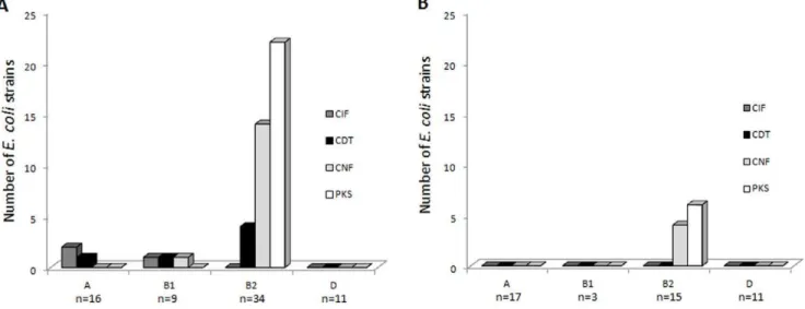
The results are shown in Table 4, Figure 2 and Tables S1, S2, S3. Twenty-five CRC specimens among 38 (65.8%) contained CM-positiveE. coliand only 6 diverticulosis specimens among 31 (19.4%), indicating that CM-harboring strains were preferentially
Table 1.Number ofE. colistrains collected from patients with CRC and diverticulosis.
Percentage (number) of samples containingE. colistrains
Number ofE. colistrains per sample Diverticulosis (n = 31) CRC (n = 38) Proximal colon cancer (n = 21) Distal colon cancer (n = 17)
0 19.4 (6) 2. 6 (1) 4.8 (1) 0.0 (0)
1 25.8 (8) 42.1 (16) 47.6 (10) 35.2 (6)
.1 54.8 (17) 55.3 (21) 47.6 (10) 64.7 (11)
associated with CRC (p,0.01). This difference was associated with a high number of CM-positive B2 E. coli in CRC specimens compared to that observed in diverticulosis specimens (Figure 2). Accordingly, strains harboring colibactin-encodingpksisland were present in 55.3% of CRC specimens (n = 21/38) and only in 19.3% of diverticulosis specimens (n = 6/31), indicating that pks
positive strains were significantly (p,0.01) more prevalent in CRC. To a lesser extent, cnf and cdt positive E. coli were significantly (p#0.02) more prevalent in the CRC than in the diverticulosis samples. These differences were mainly due to the high prevalence ofpks-,cnf- andcdt-positiveE. coliin distal CRC specimens compared to that in diverticulosis specimens (p#0.01). Altogether, these results indicate that CM-positive E. coli
distribution differs according to specimen origin.
Genotoxicity ofE. colidevoid of CM-encoding genes
We investigated whetherE. colidevoid of known CM-encoding genes can induce DNA damage in host cells. Using HeLa cultured cells, the genotoxicity of strains was investigated by single-cell gel electrophoresis assay (or comet assay), the state-of-the-art tech-nique for detecting DNA damage caused by chemical genotoxins. A total of 76 clinicalE. colistrains were investigated; 15 originated from proximal colon cancer, 21 from distal colon cancer and 40 from diverticulosis. These strains belonged to A (n = 29), D (n = 21) and B2 (n = 17) phylogroups. Interestingly, 27.6% (n = 21/76) of
E. colistrains devoid of known CM-encoding genes induced the formation of comets, which are typical of host cell DNA damage (Figure 3 and Tables S1, S2, S3). In addition, in contrast to CM-producing E. coli strains, which belong mainly to the B2 phylogroup, these comet positive strains belonged mainly to A (52%, n = 11/21) and D (29%, n = 6/21) phylogroups. Comets were observed with 13 strains (32.5%) among 40 isolated from patients with diverticulosis, and with 8 strains (22%) among 36 in
patients with CRC. The distribution of these putative genotoxic strains in the specimens was therefore different from that of CM-encoding strains.
Bacterial adhesion to intestinal epithelial cells
The ability ofE. colistrains to adhere to Int-407 epithelial cells was compared according to the origin of the strains, their phylogroup and their ability to produce cyclomodulin(s) or unknown genotoxin (positive comet assay). As shown in Figures 4A and B, highly adherentE. colistrains mostly belonged to A and D phylogroups, irrespective of the origin of the strains (CRC or
Table 2.Colonization of diverticulosis and CRC samples byE. coliphylogroups (A, B1, B2 and D).
Percentage (number) of samples containingE. coli
A B1 B2 D
Diverticulosis (n = 31) 41.9 (13) 6.5 (2) 41.9 (13) 32.2 (10)
CRC (n = 38) 28.9 (11) 23.7 (9) 73.7 (28) 26.3 (10)
Distal colon cancer (n = 17) 29.4 (5) 17.6 (3) 82.3 (14) 41.1 (7)
Proximal colon cancer (n = 21) 28.5 (6) 28.6 (6) 66.7 (14) 14.3 (3)
doi:10.1371/journal.pone.0056964.t002
Table 3.Distribution of CM-encoding genes inE. colistrains according to phylogroups (A, B1, B2 and D).
Percentage (number) ofE. colibelonging to the phylogroup
A (n = 33) B1 (n = 12) B2 (n = 49) D (n = 22) All (n = 116)
pks 0.0 (0) 0.0 (0) 57.1 (28) 0.0 (0) 24.1 (28)
cnf 0.0 (0) 8.3 (1) 36.7 (18) 0.0 (0) 16.4 (19)
cdt 3.0 (1) 8.3 (1) 8.2 (4) 0.0 (0) 5.2 (6)
cif 6.1 (2) 8.3 (1) 0.0 (0) 0.0 (0) 2.6 (3)
cm1 9.1 (3) 16.7 (2)2 63.3 (31)2 0.0 (0) 30.2 (36)2 1, cyclomodulin-encoding gene;
2, some
E. colistrains harbored more than one CM-encoding genes. doi:10.1371/journal.pone.0056964.t003
diverticulosis). Unexpectedly, mostE. colistrains belonging to B2 phylogroup displayed very low levels of adhesion. Analysis of the adhesive properties of A and DE. colistrains according to their ability to produce cyclomodulin(s) or unknown genotoxin (positive comet assay) showed that there was no significant difference (Figure 4B).
Discussion
The study of colonic mucosa-associated E. coli from patients with CRC or diverticulosis indicated that (i) E. coli strains belonging to the B2 phylogroup colonized colon cancers more frequently than they did diverticulosis samples, (ii) the CM-encoding genes were overrepresented in colon cancers, especially colibactin-encodingpksisland,cnf1andcdtgene, and (iii) the distal colon cancers were more frequently colonized by B2 E. coli
producing CMs than were the diverticulosis samples.
TheE. colistrains of the B2 phylogroup are mostly involved in extra-intestinal infections, such as urinary tract infections. They produce numerous virulence factors, notably pili adhesins, that favor colonization of the urinary tract. We can speculate that such adhesins specifically found in B2E. colialso favor the colonization of colon cancer. Bronowskiet al.observed UPEC-associated genes encoding adhesins among a panel of mucosa-associated E. coli
isolated from colon cancer, and Martin etalreported thatE. coli
strains isolated from colorectal tumors frequently expressed
hemagglutinins, which favor adhesion to intestinal epithelial Int-407 and HT29 cells [7,43]. One explanation of the high prevalence of B2 E. coli in CRC could be that changes in the host mucosa receptor repertoire have an effect on the bacterial population associated with mucosa. A higher level of colonization with B2 E. coli has also been observed in inflammatory bowel disease (IBD) [44]. In Crohn’s disease patients, such colonization was accompanied by increased ileal expression of the glycoprotein CEACAM6, which acts as a receptor for type 1 pili produced by
E. coli[45,46]. Interestingly, Crohn’s disease-associated B2E. coli
strains can induce the expression of CEACAM6 in intestinal epithelial cells, and yet CEACAM6 is a human tumor maker, whose overexpression has been observed in colonic tumors [47,48].
In this study, the CM-encoding genes were overrepresented in CRC compared with diverticulosis samples. Similar results were obtained by Arthur et al. when comparing the prevalence of colibactin-producingE. coliin CRC patients compared to control patients with diverticulosis, sporadic polyposis, irritable bowel syndrome or hemorrhoids [49]. Our data, which showed differences in prevalence of CM-producingE. colistrains belonging to B2 phylogroup, may be underestimated due to our experimen-tal procedure of preparing colonic specimens. We removed most fecal bacteria by washing the specimens thoroughly three times in PBS, but some mucus-associated bacteria were still present. Two studies, by Swidsinski et al. and Martin et al., reported higher
Figure 2. Distribution ofE. colistrains producing various cyclomodulins according to phylogroups and specimen origins.A,E. coli strains (n = 70) isolated from CRC samples (n = 38), and B,E. colistrains (n = 46) isolated from diverticulosis samples (n = 31).
doi:10.1371/journal.pone.0056964.g002
Table 4.CM-encoding gene content of CRC and diverticulosis samples.
Number (percentage) of specimens exhibiting CM-encoding genes
pks cnf cdt cif cm1,2
Diverticulosis (n = 31) 6 (19.3) 4 (12.9) 0 (0.0) 0 (0.0) 6 (19.4)
CRC (n = 38) 21 (55.3) 15 (39.5) 6 (15.8) 3 (7.9) 25 (65.8)
Distal colon cancer (n = 17) 11 (64.7) 9 (52.9) 4 (23.5) 1 (5.9) 13 (76.5)
Proximal colon cancer (n = 21) 10 (47.8) 6 (28.6) 2 (9.5) 2 (9.5) 12 (57.1)
1, cyclomodulin-encoding gene; 2, some
numbers ofE. coliin colon cancer samples than in controls when biopsy samples were studied after surface mucus removal, indicating that higher numbesr of E. coli are in very close association with the mucosa in CRC samples compared to those of controls. The CM-encoding genes identified werepksisland,cnf1
and cdt gene. Accordingly, Bronowski et al. have previously identified among a panel of 10 E. coli isolated from biopsies of colon cancers, 3 strains harboring cnf1 genes and 4 strains harboring a polyketide synthase-encoding gene belonging to pks
island [7]. The high prevalence of CM-encoding genes in CRC suggests a possible role of CM-encodingE. coliin the development of malignant colon tumors. It is well established that colibactin-encodingpks-harboringE. colistrains are mutagenic and genotoxic
in vitro and in vivo [22,23]. They can promote CRC without
affecting intestinal inflammation [50]. In addition, transient infection of human cell lines by such strains induces anchorage-independent colony formation [23], a process involved in metastases. Because of these mutagenic and cell transformation activities, E. coli strains harboring the colibactin-encoding pks
island may affect carcinogenesis at different stages. Cdt also induces DNA breaks in eukaryotic cells [26]. The presence of bacteria harboring cdt have already been associated with some lymphomas of the small intestine and can promote the develop-ment of hepatic and colon tumors [25,51–54]. CNF promotes cell proliferation by activating the Rho-GTPases and stimulates the transition from G1 to S phases [55]. In addition, CNF1 inhibits
apoptosis and alters tight junction structure and epithelial barrier function [56–58], processes that could favor carcinogenesis. Maddockset al.have shown that EPEC are more frequent in the colon adenocarcinoma tissues than in matched normal colon tissues, with a prevalence of 25% and 0%, respectively [59]. EPEC inject into host cells bacterial effectors, which have an effect on DNA damage repair and the cytoskeleton [59,60]. One effector is the product of thecifgene, which alters the ubiquitination process
and thus the degradation of proteins involved in many cellular processes such as cell cycle regulation and cytoskeleton [21,61]. In addition, the murine EPEC-like pathogen, Citrobacter rodentium, which harbors the LEE locus, is the cause of transmissible colonic hyperplasia [62], reduces the latency period of chemically induced tumors [63] and promotes colonic adenoma formation in APC/ Min mice [64].
The distal colon cancers were more frequently colonized by B2
E. coli encoding CMs than the proximal colon cancers or
diverticulosis samples, suggesting that the production of CMs directly or indirectly can provide a selective advantage for the colonization of distal colon cancers. This finding could be explained by major differences in the embryologic origin, bacterial flora composition, and physiology of the distal and proximal colons. The proximal colon derives from the midgut whereas the distal colon develops from the hindgut. The epithelial metabolism in the distal colon mainly involves butyrate, and in the proximal epithelium, acetate. Bacterial populations are qualitatively and quantitatively different in the lumen of proximal and distal colons [65], but studies showed that in a given patient similar populations of mucosa-associated bacteria are accommodated along the different parts of the colon irrespective of differences in the luminal content [66–68]. Most CM-producing strains harbor colibactin-encodingpksisland or the combination ofcnfgenes with
pksisland (79% of CM-producingE. coli), suggesting an important role of pks island in the distribution of CM-producing E. coli. Nougayre`de et al. proposed that the presence of colibactin-encodingpkscan favor the colonization of the intestinal tract [22]. As a PK-NRP-type compound, colibactin requires specific precursors belonging to the bacterial secondary metabolism. The physiological features of the distal and proximal colons could modifyE. colimetabolism and therefore the synthesis of PK-NRP compounds such as colibactin. Consequently, the efficiency of colibactin in promoting colonization may be affected and play a role in the distribution of CM-producingE. coli.
Interestingly, the proximal and distal colons also differ by certain aspects of their molecular physiopathology. The genomic instability associated with the microsatellite instability (MSI) phenotype is a hallmark of proximal colon cancer [69]. In contrast, chromosomal instability (CIN) is a major feature of distal colon cancers [69]. The pks-harboring E. coli induce major chromosomal damage and genetic instability [23]. Moreover, the CNF1-encoding gene, which is frequently associated with pks
island, induces abnormal chromosome segregation. These CM-encoding E. coli strains may therefore have an impact on the development of colon cancers.
The B2 phylogenetic group of E. coli is attracting attention because it contains strains responsible for severe infections, and their genetic background is adapted to the acquisition and/or maintenance of numerous virulence factors [19]. However, theE. colistrains belonging to the A and D phylogroups account for a large proportion (40% to 55%) of theE. colistrains in the intestinal tract [17,70,71]. Surprisingly, we observed that E. coli strains belonging to A and D phylogroups, irrespective of the origin of the strains (CRC or diverticulosis), were highly adherent to intestinal epithelial cells compared to B2E. colistrains, which displayed very low levels of adhesion. In addition, someE. colistrains belonging to A and D phylogroups in addition to having adhesion ability were able to induce DNA damage, suggesting that these strains have acquired unknown genotoxin(s). The distribution of these strains in CRC and diverticulosis specimens was not significantly different. The genotoxicE. colistrains of A and D subgroups, in contrast to B2 CM- producingE. coli, are therefore not specifically associated
Figure 3. DNA damage was detected by the comet assay in HeLa cells exposed toE. colifor 3 h.DNA damage was not detected in HeLa cells infected with theE. colistrain IHE3034DclbPharboring a defectivepksisland (A). Comet assay was positive in HeLa cells infected with theE. colistrain IHE3034 harboringpksisland (B) or withE. coli devoid of known cyclomodulin-encoding genes (C).
with CRC. However, their presence in close contact with colon tumors may still have an influence on the evolution of CRC.
The mechanism by which CM-producing E. coli strains can promote carcinogenesis is probably not due to the bacteria alone, and very likely involves numerous factors such as host suscepti-bility. DNA repair plays a pivotal role in maintaining genomic integrity with over 130 genes involved in various repair pathways that include base excision repair, nucleotide excision repair, double strand break repair and DNA mismatch repair [72]. In addition, polymorphisms within repair process genes are widely reported to be associated with an extensive range of malignancies that include CRC [73–75]. These polymorphisms can decrease their efficiency and explain susceptibility to the repeated aggressions of host DNA by residentE. coliproducing genotoxins. Finally, on the assumption that bacteria are associated with CRC, bacterial strains present at the origin of the cancer may disappear and be replaced by other bacteria better adapted to the cancer environment. In consequence, it remains difficult to determine whether the increase in specific bacteria is the consequence of the presence of malignant tissues or the cause of the cancer. However, these CM-producing bacteria, which colonize the malignant tumors, probably have an impact on the evolution of CRC.
In conclusion, our study showed a high prevalence of CM-producing B2E. coliin biopsies of colon cancers, especially at the distal part. It suggests therefore a possible role of CM-producingE. coliin colon cancers. This could be investigated in a longitudinal observational study to clarify the role of CMs in colorectal carcinogenesis. It also emerged that the different endogenous features of the proximal and distal colons and their different responsiveness to exogenous factors probably lead to the emergence of specific bacterial populations which can affect carcinogenesis. As previously reported for ExPEC, cyclomodulins are mostly found inE. coli of the B2 phylogroup. However, we observed that colonic mucosa-associatedE. colibelonging to the phylogroups A and D and devoid of known CM exhibit host cell genotoxic activity and should be considered as potentially harmful.
Supporting Information
Table S1 E. coli strains isolated from distal colonic
cancers.
(DOCX)
Table S2 E. coli strains isolated from proximal colon
cancers.
(DOCX)
Figure 4. Adhesion ability ofE. colistrains isolated from diverticulosis and CRC (proximal and distal) samples to Int-407 intestinal epithelial cells.A, adhesion ability according to the specimen origin and E. coli phylogroup. B, Adhesion ability ofE. colistrains isolated from diverticulosis and CRC (proximal and distal) samples and belonging to A and D phylogroups according to their ability to produce or not a cyclomodulin/genotoxin. The results are expressed as number of adherent bacteria per cell after 3 h infection period. Data are means+/2SEM for at least 3 independent experiments.
Table S3 E. colistrains isolated from diverticulosis.
(DOCX)
Table S4 Primers used in this study.
(DOCX)
Table S5 Archetypal E. coli control strains used in this
study.
(DOCX)
Acknowledgments
We thank Eric Oswald, Jose´ Antonio Orden, Philippe Bidet for kindly providing us with the strains IHE3034DClbP and DH10bpBACpks, C48a and RS218 respectively. We thank Rolande Perroux and Marle`ne Jan for helpful technical assistance.
Author Contributions
Conceived and designed the experiments: RB DP ADM EB DD. Performed the experiments: DD EB PS JR JD. Analyzed the data: DD EB PS JR DP ADM RB. Contributed reagents/materials/analysis tools: DP RB ADM. Wrote the paper: DD EB ADM RB.
References
1. Karsa LV, Lignini TA, Patnick J, Lambert R, Sauvaget C (2010) The dimensions of the CRC problem. Best Pract Res Clin Gastroenterol 24: 381– 396.
2. Kwak EL, Chung DC (2007) Hereditary colorectal cancer syndromes: an overview. Clin Colorectal Cancer 6: 340–344.
3. Half E, Bercovich D, Rozen P (2009) Familial adenomatous polyposis. Orphanet Journal of Rare Diseases 4: 1–23.
4. Collins D, Hogan AM, Winter DC (2011) Microbial and viral pathogens in colorectal cancer. Lancet Oncol 12: 504–512.
5. Swidsinski A, Khilkin M, Kerjaschki D, Schreiber S, Ortner M, et al. (1998) Association between intraepithelial Escherichia coli and colorectal cancer. Gastroenterology 115: 281–286.
6. Martin HM, Campbell BJ, Hart CA, Mpofu C, Nayar M, et al. (2004) Enhanced Escherichia coli adherence and invasion in Crohn’s disease and colon cancer. Gastroenterology 127: 80–93.
7. Bronowski C, Smith SL, Yokota K, Corkill JE, Martin HM, et al. (2008) A subset of mucosa-associatedEscherichia coliisolates from patients with colon cancer, but not Crohn’s disease, share pathogenicity islands with urinary pathogenic E. coli. Microbiology 154: 571–583. doi:10.1099/mic.0.2007/ 013086-0.
8. Nougayrede JP, Taieb F, De Rycke J, Oswald E (2005) Cyclomodulins: bacterial effectors that modulate the eukaryotic cell cycle. Trends Microbiol 13: 103–110. 9. Wu S, Morin PJ, Maouyo D, Sears CL (2003) Bacteroides fragilis enterotoxin induces c-Myc expression and cellular proliferation. Gastroenterology 124: 392– 400.
10. Wu S, Rhee KJ, Albesiano E, Rabizadeh S, Wu X, et al. (2009) A human colonic commensal promotes colon tumorigenesis via activation of T helper type 17 T cell responses. Nat Med 15: 1016–1022.
11. Leser TD, Molbak L (2009) Better living through microbial action: the benefits of the mammalian gastrointestinal microbiota on the host. Environ Microbiol 11: 2194–2206.
12. Chassaing B, Darfeuille-Michaud A (2011) The commensal microbiota and enteropathogens in the pathogenesis of inflammatory bowel diseases. Gastroen-terology 140: 1720–1728.
13. Croxen MA, Finlay BB (2009) Molecular mechanisms of Escherichia coli pathogenicity. Nat Rev Microbiol 8: 26–38.
14. Kaper JB, Nataro JP, Mobley HL (2004) Pathogenic Escherichia coli. Nat Rev Microbiol 2: 123–140. doi:10.1038/nrmicro818.
15. Clermont O, Bonacorsi S, Bingen E (2000) Rapid and simple determination of the Escherichia coli phylogenetic group. Appl Environ Microbiol 66: 4555– 4558.
16. Herzer PJ, Inouye S, Inouye M, Whittam TS (1990) Phylogenetic distribution of branched RNA-linked multicopy single-stranded DNA among natural isolates of Escherichia coli. J Bacteriol 172: 6175–6181.
17. Picard B, Garcia JS, Gouriou S, Duriez P, Brahimi N, et al. (1999) The link between phylogeny and virulence in Escherichia coli extraintestinal infection. Infect Immun 67: 546–553.
18. Escobar-Pa´ramo P, Sabbagh A, Darlu P, Pradillon O, Vaury C, et al. (2004) Decreasing the effects of horizontal gene transfer on bacterial phylogeny?: the Escherichia coli case study. Molecular Phylogenetics and Evolution 30: 243–250. doi:10.1016/S1055-7903(03)00181-7.
19. Escobar-Pa´ramo P, Clermont O, Blanc-Potard A-B, Bui H, Le Bougue´nec C, et al. (2004) A specific genetic background is required for acquisition and expression of virulence factors in Escherichia coli. Molecular biology and evolution 21: 1085–1094. doi:10.1093/molbev/msh118.
20. Taieb F, Nougayrede JP, Watrin C, Samba-Louaka A, Oswald E (2006) Escherichia coli cyclomodulin Cif induces G2 arrest of the host cell cycle without activation of the DNA-damage checkpoint-signalling pathway. Cell Microbiol 8: 1910–1921.
21. Jubelin G, Taieb F, Duda DM, Hsu Y, Samba-Louaka A, et al. (2010) Pathogenic bacteria target NEDD8-conjugated cullins to hijack host-cell signaling pathways. PLoS Pathog 6: e1001128.
22. Nougayre`de J-P, Homburg S, Taieb F, Boury M, Brzuszkiewicz E, et al. (2006) Escherichia coli induces DNA double-strand breaks in eukaryotic cells. Science 313: 848–851. doi:10.1126/science.1127059.
23. Cuevas-Ramos G, Petit CR, Marcq I, Boury M, Oswald E, et al. (2010) Escherichia coli induces DNA damage in vivo and triggers genomic instability in mammalian cells. Proc Natl Acad Sci U S A 107: 11537–11542.
24. De Rycke J, Oswald E (2001) Cytolethal distending toxin (CDT): a bacterial weapon to control host cell proliferation? FEMS microbiology letters 203: 141– 148.
25. Ge Z, Rogers AB, Feng Y, Lee A, Xu S, et al. (2007) Bacterial cytolethal distending toxin promotes the development of dysplasia in a model of microbially induced hepatocarcinogenesis. Cell Microbiol 9: 2070–2080. 26. Ge Z, Schauer DB, Fox JG (2008) In vivo virulence properties of bacterial
cytolethal-distending toxin. Cell Microbiol 10: 1599–1607.
27. Versalovic J, Koeuth T, Lupski JR (1991) Distribution of repetitive DNA sequences in eubacteria and application to fingerprinting of bacterial genomes. Nucleic Acids Res 19: 6823–6831.
28. Wang G, Whittam TS, Berg CM, Berg DE (1993) RAPD (arbitrary primer) PCR is more sensitive than multilocus enzyme electrophoresis for distinguishing related bacterial strains. Nucleic Acids Res 21: 5930–5933.
29. Bidet P, Metais A, Mahjoub-Messai F, Durand L, Dehem M, et al. (2007) Detection and identification by PCR of a highly virulent phylogenetic subgroup among extraintestinal pathogenic Escherichia coli B2 strains. Appl Environ Microbiol 73: 2373–2377.
30. Dubois D, Delmas J, Cady A, Robin F, Sivignon A, et al. (2010) Cyclomodulins in urosepsis strains of Escherichia coli. J Clin Microbiol 48: 2122–2129. 31. Peres SY, Marches O, Daigle F, Nougayrede JP, Herault F, et al. (1997) A new
cytolethal distending toxin (CDT) from Escherichia coli producing CNF2 blocks HeLa cell division in G2/M phase. Mol Microbiol 24: 1095–1107.
32. Boudeau J, Barnich N, Darfeuille-Michaud A (2001) Type 1 pili-mediated adherence of Escherichia coli strain LF82 isolated from Crohn’s disease is involved in bacterial invasion of intestinal epithelial cells. Mol Microbiol 39: 1272–1284.
33. Sert V, Cans C, Tasca C, Bret-Bennis L, Oswald E, et al. (1999) The bacterial cytolethal distending toxin (CDT) triggers a G2 cell cycle checkpoint in mammalian cells without preliminary induction of DNA strand breaks. Oncogene 18: 6296–6304. doi:10.1038/sj.onc.1203007.
34. Orden JA, Dominguez-Bernal G, Martinez-Pulgarin S, Blanco M, Blanco JE, et al. (2007) Necrotoxigenic Escherichia coli from sheep and goats produce a new type of cytotoxic necrotizing factor (CNF3) associated with the eae and ehxA genes. Int Microbiol 10: 47–55.
35. Johnson JR, Johnston B, Kuskowski MA, Nougayrede JP, Oswald E (2008) Molecular epidemiology and phylogenetic distribution of the Escherichia coli pks genomic island. J Clin Microbiol 46: 3906–3911.
36. De Rycke J, Milon A, Oswald E (1999) Necrotoxic Escherichia coli (NTEC): two emerging categories of human and animal pathogens. Vet Res 30: 221–233. 37. Johnson TJ, DebRoy C, Belton S, Williams ML, Lawrence M, et al. (2010)
Pyrosequencing of the Vir plasmid of necrotoxigenic Escherichia coli. Vet Microbiol 144: 100–109.
38. Pandey M, Khan A, Das SC, Sarkar B, Kahali S, et al. (2003) Association of cytolethal distending toxin locus cdtB with enteropathogenic Escherichia coli isolated from patients with acute diarrhea in Calcutta, India. J Clin Microbiol 41: 5277–5281.
39. Bielaszewska M, Fell M, Greune L, Prager R, Fruth A, et al. (2004) Characterization of cytolethal distending toxin genes and expression in shiga toxin-producing Escherichia coli strains of non-O157 serogroups. Infect Immun 72: 1812–1816.
40. Orth D, Grif K, Khan AB, Naim A, Dierich MP, et al. (2007) The Shiga toxin genotype rather than the amount of Shiga toxin or the cytotoxicity of Shiga toxin in vitro correlates with the appearance of the hemolytic uremic syndrome. Diagn Microbiol Infect Dis 59: 235–242.
41. Loukiadis E, Nobe R, Herold S, Tramuta C, Ogura Y, et al. (2008) Distribution, functional expression, and genetic organization of Cif, a phage-encoded type III-secreted effector from enteropathogenic and enterohemorrhagic Escherichia coli. J Bacteriol 190: 275–285.
43. Martin HM, Campbell BJ, Hart CA, Mpofu C, Nayar M, et al. (2004) Enhanced Escherichia coli adherence and invasion in Crohn’s disease and colon cancer. Gastroenterology 127: 80–93. doi:10.1053/j.gastro.2004.03.054.
44. Kotlowski R, Bernstein CN, Sepehri S, Krause DO (2007) High prevalence of Escherichia coli belonging to the B2+D phylogenetic group in inflammatory bowel disease. Gut 56: 669–675.
45. Barnich N, Carvalho FA, Glasser AL, Darcha C, Jantscheff P, et al. (2007) CEACAM6 acts as a receptor for adherent-invasive E. coli, supporting ileal mucosa colonization in Crohn disease. J Clin Invest 117: 1566–1574. 46. Carvalho FA, Barnich N, Sivignon A, Darcha C, Chan CH, et al. (2009)
Crohn’s disease adherent-invasive Escherichia coli colonize and induce strong gut inflammation in transgenic mice expressing human CEACAM. J Exp Med 206: 2179–2189.
47. Scholzel S, Zimmermann W, Schwarzkopf G, Grunert F, Rogaczewski B, et al. (2000) Carcinoembryonic antigen family members CEACAM6 and CEACAM7 are differentially expressed in normal tissues and oppositely deregulated in hyperplastic colorectal polyps and early adenomas. Am J Pathol 156: 595–605. 48. Jantscheff P, Terracciano L, Lowy A, Glatz-Krieger K, Grunert F, et al. (2003) Expression of CEACAM6 in resectable colorectal cancer: a factor of independent prognostic significance. J Clin Oncol 21: 3638–3646.
49. Arthur JC, Perez-Chanona E, Mu¨hlbauer M, Tomkovich S, Uronis JM, et al. (2012) Intestinal Inflammation Targets Cancer-Inducing Activity of the Microbiota. Science 338: 120–123.
50. Janelle AC, Tomkovich SE, Muehlbauer M, Ting-Jia F, Dogan B, et al. (2012) The E. coli Genotoxic Island Pks Promotes Colorectal Cancer (CRC) Without Impacting Intestinal Inflammation. Gastroenterology 142: S–639.
51. Lecuit M, Abachin E, Martin A, Poyart C, Pochart P, et al. (2004) Immunoproliferative small intestinal disease associated with Campylobacter jejuni. N Engl J Med 350: 239–248.
52. Nagamine CM, Sohn JJ, Rickman BH, Rogers AB, Fox JG, et al. (2008) Helicobacter hepaticus infection promotes colon tumorigenesis in the BALB/c-Rag2(-/-) Apc(Min/+) mouse. Infect Immun 76: 2758–2766.
53. Nagamine CM, Rogers AB, Fox JG, Schauer DB (2008) Helicobacter hepaticus promotes azoxymethane-initiated colon tumorigenesis in BALB/c-IL10-defi-cient mice. Int J Cancer 122: 832–838.
54. Fox JG, Feng Y, Theve EJ, Raczynski AR, Fiala JL, et al. (2010) Gut microbes define liver cancer risk in mice exposed to chemical and viral transgenic hepatocarcinogens. Gut 59: 88–97.
55. Lemonnier M, Landraud L, Lemichez E (2007) Rho GTPase-activating bacterial toxins: from bacterial virulence regulation to eukaryotic cell biology. FEMS Microbiol Rev 31: 515–534.
56. Fiorentini C, Matarrese P, Straface E, Falzano L, Fabbri A, et al. (1998) Toxin-induced activation of Rho GTP-binding protein increases Bcl-2 expression and influences mitochondrial homeostasis. Exp Cell Res 242: 341–350.
57. Hopkins AM, Walsh SV, Verkade P, Boquet P, Nusrat A (2003) Constitutive activation of Rho proteins by CNF-1 influences tight junction structure and epithelial barrier function. J Cell Sci 116: 725–742.
58. Malorni W, Fiorentini C (2006) Is the Rac GTPase-activating toxin CNF1 a smart hijacker of host cell fate? Faseb J 20: 606–609.
59. Maddocks OD, Short AJ, Donnenberg MS, Bader S, Harrison DJ (2009) Attaching and effacing Escherichia coli downregulate DNA mismatch repair
protein in vitro and are associated with colorectal adenocarcinomas in humans. PLoS One 4: e5517.
60. Vogelmann R, Amieva MR (2007) The role of bacterial pathogens in cancer. Curr Opin Microbiol 10: 76–81.
61. Marche`s O, Ledger TN, Boury M, Ohara M, Tu X, et al. (2003) Enteropathogenic and enterohaemorrhagic Escherichia coli deliver a novel effector called Cif, which blocks cell cycle G2/M transition. Molecular Microbiology 50: 1553–1567. doi:10.1046/j.1365-2958.2003.03821.x. 62. Schauer DB, Zabel BA, Pedraza IF, O’Hara CM, Steigerwalt AG, et al. (1995)
Genetic and biochemical characterization of Citrobacter rodentium sp. nov. J Clin Microbiol 33: 2064–2068.
63. Barthold SW, Jonas AM (1977) Morphogenesis of early 1, 2-dimethylhydrazine-induced lesions and latent period reduction of colon carcinogenesis in mice by a variant of Citrobacter freundii. Cancer Res 37: 4352–4360.
64. Newman JV, Kosaka T, Sheppard BJ, Fox JG, Schauer DB (2001) Bacterial infection promotes colon tumorigenesis in Apc(Min/+) mice. J Infect Dis 184: 227–230.
65. Bleday R, Braidt J, Ruoff K, Shellito PC, Ackroyd FW (1993) Quantitative cultures of the mucosal–associated bacteria in the mechanically prepared colon and rectum. Dis Colon Rectum 36: 844–849.
66. Poxton IR, Brown R, Sawyerr A, Ferguson A (1997) The mucosal anaerobic gram-negative bacteria of the human colon. Clin Infect Dis 25 Suppl 2: S111– 113.
67. Eckburg PB, Bik EM, Bernstein CN, Purdom E, Dethlefsen L, et al. (2005) Diversity of the human intestinal microbial flora. Science 308: 1635–1638. doi:10.1126/science.1110591.
68. Green GL, Brostoff J, Hudspith B, Michael M, Mylonaki M, et al. (2006) Molecular characterization of the bacteria adherent to human colorectal m uc osa . J A ppl M ic robio l 1 00 : 46 0– 4 69 . doi:10 .11 11 /j.13 65 -2672.2005.02783.x.
69. Gervaz P, Cerottini JP, Bouzourene H, Hahnloser D, Doan CL, et al. (2002) Comparison of microsatellite instability and chromosomal instability in predicting survival of patients with T3N0 colorectal cancer. Surgery 131: 190–197.
70. Duriez P, Clermont O, Bonacorsi S, Bingen E, Chaventre´ A, et al. (2001) Commensal Escherichia coli isolates are phylogenetically distributed among geographically distinct human populations. Microbiology 147: 1671–1676. 71. Zhang L, Foxman B, Marrs C (2002) Both urinary and rectal Escherichia coli
isolates are dominated by strains of phylogenetic group B2. J Clin Microbiol 40: 3951–3955.
72. Wood RD, Mitchell M, Sgouros J, Lindahl T (2001) Human DNA repair genes. Science 291: 1284–1289.
73. An Y, Jin G, Wang H, Wang Y, Liu H, et al. (2008) Polymorphisms in hMLH1 and risk of early-onset lung cancer in a southeast Chinese population. Lung Cancer 59: 164–170.
74. Hirata H, Hinoda Y, Kawamoto K, Kikuno N, Suehiro Y, et al. (2008) Mismatch repair gene MSH3 polymorphism is associated with the risk of sporadic prostate cancer. J Urol 179: 2020–2024.