The e ffe ct o f po rphyrins o n no rm al
and transfo rm e d m o use ce ll line s in
the pre se nce o f visible light
Departamento de Q uímica e Física Molecular, Instituto de Q uímica de São Carlos, Universidade de São Paulo, São Carlos, SP, Brasil
S.P.S. Tita and J.R. Perussi
Abstract
Photodynamic therapy consists of the uptake of a photosensitizing dye, often a porphyrin, by tumor tissue and subsequent irradiation of the tumor with visible light of an appropriate wavelength matched to the absorption spectrum of the photosensitizing dye. This class of molecules produces reactive oxygen species when activated by light, resulting in a direct or indirect cytotoxic effect on the target cells. Photodynamic therapy has been used in the treatment of cancer but the technology has a potential for the treatment of several disease condi-tions mainly because of its selectivity. However, it is not clear why the porphyrins are retained preferentially by abnormal tissue. This paper describes a study of the effect of the association of porphyrin and visible light on two mouse fibroblast cell lines: A31, normal cells and B61, an EJ-ras transformed variant of A31. Two water-soluble por-phyrins were used, a positively charged one, tetra(N-methyl-4-pyridyl)porphyrin chloride, and a negatively charged one, tetra(4-sulfonatophenyl)porphyrin-Na salt (TPPS4) in order to assess the effect on cell survival. The results suggest that the B61 cell line is more sensitive to incubation with the anionic porphyrin (TPPS4) followed by light irradiation and that the anionic porphyrin is more efficient in killing the cells than the cationic porphyrin.
Co rre spo nde nce
J.R. Perussi
Departamento de Q uímica e Física Molecular, IQ SC, USP Av. Dr. Carlos Botelho, 1465 13560-970 São Carlos, SP Brasil
Fax: + 55-16-273-9985 E-mail: janice@ iqsc.sc.usp.br Research supported by FAPESP and CNPq. S.P. Silva was the recipient of CNPq and FAPESP undergraduate fellowships.
Received September 11, 2000 Accepted June 18, 2001
Ke y wo rds
·Water-soluble porphyrins ·Mouse fibroblast lines
A31 and B61
·Photodynamic therapy ·Visible light
Intro ductio n
Photodynamic therapy (PDT) is a rela-tively new modality used for cancer treat-ment, consisting of the combined use of sys-temically administered photosensitizing por-phyrins and local applications of light emit-ted by lamps or lasers in the presence of oxygen. The technique is based on the pref-erential accumulation of porphyrin in tumor tissue with subsequent cytotoxicity medi-ated by singlet oxygen production (1). Its therapeutic usefulness is due to selective and rapid tumor necrosis after illumination of the
be administered at high doses, having no therapeutic value by itself for treating can-cer. However, this dye has photodynamic properties, i.e., in the presence of oxygen and visible light it becomes highly destruc-tive to its immediate environment (5). Por-phyrin has been the preferred drug used as a photosensitizing substance due to its high affinity for the tumor cell and its strong effect under irradiation with light (6). Differ-ent photosensitizers have been tested for localization in human tumor tissues, includ-ing fluorescein, cyanine dyes, porphyrin com-pounds, eosin, tetracycline, acridine orange and Rhodamine-123 (7). In vivo,the
photo-sensitizer mainly accumulates in organs of the reticuloendothelial system (liver, spleen, kid-ney, and lungs), atherosclerotic plaques, ar-eas of inflammation and healing wounds (8). The cationic, meso-substituted porphy-rin tetra(N-methyl-4-pyridyl)porphyporphy-rin chlo-ride (TMPyP) has been described as a potent photosensitizer in vivo (9) and in vitro (10). This porphyrin is a DNA intercalator in vitro
(11) and can photoinduce genotoxic effects in tumor cells in culture (10). Many water-soluble porphyrins form complexes with al-bumins, the main protein of blood plasma (12,13). Water-soluble porphyrins compete with hydrophobic species for binding sites in albumin, thus increasing the binding of wa-ter-soluble porphyrins to membranes and consequently the effectiveness of PDT (14, 15). Moreover, the high affinity and photo-toxicity of water-soluble sulfonated meso
-tetraphenylporphyrins (TPPS4) themselves for tumor cells allow their consideration as promising compounds for use in PDT (16).
This paper presents the results of a study using two water-soluble porphyrins associ-ated with visible light in two cell lines in order to examine their effect on cell survival. A cationic porphyrin (TMPyP) and an an-ionic porphyrin (TPPS4) were used in their free base form on a normal cell line originat-ing from mouse fibroblasts (A31) and a trans-formed cell line originating from A31 by
transfection (B61). The choice of this class of porphyrins as models is justifiable for this study since one can compare the effect of the charges on the molecules on their cytotoxic effect on cells.
Mate rial and Me tho ds
Che micals
Tetra(N-methyl-4-pyridyl)porphyrin chloride (TMPyP) and tetra(4-sulfona-tophenyl)porphyrin-Na salt (TPPS4) were purchased from Mid Century Chemicals (Posen, IL, USA). Stock solutions were pre-pared in water and diluted in Dulbeccos modified Eagles medium (DMEM) with 1% FBS just before use.
Ce ll line s
The normal cell line used was A31, origi-nating from genetically pure BALBc/3T3 mice (17) and consisting of embryo fibro-blasts. The transformed cell line B61, also originating from BALBc mice, was obtained from A31 by transfection of the human EJ-ras oncogene; B61 cells are transformed and tumorigenic (18). The cells were grown in 25-cm2
flasks containing DMEM supple-mented with 10% FBS, streptomycin and ampicillin. The cells were kept at 37o
C and 5% CO2 and harvested with 0.2% trypsin + 0.02% EDTA. Cell viability was assessed by the Trypan blue method.
Cyto to xic assays
The cells were plated at a density of 102 cells/ml with DMEM + 10% FBS onto 35-mm polypropylene Petri dishes, grown at 37o
C and 5% CO2 for 24 h, washed with PBS, and treated with cationic or anionic porphyrin at concentrations ranging from 0 to 30 µM for 1 h at 37o
supplemented medium until macroscopic colonies could be observed. The cells were then stained with Giemsa and the colonies counted directly on the plates or on a com-puter screen after scanning the plates.
Effect of the combination of porphyrin and light
The cells were plated in triplicate onto 35-mm polypropylene Petri dishes contain-ing DMEM + 10% FBS at a density of 5 x 10⁴ cells/ml. Cells were grown at 37oC for about 48 h in order to reach a density of about 105 cells/ml. Cells were harvested and counted every day. Immediately before drug addi-tion, three plates were counted. Then the monolayer was washed with PBS and treated with 9 µM porphyrin in DMEM + 1% FBS for 20 min. The cells were washed three times with DMEM + 1% FBS and ordinary supplemented medium was added. The cells were then exposed to visible light going through a 3-cm high layer of water in order to avoid cell heating. The medium tempera-ture was monitored with a thermocouple. The distance between the cells and the 150-W lamp was 13 cm and the illumination time was 45 min. A blue filter was used to select the wavelength in the range of 500 to 650 nm. The cells were then incubated in tripli-cate and counted after 30 min, 2 h, 4 h and then every day until the time when they were trypsinized, stained with Trypan blue and counted in a hemocytometer. Viability was calculated and viability curves as a function of time were obtained.
Re sults
The cytotoxic assays performed with the B61 cell line allowed the determination of the IC50 values for the porphyrins. The IC50 for porphyrins in B61 cells were 2 ± 1 µM (N = 3) and 5 ± 1 µM (N = 2) for TMPyP and TPPS4, respectively, at pH 7.0 in PBS. The results suggest that the cationic porphyrin is more cytotoxic for this transformed cell line,
since its IC50 was about 2.5-fold lower than the one obtained for the anionic porphyrin.
In order to assess the effect of the combi-nation of porphyrin and light, the growth curves were monitored in the presence of porphyrin with and without illumination. It should be mentioned that, as a control, in all experiments the cells were exposed to light under the same conditions and for the same period of time in the absence of a photosen-sitizer. No alteration was observed in any of the parameters of the growth curves for ei-ther cell line. This was seen as an indication that light by itself does not affect the cells in the absence of a photosensitizer.
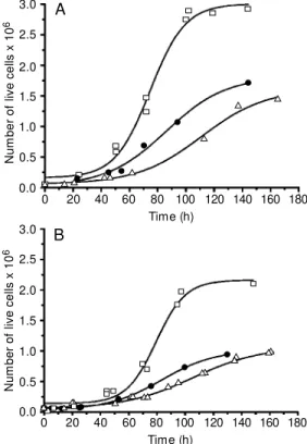
Figure 1 presents the growth curves, i.e., the number of cells as a function of time for B61 (A) and A31 (B) cells alone, in the presence of anionic porphyrin and anionic porphyrin associated with light. It was ob-served that all the parameters of the curve were changed. From the figure, we may as-sume that the B61 cells will take more time to become confluent after being treated with the drug, a fact that was still more
pro-N
u
m
b
e
r
o
f
liv
e
c
e
lls
x
1
0
6
3.0
2.5
2.0
1.5
1.0
0.5
0.0
3.0
2.5
2.0
1.5
1.0
0.5
0.0
N
u
m
b
e
r
o
f
liv
e
c
e
lls
x
1
0
6
0 20 40 60 80 100 120 140 160 180
Time (h)
0 20 40 60 80 100 120 140 160 180
Time (h) B
A Figure 1. Grow th curves for B61
cells (A) and for A31 cells (B) treated w ith 9 µM of anionic
por-phyrin (TPPS4). Control: cells
nounced in the presence of light. The values for the doubling time were affected after incubation with TPPS4 alone (a 66% in-crease for B61 and a 54% inin-crease for A31) and with light (89 and 87% increases for B61 and A31, respectively). Viability determined on the basis of the time the cells reached saturation density decreased from 100 to about 66% in the presence of TPPS4 alone and to 52% in the presence of TPPS4 plus light for B61. It can be seen that incubation with porphyrin killed about 34% of B61 cells and incubation with porphyrin plus light killed as much as 48% of the cells. We may suggest that the toxicity of irradiation not only affected the population of living
cells but also that the damage continued during growth, preventing the recovery of normal metabolism. These results support the idea that photochemical treatment of the cells greatly increases the efficiency of por-phyrin in the control of cell number in a tumor. For the normal A31 cells, cell viabil-ity remained high (around 95%) before and after the treatment and irradiation.
Figure 2 presents cell viability as a func-tion of time for both cell lines after treatment with the anionic porphyrin (TPPS4) and light irradiation. From the figure one can see that in fact the cells can recover from treatment to a different extent, but it is quite clear that B61 cells are much more affected and can-not reach more than 50% of viability even after a long time, while A31 can recover quite well.
Figure 3 shows the effects of the posi-tively charged TMPyP on the growth of B61 cells. These curves suggest that these cells are not very much sensitive to the presence of this porphyrin both as a drug itself or as a photosensitizer, i.e., after irradiation. In the presence of this porphyrin, the log phase had not suffered alteration and the doubling time presented an increase of just 21% after incu-bation with porphyrin and about 36% with light exposure. The viability of B61 cells decreased by 23% after TMPyP treatment and by 32% when treatment was combined with light irradiation. Thus, the results show that cationic porphyrin is less cytotoxic than anionic porphyrin both in the dark and after light irradiation.
D iscussio n
Both porphyrins used in this study pre-sented a slight toxicity for both cell lines in the dark, i.e., the ability to kill cells in the absence of light when used at therapeutic doses. A relatively low porphyrin concentra-tion was used, 9 µM (which corresponds to 7 mg/ml for cationic porphyrin and 11 mg/ml for anionic porphyrin) and the exposure time
V
ia
b
ili
ty
(
%
)
100
80
60
40
20
0
0 20 40 60 80 100 120 140
Time (h)
3.0
2.5
2.0
1.5
1.0
0.5
0.0
N
u
m
b
e
r
o
f
liv
e
c
e
lls
x
1
0
6
0 20 40 60 80 100 120 140 160 180
Time (h) Figure 2. Viability as a function
of time for A31 (squares) and B61 cells (circles) after treat-ment w ith the anionic porphyrin
(TPPS4) and light irradiation.
to the porphyrins was only 20 min. It is desirable that the photosensitizer have a high toxicity just after illumination, a parameter that defines the therapeutic effectiveness of the compound. Both porphyrins also meet the conventional requirement for photosen-sitizers to have a homogeneous chemical com-position and high solubility in water, facilitat-ing intravenous administration to the patient.
The viability curve as a function of time for both cell lines after treatment with the photosensitizer and light irradiation clearly showed that in fact transformed and normal cells can recover from treatment to a differ-ent extdiffer-ent. It is eviddiffer-ent that B61 cells are much more affected by treatment with the anionic porphyrin and cannot reach more than 50% viability even after a long period of time, while A31 can recover completely.
We may conclude that in the experiments performed with the anionic porphyrin (TPPS4) with no light irradiation the more affected cell line was B61 since the doubling time was more affected but cell viability was the parameter showing more sensitivity to the presence of this anionic porphyrin. In the experiments where light exposure was used after porphyrin incubation, B61 was also the more affected cell line.
In the experiments with the cationic por-phyrin, the cell line B61 presented only a slight increase in doubling time (36%) com-pared to the results obtained for the anionic porphyrin (89%) after visible light irradia-tion. The viability determined by the time the cells reached saturation density was about 68%, higher than for TPPS4 in this cell line (52%). Thus, these numbers suggest that the B61 cell line is more sensitive to the effects of the anionic porphyrin (TPPS4).
On the basis of the results obtained, we may conclude that, for both cell lines stud-ied, the anionic porphyrin is more efficient in killing the cells as a photosensitizer when incubated for 20 min and submitted to light irradiation for 45 min. The present results showed that, in fact, there is a considerable
decrease in the cell population after porphy-rin incubation followed by light irradiation, suggesting that this treatment can be em-ployed as a method to kill tumor cells.
It is well known that PDT leads to apop-tosis. However, this event was not investi-gated in the present study and it is important to say that the approach used here may have a limitation by underestimating the number of affected cells after treatment.
It is interesting to point out that B61 cells (transformed cells) were considerably more affected than A31 cells (normal cells), sup-porting the hypothesis that porphyrins accu-mulate rapidly and preferentially in tumor cells. This fact is extremely important for the application of PDT to the treatment of can-cer, since the porphyrin is administered in-travenously and is supposed to kill only the malignant cells after light irradiation. Thus, the therapeutic usefulness of PDT is based on the preferential accumulation of porphy-rin in tumor tissue. To the best of our knowl-edge, no report in the literature showing this enormous difference in the effect of porphyrin comparing normal and transformed or tumor cell lines has been previously presented.
Our results also suggest that important membrane alterations should take place in the tumor cells causing the selectivity of cell accumulation of the photosensitizing dye that will generate the toxic species leading to cell death.
The results presented here concerning the higher effectiveness of the anionic por-phyrin (TPPS4) compared with the cationic one (TMPyP) have been confirmed in our laboratory using a human tumor cell line irradiated with laser light.
Ackno wle dgm e nts
Re fe re nce s
1. Karrer S, Szeimies R-M , Abels C & Land-thaler M (1998). The use of photodynamic therapy for skin cancer. Onkologie, 21: 20-27.
2. Dougherty TJ, Potter WR & Weishaupt KR (1984). The structure of the active components of haematoporphyrin deriva-tive. In: Andreoni A & Cubedda R
(Edi-tors), Porphyrins in Tumour Phototherapy.
Plenum Press, New York, 23-35. 3. Sibata CH, Colussi VC, Oleinick NL &
Kinsella TJ (2000). Photodynamic therapy: a new concept in medical treatment. Bra-zilian Journal of M edical and Biological Research, 33: 869-880.
4. Schuitmaker JJ, Baas P, van Leengoed HLLM , van der M eulen FW, Star WM & van Zandw ijk N (1996). Photodynamic therapy: a promising new modality for treatment of cancer. Journal of Photo-chemistry and Photobiology. B: Biology, 34: 3-12.
5. Sternberg ED, Dolphin D & Brucker C (1998). Porphyrin-based photosensitizers for use in photodynamic therapy. Tetrahe-dron, 54: 4151-4202.
6. Sekiya S, Kubota K, Kasai T, Iw asaki H, Yamauchi K, Takamizaw a H & Tenjin Y (1988). Cytocidal effects of hematopor-phyrin derivative and argon dye laser on human gynecologic tumor cells in vitro. International Journal of Gynecology and Obstetrics, 26: 151-158.
7. Haghighat S, Cast ro DJ, Luf kin RB, Fetterman HR, Castro DJ, Soudant J, Ward PH & Saxton RE (1992). Laser dyes for experimental phototherapy of human cancer: comparison of three rhodamines. Laryngoscope, 102: 81-87.
8. Hamblin M R & New man EL (1994). On the mechanism of the tumor localizing effect in photodynamic therapy. Journal of Photochemistry and Photobiology. B: Biology, 23: 3-8.
9. Villanueva A & Jori G (1993). Pharmacoki-netic and tumour-photosensitizing prop-erties of the cationic porphyrin meso-tetra (4N-methylpyridyl) porphine. Cancer Let-ters, 73: 59-64.
10. Villanueva A, Juarranz A, Díaz V, Gomez J & Cañete M (1992). Photodynamic effects of a cationic mesosubstituted porphyrin in cell cultures. Anti-Cancer Drug Design, 7: 297-303.
11. Fiel RJ, DattaGupta N, M ark EH & How -ard JC (1979). Interaction of DNA w ith a porphyrin ligand: evidence for intercala-tion. Nucleic Acids Research, 6: 3093-3118.
12. Lamola AA, Asher I, M uller-Eberhard U & Poh-Fitzpatrick M (1981). Fluorimetric study of the binding of protoporphyrin to hem opexin and album in. Biochem ical Journal, 63: 693-698.
13. Datta-Gupta N, M alakar D & Dozier J (1989). Binding studies of 4 free base
por-phyrins and 6 iron (+3) porpor-phyrins w ith human-serum albumin. Research Com-munications in Chemical Pathology and Pharmacology, 63: 289-292.
14. El-Far M A & Pimstone NR (1986). Selec-tive in vivo tumor-localization of uropor-phyrin isomer-I in mouse mammary carci-noma - superiority over other porphyrins in a comparative study. Cancer Research, 46: 4390-4394.
15. El-Far M A, El-Hamil NA & Ghoneim M (1988). Selective in vivo localization of heptacarboxylic porphyrin isomer in a bladder tumor model - a novel technique to modulate porphyrin localization. Bio-chemistry, 70: 1379-1384.
16. Berg K, Bommer JC, Winkelman JW & M oan J (1990). Cellular uptake and rela-tive efficiency in cell inactivation by photo-activated sulfonated
mesa-tetraphenyl-porphines. Photochemistry and
Photobi-ology, 52: 775-781.
17. Aaronson SA & Todaro GJ (1968). Devel-opment of 3T3-like lines from BALB-c mouse embryo cultures: transformation susceptibility to SV-40. Journal of Cellular Physiology, 72: 141-148.