A pulsatile flo w m o de l fo r
in vitro
quantitative e valuatio n o f pro sthe tic
valve re gurgitatio n
1Divisão de Cardiologia, Departamento de Clínica Médica,
Faculdade de Medicina de Ribeirão Preto and 2Departamento de Física, Faculdade de Filosofia,
Ciências e Letras de Ribeirão Preto, Universidade de São Paulo, Ribeirão Preto, SP, Brasil
S. Giuliatti2, L. Gallo Jr.1, O .C. Almeida-Filho1, A. Schmidt1, J.A. Marin-Neto1, C.A. Pelá2 and B.C. Maciel1
Abstract
A pulsatile pressure-flow model was developed for in vitro quantita-tive color Doppler flow mapping studies of valvular regurgitation. The flow through the system was generated by a piston which was driven by stepper motors controlled by a computer. The piston was connected to acrylic chambers designed to simulate ventricular and atrial heart chambers. Inside the ventricular chamber, a prosthetic heart valve was placed at the inflow connection with the atrial chamber while another prosthetic valve was positioned at the outflow connec-tion with flexible tubes, elastic balloons and a reservoir arranged to mimic the peripheral circulation. The flow model was filled with a 0.25% corn starch/water suspension to improve Doppler imaging. A continuous flow pump transferred the liquid from the peripheral reservoir to another one connected to the atrial chamber. The dimensions of the flow model were designed to permit adequate imaging by Doppler echocardiography. Acoustic windows allowed placement of transducers distal and perpendicular to the valves, so that the ultrasound beam could be positioned parallel to the valvular flow. Strain-gauge and electromagnetic transducers were used for measure-ments of pressure and flow in different segmeasure-ments of the system. The flow model was also designed to fit different sizes and types of prosthetic valves. This pulsatile flow model was able to generate pressure and flow in the physiological human range, with independent adjustment of pulse duration and rate as well as of stroke volume. This model mimics flow profiles observed in patients with regurgitant prosthetic valves.
Co rre spo nde nce
B.C. Maciel
Departamento de Clínica Médica FMRP, USP
Av. Bandeirantes, 3900 14048-900 Ribeirão Preto, SP Brasil
Fax: + 55-16-633-0869
E-mail: bcmaciel@ fmrp.usp.br
Research partially supported by FAPESP (No. 89/0528-4) and CNPq (No. 830025/92-8).
Received August 2, 1999 Accepted January 10, 2000
Ke y wo rds
·Flow model
·Echocardiography
·Valvular regurgitation
Spectral and especially color Doppler flow mapping techniques have been used exten-sively in the clinical setting to estimate non-invasively the severity of valvular regurgita-tion (1-5). A number of different Doppler methods for quantitation of valve regurgita-tion have been described, including meas-urement of the spatial distribution of
regur-gitant jets in the receiving chamber (3,6), quantitation of regurgitant flow rate using flow convergence proximal to the orifice (7,8), momentum flux analysis (9), and di-mensions of vena contracta(10,11).
limi-tations of these methods for grading the se-verity of regurgitation. Technical factors re-lated either to the instrumentation or to the physics of ultrasound and a variety of physi-ological factors in addition to the volume of regurgitation and the entrainment of the sur-rounding fluid in the receiving chamber can influence the quantitation of valvular insuf-ficiency by the color Doppler techniques (10,12-15).
The evaluation of the relative influence of these multiple and interrelated factors for quantitative evaluation of valvular regurgi-tation cannot be adequately performed in the clinical setting. Therefore, in vitro models of
valve regurgitation, by producing strict and simultaneous control of multiple variables, play an important role for the methodologi-cal validation of Doppler methods for esti-mating the severity of valvular insufficiency. Several types of in vitro flow models have
been designed for Doppler echocardiographic studies (12,16-19). Pulsatile flow models can provide better simulation of the physi-ological conditions observed in the human cardiovascular system.
We describe here an in vitro pulsatile
pressure-flow model designed to simulate pressure and flow patterns documented in the human heart and to provide adequate conditions for quantitative evaluation of val-vular regurgitation by color Doppler flow mapping in prosthetic valves strategically placed at the inflow and outflow portions of the flow model. In addition, the system was designed to permit controlled variations of peripheral circulation resistance and cham-ber compliance.
The flow simulator, depicted in Figure 1, was composed of 1) a computerized pulse generation system controlling a piston driven by stepper motors; 2) acrylic chambers de-signed to simulate heart chambers, and 3) flexible tubes, elastic balloons and reser-voirs arranged to mimic the peripheral circu-lation.
The flow model was filled with a 0.25% corn starch/water suspension to improve Dop-pler imaging. The pulsatile flow through the system was produced by a piston driven by two stepper motors (Miniangle stepper, 34PM - CO41 model; Minebea Co.) controlled by an IBM compatible personal computer. Software was developed using Quick Basic 40 to
gener-Stepper motors
Interface Pow er
supply Computer
Air pump
Continuous flow pump Reservoir A
Reservoir B Piston
6 4 5
1
2 3
1
1
3
1
7 10
11
11
11 9
8
1. Pressure transducer 7. Acrylic cylinder
2. M echanic valve 8. M anometer
3. Biological valve 9. Solenoid valve
4. Ventricular chamber 10. Window s for the ultrasound transducer
5. Atrial chamber 11. Flexible tube
6. Infrared sensor
10 Figure 1 - Schematic diagram of
ate a pulsatile function with variable and ad-justable profile, amplitude and rate. This soft-ware was also used to control the opening of a solenoid valve which was the part of the sys-tem designed to mimic atrial contraction. An electronic interface between the computer and the stepper motors was used to meet compat-ible voltage and current requirements.
The piston was built inside an acrylic cham-ber which had two connections: at one end it was connected by a non-valved tube to the acrylic chambers designed to simulate ven-tricular and atrial heart chambers; at the other end, a valved tube connected the piston to a flexible tube and a reservoir (reservoir B). During forward movement of the piston this valve did not permit flow directed to the reser-voir, but during the piston backward move-ment the chamber was filled with liquid from reservoir B. This mechanism reduced the re-sistance during the excursion of the piston. Inside the ventricular chamber, a replace-able prosthetic heart valve (biological or me-chanical) of variable size could be placed at the inflow connection with the atrial cham-ber while another prosthetic valve was posi-tioned at the outflow connection with the flex-ible tubes, elastic balloons and a reservoir arranged to mimic the peripheral circulation. The balloons were arranged according to the Windkessel model to simulate variable arterial compliances. Fluid level at the atrial reservoir was maintained constant by a continuous flow pump (VEB MLW Prüfgeräte-Werk, UH4 mo-del) which transferred liquid from the periph-eral reservoir B to another one connected to the atrial chamber (reservoir A). The height of reservoir A could be changed to obtain different atrial pressures.
The compliance of the acrylic atrial chamber was modified by connecting it to an elastic balloon filled with the same corn starch/water suspension. To simulate atrial contraction, this balloon was positioned in-side another acrylic cylinder filled with at-mospheric air. This cylinder completely sur-rounded the balloon. A solenoid valve was
connected to the cylinder and to an air pump. An infrared sensor located in the piston was used to synchronize the opening of the sole-noid valve during a controlled period of time with the piston movement, allowing atmo-spheric air to enter the cylinder and to com-press the balloon, increasing atrial pres-sure and simulating atrial contraction.
The dimensions of the flow model were designed to permit adequate imaging by Doppler echocardiography. Acoustic win-dows allowed placement of transducers dis-tal and perpendicular to the valves, so that the ultrasound beam could be positioned parallel to the valvular flow. A commer-cially available ultrasound system (Hewlett-Packard Sonos 1000, Waltham, MA, USA) with 3.5- or 5.0-MHz phased-array trans-ducers was used for imaging flow through the prosthetic valves. Strain-gauge (Statham, P23Ab) and electromagnetic transducers (Electromagnetic Blood Flowmeter, SP2200 model) were used for measurements of pres-sure and flow in different segments of the system. An 8-channel physiologic recorder (Hewlett-Packard 4588A) was used to record these variables. The flow model was also designed to fit different sizes and types of prosthetic valves.
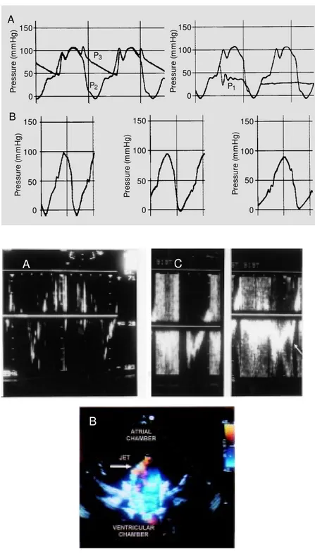
Pressure curves similar to those observed in human ventricular and atrial chambers as well as in the peripheral arterial system were observed in the flow model as shown in Figure 2. Even a dicrotic notch similar to that seen in the aortic pressure curve was ob-tained with this model. A negative early diastolic ventricular pressure varying from -10 to -30 mmHg, probably related to the backward movement of the piston and to the system (tube plus gauge) impedance, was frequently documented. During the dia-stolic period, the backward movement of the piston also influenced the shape of the pulsatile atrial pressure curve. However, the mean atrial pressure was similar to that observed under physiological conditions.
movement it was possible to modify pres-sure and flow generated by the pulsatile flow model within a physiological range (90 to 120 ml/beat). Increasing impedance by lo-calized constriction in the peripheral circu-lation progressively increased pulse pres-sure. The flow model allowed variation of pulse rate from 35 to 54 beats/min and also independent control of the rate of pressure rise during forward movement of the piston, keeping constant pulse rate and amplitude (Figure 2).
Flow velocities through mechanical and biological prosthetic valves were documented by color Doppler flow mapping and pulsed Doppler. The patterns of normal and regur-gitant flows (Figure 3) obtained with this mo-del were similar to those obtained in patients with prosthetic valves (Figure 3). Quantifica-tion of the magnitude of valvular regurgitaQuantifica-tion can be obtained by evaluating flow through prosthetic valves and peripheral circulation with electromagnetic flow meters. When the atrial contraction was synchronized with piston movement the velocity profile obtained mimicked the diastolic flow documented at the ventricular inflow (Figure 3).
The in vitro pulsatile flow system
de-scribed in this investigation was suitable for simulating pressure and flow conditions simi-lar to those observed in the human circula-tion under physiological condicircula-tions. In addi-tion, it allowed a strictly controlled and inde-pendent variation of a number of different variables which can influence the magnitude of valvular regurgitation as evaluated by color Doppler flow mapping. Therefore, repro-ducible right and left human ventricular flow and pressure conditions could be simulated
Figure 2 - Panel A, Pressure curves w ere recorded in the “ atrial” (P1) and “ ventricular” (P2) chambers and
also at the “ peripheral circulation” (P3) level of the flow
model. Panel B, Pressure curves w ere recorded at the “ ventricular” chamber level w hen the rate of pressure increase during forw ard movement of the piston w as modified independently.
Figure 3 - Panel A, Pulsed Doppler velocities recorded at the outflow of the “ ventricular” chamber show ing a flow profile similar to that observed in the human left ventricular outflow tract. Panel B, Regurgitant jet as documented by color Doppler flow mapping in a biological prosthetic valve positioned at the “ ventricular” inflow level. Panel C, Pulsed Doppler velocities recorded at the inflow of the “ ventricular” chamber show ing aliasing related to the regurgitant jet (produced in a biological prosthetic valve) and the inflow velocities w ithout (left) and w ith (right) simulation of “ atrial” contraction (arrow represents velocities related to “ atrial contraction” ).
P
re
s
s
u
re
(
m
m
H
g
) 150
P
re
s
s
u
re
(
m
m
H
g
) 150
100
50
0 100
50
0
P
re
s
s
u
re
(
m
m
H
g
)
150
P
re
s
s
u
re
(
m
m
H
g
)
150
100
50
0 100
50
0
P
re
s
s
u
re
(
m
m
H
g
)
150
100
50
0 A
B
P3
P2 P1
A C
with independent adjustments of pulse dura-tion and rate, rate of pressure generadura-tion, stroke volume, system impedance and cham-ber compliances. The flow system was also designed and effectively used to evaluate normal and dysfunctional (regurgitant or stenotic) prostheses of variable types and sizes. The flow model was also able to mimic atrial contraction synchronized with piston movement, thereby producing a ventricu-lar chamber filling pattern similar to the human physiological pattern. A cycle-to-cycle monitoring of pressure and flow curves in different segments of the flow model as-sured continuous quantitation of pressure gradients and flow conditions. Considering that this flow model was specifically de-signed for Doppler echocardiography stud-ies, acoustic windows were strategically po-sitioned to provide adequate visualization of the flow through the prosthetic valves.
Although this flow simulator has been designed for quantitative evaluation of the magnitude of valvular regurgitation by Dop-pler echocardiographic techniques, it can also be used for a number of different types of investigations including 1) evaluation of the flow pattern through normal valve
pros-theses of variable sizes, 2) analysis of the effects of atrial or ventricular compli-ance and of atrial contraction on ventric-ular filling, and 3) quantitative evaluation of prosthetic valve regurgitation at the ven-tricular outflow and inflow levels.
Some limitations were identified in this pulsatile flow model. Stepper motor power was not able to maintain an adequate excur-sion of the piston when peripheral resistance was progressively increased. Therefore, this simulator appears to be inadequate for quan-titative studies of stenotic prosthetic valves positioned at the ventricular outflow level. In addition, only a limited range of pulse rate, varying from 35 to 54 pulses/min, could be generated using this flow model. It would be possible to correct these limitations using more powerful motors and reducing the di-mensions of the piston.
In conclusion, the pulsatile flow model described in this investigation was adequate to simulate pressure and flow conditions in the human physiological range and may thus represent an important tool for quantitative evaluation of the magnitude of valvular re-gurgitation by color Doppler flow mapping.
Re fe re nce s
1. Quinones M A, Young JB, Waggoner AD, Ostojic M C, Ribeiro LG & M iller RR (1980). Assessment of pulsed Doppler echocardiography in detection and quanti-fication of aortic and mitral regurgitation.
British Heart Journal, 44: 612-620. 2. M iyat ake K, Izum i S, Okam ot o M ,
Kinoshita N, Asonuma H, Nakagaw a H, Yamamoto K, Takamiya M , Sakakibara H & Nimura Y (1986). Semiquantitative grad-ing of severity of mitral regurgitation by real-time tw o dimensional Doppler flow imaging technique. Journal of the Ameri-can College of Cardiology, 7: 82-88. 3. Helmcke F, Nanda NC, Hsiun M C, Soto B,
Adey CK, Goyal RG & Gatew ood RP (1987). Color Doppler assessment of mi-tral regurgitation w ith orthogonal planes.
Circulation, 75: 175-183.
4. Sahn DJ & M aciel BC (1988).
Physiologi-cal valvular regurgitation-Doppler echocar-diography and the potential for iatrogenic heart disease. Circulation, 78: 1075-1077. 5. M aciel BC, Simpson IA, Valdes-Cruz LM , Recusani F, Hoit B, Dalton N, Weintraub R & Sahn DJ (1991). Color flow Doppler mapping studies of physiologic pulmonary and tricuspid regurgitation: evidence for true regurgitation as opposed to a valve closing volume. Journal of the American Society of Echocardiography, 4: 589-597. 6. Spain M G, Sm ith M D, Grayburn PA, Harlam ert EA & DeM aria AN (1989). Quantitative assessment of mitral regur-gitation by Doppler color flow imaging: angiographic and hemodynamic correla-tions. Journal of the American College of Cardiology, 13: 585-590.
7. Recusani F, Bargiggia GS, Yoganathan AP, Raisaro A, Valdes-Cruz LM , Sung HW,
Bert ucci C, Gallat i M , M oisés VA, Simpson IA, Tronconi L & Sahn DJ (1991). A new method for quantification of regur-gitant flow rate using color Doppler flow imaging of the flow convergence region proximal to a discrete orifice. An in vitro
study. Circulation, 83: 594-604.
8. Bargiggia GS, Tronconi L, Sahn DJ, Recusani F, Raisaro A, De Servi S, Valdes-Cruz LM & M ontemartini C (1991). A new method for quantification of mitral regur-gitation based on color flow Doppler im-aging of flow convergence proximal to regurgitant orifice. Circulation, 84: 1481-1489.
10. Sw itzer DF, Yoganathan AP, Nanda NC, Woo Y-R & Ridgw ay AJ (1987). Calibra-tion of color Doppler flow mapping during extreme conditions in vitro: a foundation for a reliable quantitative grading system for aortic incompetence. Circulation,75: 837-846.
11. Feshke W, Omran H, M anz M , Köhler J, Hagendorff A & Lüderitz B (1994). Color-coded Doppler im aging of t he vena contracta as a basis for quantification of pure mitral regurgitation. American Jour-nal of Cardiology, 73: 268-274.
12. M aciel BC, M oises VA, Shandas R, Simpson IA, Beltran M , Valdes-Cruz LM & Sahn DJ (1991). Effects of pressure and volume of the receiving chamber on the spatial distribution of regurgitant jets as imaged by color Doppler flow mapping: an in vitro study. Circulation, 83: 605-613. 13. Simpson IA, Valdes-Cruz LM , Sahn DJ, Nurillo A, Tamura T & Chung K-J (1989).
Doppler color flow mapping of simulated
in vitro regurgitant jets: evaluation of the effects of orifice size and hemodynamic variables. Journal of the American Col-lege of Cardiology, 13: 1195-1207. 14. Hoit BD, Jones M , Eidbo EE, Elias W &
Sahn DJ (1989). Sources of variability for Doppler color flow mapping of regurgitant jets in an animal model of mitral regurgita-tion. Journal of the American College of Cardiology, 13: 1631-1636.
15. Ut sunom iya T, Ogaw a T, King SW , Sunada E, M oore GW, Henry WL & Gardin JM (1990). Effect of machine parameters on variance display in Doppler color flow mapping. American Heart Journal, 120: 1395-1402.
16. Yearw ood TL & Chandran KB (1980). Ex-perimental investigation of steady flow through a model of the human aortic arch.
Journal of Biomechanics, 13: 1075-1088. 17. Hasenkam JM , W estphal D, Reul H,
Gorm sen J, Giersiepen M , Stodkilde-Jorgensen H & Paulsen PK (1987). Three-dimensional visualisation of axial velocity dow nstream of six different mechanical aortic valve prostheses measured w ith a hot-film anemometer in a steady state flow model. Journal of Biomechanics, 20: 353-364.
18. Ojha M , Johnston KW, Cobbold RSC & Hummel RL (1989). Potential limitations of center-line pulsed Doppler recordings: an in vitro flow visualisation study. Jour-nal of Vascular Surgery, 9: 520-525. 19. Simpson IA, Fisher J, Reece IJ, Houston