He art rate variability unde r re sting
co nditio ns in po stm e no pausal and
yo ung wo m e n
1Laboratório de Fisioterapia Cardiovascular, Departamento de Fisioterapia, 2Departamento de Ciências Fisiológicas and 3Departamento de Física,
Universidade Federal de São Carlos, São Carlos, SP, Brasil
4Divisão de Cardiologia, Departamento de Clínica Médica, and 5Departamento de Ginecologia e O bstetrícia,
Faculdade de Medicina de Ribeirão Preto, Universidade de São Paulo, Ribeirão Preto, SP, Brasil
T.F. Ribeiro1,4, G.D. Azevedo5,
J.C. Crescêncio4,
V.R.F.S. Marães1,
V. Papa1, A.M. Catai1,
R.M.M. Verzola2,
L. O liveira3,
M.F. Silva de Sá5,
L. Gallo Jr.4 and E. Silva1
Abstract
The aim of the present study was to compare the modulation of heart rate in a group of postmenopausal women to that of a group of young women under resting conditions on the basis of R-R interval variabil-ity. Ten healthy postmenopausal women (mean ± SD, 58.3 ± 6.8 years) and 10 healthy young women (mean ± SD, 21.6 ± 0.82 years) were submitted to a control resting electrocardiogram (ECG) in the supine and sitting positions over a period of 6 min. The ECG was obtained from a one-channel heart monitor at the CM5 lead and processed and stored using an analog to digital converter connected to a microcom-puter. R-R intervals were calculated on a beat-to-beat basis from the ECG recording in real time using a signal-processing software. Heart rate variability (HRV) was expressed as standard deviation (RMSM) and mean square root (RMSSD). In the supine position, the postmeno-pausal group showed significantly lower (P<0.05) median values of RMSM (34.9) and RMSSD (22.32) than the young group (RMSM: 62.11 and RMSSD: 49.1). The same occurred in the sitting position (RMSM: 33.0 and RMSSD: 18.9 compared to RMSM: 57.6 and RMSSD: 42.8 for the young group). These results indicate a decrease in parasympathetic modulation in postmenopausal women compared to young women which was possibly due both to the influence of age and hormonal factors. Thus, time domain HRV proved to be a nonin-vasive and sensitive method for the identification of changes in autonomic modulation of the sinus node in postmenopausal women.
Co rre spo nde nce
E. da Silva
Laboratório de Fisioterapia Cardiovascular
Departamento de Fisioterapia, UFSCar Rodovia Washington Luís, km 235 13565-905 São Carlos, SP Brasil
E-mail: esters@ power.ufscar.br
Research supported by CAPES, CNPq (Nos. 300528/85 and 520686/95) and FAPESP (No. 98/13257-8).
Received May 15, 2000 Accepted March 2, 2001
Ke y wo rds
·Heart rate variability ·Menopause
·Autonomic nervous system ·Time domain indexes
Intro ductio n
The heart is an organ under the influence of the autonomic nervous system for the maintenance of homeostasis, and, in this re-spect, one of its main characteristics is the constant modification of its rate on beat-to-beat basis (1,2).
recordings (3-5). This method has proved to be of great clinical usefulness to evaluate the balance of sympathetic and parasympathetic regulation in several pathological conditions (6-12). HRV has proved to be a more sensi-tive tool for the detection of autonomic bal-ance than mean heart rate (HR) (9).
The so-called measures in the time do-main are indexes obtained from a continu-ous ECG recording, which are used to deter-mine the dispersion of the duration of the R wave intervals among its mean or a reference value. The various indexes proposed for the measurement of HRV in the time domain can be divided into three types: arithmetic, geometric or statistical calculations (2,10,11). A study of healthy volunteers using phar-macological blockade with atropine docu-mented a statistically significant reduction in some HRV indexes in the time domain, a fact that was not observed after blockade with propranolol (13). These investigators concluded that there is a correlation between temporal variability indexes and vagal tonus acting on the sinus node.
On the other hand, in patients with heart disease, reduced HRV has been associated with the occurrence of ventricular arrhyth-mias, such as ventricular fibrillation and in-creased mortality (9,10). The prevalence of cardiovascular diseases increases with ad-vancing age in women, especially after meno-pause and among those with a sedentary life style (14-16). Few studies have evaluated neurocardiac parameters during the various phases of womens life for the determination of risk factors, a procedure that would be useful and highly relevant for cardiovascular evaluation of women at higher risk to de-velop heart disease, thus permitting early intervention.
Analysis of the HRV response can be used to evaluate the adaptations of the auto-nomic nervous system in women related to changes due to aging and to the decreased beneficial effects of natural estrogen on the heart.
The objective of the present study was to compare time domain HRV between post-menopausal and young women under supine and sitting resting conditions.
Me tho ds
The study was conducted on 10 young volunteers and 10 postmenopausal volun-teers who were not on hormone replacement therapy. Their anthropometric characteris-tics, reported as mean and standard devia-tions, were: age 21.6 ± 0.82 years, weight 56.3 ± 6.62 kg, and height 161.6 ± 4.74 cm, and age 58.3 ± 6.8 years, weight 65.2 ± 12.11 kg, and height 156.3 ± 5.51 cm, respectively. Both groups were composed of healthy non-smoking women with a sedentary life style; the volunteers were not taking any medica-tions. Before the study the volunteers were submitted to clinical and laboratory exami-nations, including a resting ECG and dy-namic exercise seated on a cycle ergometer until they reached a target heart rate cor-rected for age; this last test was used to include in the study only women without ischemic heart disease.
The study was approved by the Ethics Committee of Universidade Federal de São Carlos and the volunteers gave written in-formed consent to participate in the study.
The experiments were carried out in a specially acclimatized room at 22oC and 60%
basis from the ECG using a signal-process-ing software (5). Systemic arterial pressure was measured using a mercury column sphyg-momanometer under resting conditions in the supine and sitting positions. For the young group, the recordings were always obtained during the same period of the menstrual cycle, i.e., between the 8th and the 10th day of the cycle. R-R intervals were re-corded over a period of 6 min under resting conditions in the supine and sitting positions and are shown in Figures 1 and 2, respec-tively.
Two indexes were used for the measure-ment of HRV, i.e., the standard deviations of R-R intervals (RMSM or SDNN), and the square root of the mean of the squared differ-ences between adjacent normal R-R inter-vals (RMSSD). RMSM corresponds to the square root of the sum of squares of the differences of individual values in relation to the mean value divided by the number of R-R intervals within a given time (Equation 1). RMSSD corresponds to the square root of the mean sum of squares of the differ-ences between the adjacent normal R-R in-tervals in the record divided by the number of R-R intervals within a given time minus one R-R interval (Equation 2) (17).
(
)
N 2
i i 1
RR RR
RMSM
N
=
-=
å
Eq. 1(
)
N 1
2
i i 1
i 1
RR RR
RMSSD
N 1
-+ =
-=
-å
Eq. 2where N = number of R-R intervals in the series of selected data.
The Graphic Electrocardiogram Analyzer, an application developed at the Laboratório de Fisioterapia Cardiovascular, Universida-de FeUniversida-deral Universida-de São Carlos, was used to obtain the variability indexes.
R
-R
i
n
te
rv
a
l
(m
s
)
1250
Young
Postmenopausal 1150
1050
950
850
750
650
0 60 120 180 240 300 360 Time (s)
Figure 1. R-R interval, in ms, for the young (gray) and postmenopausal (black) volunteers under resting conditions in the supine position (N = 2).
Figure 2. R-R interval, in ms, for the young (gray) and postmenopausal (black) volunteers under resting conditions in the sitting position (N = 2).
R
-R
i
n
te
rv
a
l
(m
s
)
1250
Young
Postmenopausal 1150
1050
950
850
750
650
0 60 120 180 240 300 360 Time (s)
Re sults
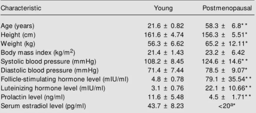
Table 1 shows the anthropometric char-acteristics, body mass index, and clinical and hormonal determinations for the studied volunteers.
median, 1st and 3rd quartiles and outliers. Data were analyzed statistically by the non-parametric Mann-Whitney test for intergroup comparison and by the Wilcoxon rank test for intragroup analysis, with the level of significance set at P<0.05. The analysis was carried out using the Statistic for Windows software, release 5.1. StatSoft, Inc., 1994-1996.
Figures 3 and 4 illustrate the analysis of HRV expressed as RMSM and RMSSD in-dexes of R-R interval variability obtained for the groups of postmenopausal and young volunteers under resting conditions in the supine and sitting positions. Intergroup anal-ysis showed significant differences (P<0.05), with lower RMSM and RMSSD indexes in the menopausal group compared to the young one. However, intragroup analysis showed no significant differences (P>0.05) in HRV indexes (RMSM and RMSSD) in the supine or seated position. Figure 5 shows that there was no significant difference (P>0.05) in mean HR between the groups.
D iscussio n
Autonomic modulation is the main mech-anism responsible for HR control in normal volunteers. In this context, stimulation of the parasympathetic nerves is associated with a decrease in HR and an increase in its vari-ability, whereas stimulation of the sympa-thetic system is associated with an increase in HR and a decrease in HR variability (18,19). Under resting conditions, both the sympathetic and parasympathetic systems are tonically active, with a predominant va-gal effect (1,2,11,18,19).
The literature has reported the use of a great variety of different HRV indexes. How-ever, it has not been definitely proved that any one of these indexes is superior to an-other (1,2,11,17).
The selection of the RMSM and RMSSD in this study was based on the relatively high sensitivity of these indexes due to the use of
Table 1. Anthropometric, clinical and hormonal characteristics of the groups studied.
Characteristic Young Postmenopausal
Age (years) 21.6 ± 0.82 58.3 ± 6.8* * Height (cm) 161.6 ± 4.74 156.3 ± 5.51* Weight (kg) 56.3 ± 6.62 65.2 ± 12.11* Body mass index (kg/m2) 21.4 ± 1.43 23.2 ± 6.42 Systolic blood pressure (mmHg) 108.2 ± 8.45 124.6 ± 14.6* * Diastolic blood pressure (mmHg) 71.4 ± 7.44 78.5 ± 9.07* Follicle-stimulating hormone level (mIU/ml) 4.8 ± 0.78 79.1 ± 35.54* * Luteinizing hormone level (mIU/ml) 3.1 ± 0.76 22.1 ± 10.66* * Prolactin level (ng/ml) 11.6 ± 5.48 4.5 ± 1.71* * Serum estradiol level (pg/ml) 43.7 ± 8.23 <20a*
Values are reported as means ± SD. aLow er concentration of assay standard curve. N = 10 for each group. * P<0.05 and * * P<0.001 compared to young volunteers (M ann-Whitney test).
Figure 3. RM SM indexes, the standard deviation of R-R inter-vals, in ms, for the young group (N = 10) and the postm eno-pausal group (N = 10) under rest-ing conditions in the supine and sitting positions. 12345 12345 12345 12345 12345 12345 12345 12345 12345 12345 12345 12345 12345 12345 12345 12345 12345 12345 12345 12345 12345 12345 12345 12345 12345 12345 12345 12345 R M S M o f R -R i n te rv a ls ( m s ) 120 P<0.05 P<0.05 100 80 60 40 20 0
Supine Sitting Supine Sitting Young Postmenopausal Non-outlier max Non-outlier min 12345 12345 12345 12345 75% 25% M edian Outliers
Figure 4. RM SSD indexes, the square root of the mean of the squared differences betw een adjacent normal R-R intervals, in ms, for the young group (N = 10) and the postmenopausal group (N = 10) under resting conditions in the supine and sitting posi-tions. 12345 12345 12345 12345 12345 12345 12345 12345 12345 12345 12345 12345 12345 12345 12345 12345 12345 12345 12345 12345 12345 12345 12345 12345 12345 12345 P<0.05 P<0.05
12345 12345 12345 12345 12345 12345 12345 12345 12345 12345 12345 12345
12345 12345 12345 12345 12345 12345 12345 12345 12345 12345 12345 12345 12345
12345 12345 12345 12345 12345 12345 12345 12345 12345 12345 12345 12345
12345 12345 12345 12345 12345 12345 12345 12345
squares of the differences (17). The choice of a 6-min recording period for assessment of HRV, i.e., the so-called short-term assess-ment, was based on the fact that this time interval is sufficient to allow a full stabiliza-tion of R-R interval variability, as reported in most studies in this area (2,20,21). Fi-nally, compared to frequency domain in-dexes, time domain indexes are less restric-tive in terms of the occurrence of nonlinearity, particularly when different age groups are compared (2,11).
Several studies have shown that HRV declines with age in both sexes. The rela-tively greater high frequency component of total variability reported in women in the age range of 35 to 65 years may be responsible for the overall protection of women com-pared to men against coronary heart disease, and in particular coronary mortality and sud-den cardiac death in this age range. Also, the present study confirms the loss of heart pro-tection in postmenopausal women. A pos-sible protective mechanism in younger women could be the reduction of arrhyth-mias related to enhanced vagal activity (14,21).
However, different results related to the aging and gender effects of HRV have been reported in other studies. Some investigators (22), using a methodology similar to ours for the evaluation of young men and middle-aged men with active life styles, found no significant differences in HRV indexes (RMSM and RMSSD) related to body posi-tion, indicating that the vagal-sympathetic modulation was unchanged under these con-ditions. In another recent study (14), the authors observed that HRV (time and fre-quency domain measures) declined with ad-vancing age in healthy sedentary and physi-cally active women, suggesting that a reduc-tion in HRV with age may be an inevitable consequence of the aging process; however, HRV was significantly higher in physically active women at any age.
In the present study the decrease in HRV
P = 0.30
Supine Sitting Supine Sitting Young Postmenopausal Non-outlier max
Non-outlier min
123456 123456 123456
75% 25%
M edian Outliers
H
e
a
rt
r
a
te
(
b
p
m
)
95 90 85 80 75 70
50 65 60 55
P = 0.2701
P = 0.3150 P = 0.024
Figure 5. M ean heart rate, in bpm over a period of 6 min for the young group (N = 10) and the postmenopausal group (N = 10) under resting conditions in the supine and sitting positions.
observed in sedentary postmenopausal women was significant, reflecting the influ-ence of age, possibly associated with physi-cal inactivity and deficient hormonal pro-duction. The design of our study did not allow us to quantify the participation of each of these mechanisms in the HRV changes observed in menopause women.
Based on epidemiological data indicat-ing a significant increase in the incidence of cardiovascular disease in postmenopausal women, several studies have been conducted in order to determine the effects of hypo-estrogenism on cardiovascular physiology, as well as the probable benefits of hormonal replacement for the prevention of these dis-eases.
So far, little is known about the modula-tion of HRV by hormone replacement therapy in postmenopausal women. HRV has been shown to be increased by 17ß-estradiol in an uncontrolled clinical study (24), a finding that could at least partly explain the potential cardioprotective estrogen actions (25).
A study by Christ et al. (25) demon-strated that hormone replacement therapy containing progestins may attenuate HRV in healthy postmenopausal women. Thus, the addition of progestins to hormone replace-ment therapy may outweigh the postulated beneficial effects of estrogens on cardiovas-cular risk. These observations may explain at least in part the disappointing results of the HERS study (25).
The decrease in HRV is related to a re-duction in parasympathetic activity with ad-vancing age (21,22). Although the mechan-ism underlying this finding has not been clearly defined, there is experimental evi-dence of a protective effect of vagal stimula-tion against electrical ventricular vulnerabil-ity, in addition to other more complex mechanisms that might explain the
relation-ship between HRV and mortality due to cardiac events (10). Thus, compared with young subjects of similar activity status, post-menopausal women may be at an additional risk (i.e., beyond that due to aging itself) (14).
Considering of all these reports, time domain HRV estimated by ECG monitoring may offer important information on the au-tonomic control of the heart in addition to that obtained by a traditional evaluation of risk factors. Thus, HRV can be considered a relatively simple, noninvasive and sensitive method for studying autonomic modulation of sinus node compared to others that meas-ure only average HR values under basal conditions and in the presence of a physi-ological stimulus.
Our data indicate a decrease in HRV that seems to be an expression of a reduction in autonomic modulation in postmenopausal women compared to young women, which most likely was due to the decrease in para-sympathetic modulation in sinus node. This decrease can be the result of an age effect, possibly associated with hormonal factors.
Re fe re nce s
1. Longo A, Ferreira D & Correia M J (1995). Variabilidade da freqüência cardíaca. Re-vista Portuguesa de Cardiologia,14: 241-262.
2. Task Force of the European Society of Cardiology of the North American Society of Pacing Electrophysiology (1996). Heart rate variability standards of measurement, physiological interpretation, and clinical use. Circulation, 93: 1043-1065. 3. Pagani M , Lombardi F, Guzzetti S, Rimoldi
O, Furlan R, Pizzineli P, Sandrome G, M alfatto G, Dell’orto S, Piccaluga E, Turiel M , Baselli G & M alliani A (1986). Pow er spectral analysis of heart rate and arterial pressure variabilities as a marker of sym-pathovagal interaction in man and con-scious dog. Circulation,59: 178-193. 4. Ribbert LS, Fidler V & Visser GH (1991).
Computer-assisted analysis of normal sec-ond trimester fetal heart rate patterns.
Journal of Perinatal M edicine,19: 53-59.
5. Silva E, Catai AM , Trevelin LC, Guimarães JO, Silva Jr LP, Silva LM P, Oliveira L, M illan LA, M artins LEB & Gallo Jr L (1994). Design of a com puterized system to evaluate the cardiac function during dy-namic exercise. Physics in M edicine and Biology, 33: 409 (Abstract).
6. Tiller W A, M cCraty R & Atkinson M (1996). Cardiac coherence: a new nonin-vasive measure of autonomic nervous system order. Alternative Therapies in Health and M edicine, 2: 52-65.
7. Lindqvist A (1990). Noninvasive methods to study autonomic nervous control of cir-culation. Acta Physiologica Scandinavica, 588 (Suppl): 1-107.
8. Jesus PC (1996). Considerações metodo-lógicas e caracterização de procedimen-tos implicados nas análises temporal e espectral da variabilidade da freqüência cardíaca, para avaliação clínica do controle autonômico do coração. M aster’s thesis,
Universidade de Brasília, Brasília, DF, Bra-zil.
9. Tsuji H, Larson M G, Venditini FJ, M anders ES, Evans JC, Feldman CL & Levy D (1996). Impact of reduced heart rate vari-ability on risk for cardiac events. Circula-tion, 94: 2850-2855.
10. Reis AF, Bastos BG, M esquita ET, Romêo Filho LJM & Nóbrega ACL (1998). Dis-função parassimpática, variabilidade da freqüência cardíaca e estimulação colinér-gica após infarto agudo do miocárdio. Ar-quivos Brasileiros de Cardiologia,70: 193-200.
11. Kleiger RE, St ein PK, Bosner M S & Rottman JN (1995). Time-domain meas-urements of heart rate variability. In: M alik M & Camm AJ (Editors), Heart Rate Vari-ability. Futura Publishing Company, Inc., New York.
coro-nária sobre a modulação autonômica do coração. Arquivos Brasileiros de Cardiolo-gia, 67: 325-329.
13. Hayano J, Sakakibara Y, Yam ada A, Yam ada M , M ukai S, Fujinam i T, Yokoyama K, Watanabe Y & Takata K (1991). Accuracy of assessment of car-diac vagal tone by heart rate variability in normal subjects. American Journal of Car-diology, 67: 199-204.
14. Davy KP, DeSouza AC, Jones PP & Seals DR (1998). Elevated heart rate variability in physically active young and older adult w omen. Clinical Science,94: 579-584. 15. Psat y BM , Heckbert SR, At kins D,
Lem ait re R, Koepsell TD, W ahl PW , Siscovick DS & Wagner EH (1994). The risk of myocardial infarction associated w ith the combined use of estrogens and progestins in postmenopausal w omen.
Archives of Internal M edicine, 154: 1333-1339.
16. Hulley S, Grady D, Bush T, Furberg C, Herrington D, Riggs B & Vittinghoff E (1998). Randomized trial of estrogen plus progestin for secondary prevention of
cor-onary heart disease in postmenopausal w omen. Journal of the American M edical Association, 280: 605-613.
17. Antila K (1979). Quantitative characteriza-tion of heart rate during exercise. Scandi-navian Journal of Clinical and Laboratory Investigation, 153 (Suppl): 13-58. 18. M alik M & Camm AJ (1994). Heart rate
variability and clinical cardiology. British Heart Journal, 71: 3-6.
19. M alliani A, Pagani M , Lombardi F, Furlan R, Guzzetti S & Cerutti S (1991). Spectral analysis to assess increased sympathetic tone in arterial hypertension. Hyperten-sion,17: III-36-III-42.
20. M arks BJ & Lighfoot JT (1999). Reproduc-ibility of resting heart rate variability w ith short sampling periods. Canadian Journal of Applied Physiology,24: 337-348. 21. Sinnreich R, Kark JD, Friendlander Y,
Sapoznikov D & Luria M H (1998). Five minute recordings of heart rate variability for population studies: repeatability and age-sex characteristics. Heart, 80: 156-162.
22. M arães VRFS, Silva E, Ribeiro TF, Petto J,
M oura M AS, Catai AM , Oliveira L, Trevelin LC & Gallo Jr L (1999). Study of heart rate variability in characterization of anaerobic threshold in discontinuous dynamic exer-cise tests. Journal of Heart Disease, 1: 47 (Abstract).
23. Grady D, Rubin SM , Petitti DB, Fox CS, Black D, Ettinger B, Ernster VL & Cum-mings SR (1992). Hormone therapy to pre-vent disease and prolong life in postmeno-pausal w omen. Annals of Internal M edi-cine, 117: 1016-1033.
24. Rosano GM C, Patrizi R, Leonardo F, Ponikow ski P, Collins P, Sarrel PM & Chierchia SL (1997). Effect of estrogen replacement therapy on heart rate vari-ability and heart rate in healthy postmeno-pausal w omen. American Journal of Car-diology, 80: 815-817.
25. Christ M , Seyffart K & Wehling M (1999). Attenuation of heart-rate variability in postmenopausal w omen on progestin-containing hormone replacement therapy.