Chro nic intrathe cal cannulatio n
e nhance s no cice ptive re spo nse s
in rats
Departamento de Farmacologia, Faculdade de Medicina de Ribeirão Preto, Universidade de São Paulo, Ribeirão Preto, SP, Brasil
F.R.C. Almeida, I.R.S. Schivo, B.B. Lorenzetti and S.H. Ferreira
Abstract
The influence of a chronically implanted spinal cannula on the noci-ceptive response induced by mechanical, chemical or thermal stimuli was evaluated.The hyperalgesia in response to mechanical stimula-tion induced by carrageenin or prostaglandin E2 (PGE2) was signifi-cantly increased in cannulated (Cn) rats, compared with naive (Nv) or sham-operated (Sh) rats. Only Cn animals presented an enhanced nociceptive response in the first phase of the formalin test when low doses were used (0.3 and 1%). The withdrawal latency to thermal stimulation of a paw inflamed by carrageenin was significantly re-duced in Cn rats but not in Nv or Sh rats. In contrast to Nv and Sh rats, injection in Cn animals of a standard non-steroid anti-inflammatory drug, indomethacin, either intraperitoneally or into the spinal cord via an implanted cannula or by direct puncture of the intrathecal space significantly blocked the intensity of the hyperalgesia induced by PGE2. Cannulated animals treated with indomethacin also showed a significant inhibition of second phase formalin-induced paw flinches. Histopathological analysis of the spinal cord showed an increased frequency of mononuclear inflammatory cells in the Cn groups. Thus, the presence of a chronically implanted cannula seems to cause nociceptive spinal sensitization to mechanical, chemical and thermal stimulation, which can be blocked by indomethacin, thus suggesting that it may result from the spinal release of prostaglandins due to an ongoing mild inflammation.
Co rre spo nde nce
S.H. Ferreira
Departamento de Farmacologia FMRP, USP
Av. Bandeirantes, 3900 14049-900 Ribeirão Preto, SP Brasil
Fax: + 55-16-623-2792 E-mail: shferrei@ fmrp.usp.br
Research supported by FAPESP and CNPq.
The present address of F.R.C. Almeida is Departamento de Bioquímica e Farmacologia, CCS, UFPI, Teresina, PI, Brasil, and that of B.B. Lorenzetti is Departamento de Farmacologia, Setor de Ciências Biológicas, UFPR, Curitiba, PR, Brasil.
Received November 25, 1999 Accepted May 3, 2000
Ke y wo rds
·Hyperalgesia ·Spinal sensitization ·Intrathecal cannulation ·Nociceptive response ·Inflammation
Intro ductio n
A technique for spinal intrathecal injec-tion developed by Yaksh and Rudy (1) con-sists of the chronic cannulation of the sub-arachnoid space following puncture of the atlanto-occipital membrane. This technique has been used extensively in behavioral stud-ies of drug effects and of spinal receptors and in the evaluation of spinal mediators involved in inflammatory pain (2-8).
During an inflammatory response, pros-taglandins are released locally at the periph-ery as well as at spinal and supraspinal sites (9). Prostaglandins administered intrathecally to rats and mice cause increased behavioral nociceptive responses to mechanical, ther-mal and chemical stimulation (10-12).
and Yaksh (13,14) demonstrated an inhibi-tory effect of non-steroid anti-inflammainhibi-tory drugs on inflammatory nociception, thereby pioneering the idea that the spinal cord is an important site in the mechanism of the anal-gesic action of these drugs. Supraspinal sites also partially participate in the mechanism of action of cyclooxygenase inhibitors (15). Recently, it has been shown that the pres-ence of a catheter in the epidural space pro-duces morphological signs of inflammation (16). Thus, we decided to investigate if the presence of a chronically implanted spinal cannula would cause sensitization to noci-ception induced by a mechanical, chemical (formalin), or thermal stimulus. We have also determined the antinociceptive effect of a standard non-steroid anti-inflammatory drug, indomethacin, injected systemically (intraperitoneally, ip), via a spinal cannula, or by direct intrathecal puncture in sham (Sh), naive (Nv) or chronically implanted (cannulated, Cn) animals.
Mate rial and Me tho ds
Anim als
Male Wistar rats (280-340 g) were housed in temperature-controlled rooms (23 ± 2o
C) with water and food ad libitum. All experi-mental procedures conformed to the IASP guidelines on the use of animals in pain research. Rats were used only once.
D rugs
Drugs and chemicals used were 2% lidocaine chloridrate (Astra, São Paulo, SP, Brazil), diethylic ether (Reagen, Rio de Janeiro, RJ, Brazil), and eosin (Merck, Wil-mington, DE, USA). Carrageenin (marine colloids, Philadelphia, PA, USA), prosta-glandin E2 (PGE2; Sigma Chemical Co., St.
Louis, MO, USA), 37% formaldehyde (Reagen), 2,2,2-tribromoethanol (Aldrich Chemicals Co., Milwaukee, WI, USA), and
hematoxylin (Merck) were administered in saline solution. Indomethacin (Merck) was administered in Tris-buffer.
Chro nic intrathe cal cannulatio n
The implantation procedure was that de-scribed by Yaksh and Rudy (1). Briefly, after treatment with indomethacin (5 mg/kg, ip) and anesthesia with tribromoethanol (250 mg/kg, ip), the animals were mounted in a conventional stereotaxic instrument. After a cutaneous incision, the cisternal membrane was slit and 8.5 cm of a reduced diameter PE-10 polyethylene tube was introduced. The PE-10 polyethylene tube was connected to a metal cannula through PE-20/PE-190 tubing, which remained outside the space. The metal cannula was attached to neck muscles with a suture. Drug injections were made in a volume of 5 µl followed by 5 µl of vehicle. The animals were submitted to dif-ferent tests and treatments seven days after the surgical procedure. The sham-operated animals were submitted to the same proce-dure except for the insertion of the catheter.
D ire ct intrathe cal adm inistratio n
During the mechanical hyperalgesia ex-periments, the drugs were administered un-der light ether anesthesia according to the procedure described by Papir-Kricheli et al. (17). After a small incision in the skin, injec-tions were made in a maximum volume of 20 µl between L4 and L5 vertebrae.
No cice ptive m e tho ds
Mechanical hyperalgesia. Our modifica-tion of the Randall-Selitto rat paw pressure test was used to measure hyperalgesia (15). In this test, a pressure of 20 mmHg is con-tinuously applied to the hindpaw of the rat until the animal presents a typical freezing reaction (reaction time) characterized by a reduction in escape movements, a change in respiratory frequency and the appearance of dorsal fasciculation. After measurement of the basal reaction time (30-35 s), hyperalge-sia was induced by intraplantar injection of carrageenin or PGE2. The intensity of
hyper-algesia was quantified as the difference in reaction times (delta reaction time) calcu-lated by subtracting the reaction time meas-ured 1 or 3 h after administration of the hyperalgesic substance from the control re-action time assessed before injection of the hyperalgesic stimulus. The routes and times of administration of the various drugs are indicated in the figure legends.
Formalin test. This procedure followed that described by Wheeler-Aceto et al. (19). The animals were restrained and 50 µl of 0.3-5% formalin solution was rapidly in-jected subcutaneously into the dorsal sur-face of the right hindpaw via a 30-gauge needle. The animals were placed individu-ally in acrylic chambers and two mirrors were positioned on opposite sides of each chamber to facilitate the observation of the formalin-injected paw. Nociceptive behav-ior was quantified by counting the number of spontaneous flinches/shakes of the injected paw during phase 1 (0-10 min, at 1-min intervals) and phase 2 (10-60 min, at 5-min intervals) in the experiments with different concentrations of formalin, and during phase 1 (1-2 and 5-6 min) and phase 2 (10-40 and 40-60 min) in the experiments using in-domethacin.
Thermal hyperalgesia. This procedure fol-lowed that described by Hargreaves et al.(20). Briefly, the animals were placed in an acrylic
chamber and allowed to acclimatize for 5 min before testing. An infrared radiant heat source was then positioned under the chamber floor directly beneath the hindpaw. Each activation of this infrared source triggered an electronic timer, and when the animal removed the paw both the timer and the heat stimulus were stopped (Plantar Test, Ugo Basile; 20). The withdrawal latency was measured before and after the stimulus and the results are shown as the percent ratio between the withdrawal la-tency of the injected paw and of the control paw at each observation time.
Histo patho lo gical analysis o f
spinal co rd slice s
Following the experimental sessions, ani-mals (Nv, Sh or Cn) were randomly selected and perfused with 60 ml of 10% formalde-hyde solution via the right ventricle. The spinal cords were removed carefully and left in formaldehyde for 20-30 days before embedding in paraffin. Serial 5-mm sec-tions were cut from each level of the spinal cord, rehydrated, stained with hematoxylin and eosin and mounted for light microscopy analysis. The number of spinal cords stud-ied was 43, 27 and 25 for Cn, Sh and Nv animals, respectively. The results are re-ported as percent of the animals whose sec-tions presented mononuclear infiltrate.
Re sults
The data in Figure 1 show a dose-de-pendent mechanical hyperalgesia (P<0.05, ANOVA) in chronically cannulated, naive and sham-operated animals following intra-plantar injections of either carrageenin (meas-ured 1 and 3 h after the challenge; panels A and B, respectively) or PGE2 (measured 3 h
after the challenge; panel C).
PGE2 (2-10 ng) was observed in Cn when
compared with Nv or Sh animals (P<0.05, Duncans test). In contrast with Nv or Sh animals, for Cn animals there were no sig-nificant differences between the curves measured at one and 3 h after carrageenin injections (P>0.05, ANOVA; comparing panels A and B). The greater sensitivity of Cn animals is also illustrated by the fact that the hyperalgesic plateau was reached within 1 h as compared with Nv or Sh animals (comparing panel A with B, 100 µg of carra-geenin). This figure also shows that there was no significant difference between the hyperalgesic effect induced in Nv and Sh animals. The delta reaction times for Cn, Sh or Nv animals measured before the hyperal-gesic paw treatment with PGE2 or
carragee-nin did not differ significantly (data not shown).
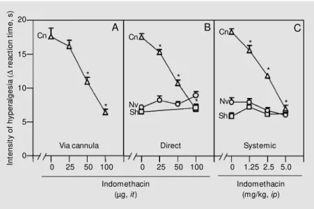
Figure 2 shows that the increased PGE2
-induced hyperalgesic effect on Cn animals was dose-dependently blocked by indometha-cin injected via a cannula (panel A = 25-100 ng), directly into the intrathecal space (panel B = 25-100 ng), or systemically (panel C = 1.25-5 mg/kg, ip) and that the drug inhibited in a dose-dependent manner the hyperalge-sic effect of 10 ng/paw of PGE2 (P>0.05,
ANOVA), an effect that was significantly different for Nv or Sh animals. These results were obtained 3 h after intraplantar PGE2
challenge because the hyperalgesic response reached a plateau only at this time. This figure also shows that intraperitoneal or di-rect intrathecal injections of indomethacin did not affect the relatively small intensity of the hyperalgesic effect induced by PGE2 in
Sh and Nv animals (no difference between doses, P>0.05).
In the second series of experiments, for-malin was used as a chemical stimulator, inducing a dose-dependent number of flinches (first phase, 0-10 min; P<0.05, ANOVA; Figure 3). This dose dependency was also observed in the second phase of the test (10-60 min; data not shown). Figure 3
Figure 2 - Effect of indomethacin on the intensity of PGE2 hyperalgesia in chronically cannulated (Cn, triangles), sham-operated (Sh, squares) and naive (Nv, circles) animals. Indomethacin w as administered via an intrathecal (it) cannula (panel A), directly into the subarachnoid space (panel B) or systemically (ip, panel C) 30 min before intraplantar injection of PGE2 (10 ng/paw ). Each point represents the mean reaction time (s) ± SEM of 5-13 animals per group measured 3 h after the hyperalgesic challenge. The data w ere analyzed by one-w ay ANOVA follow ed by Duncan’s test (asterisks indicate significant differences for each dose, P<0.05).
In
te
n
s
it
y
o
f
h
y
p
e
ra
lg
e
s
ia
(D
r
e
a
c
ti
o
n
t
im
e
,
s
)
30
25
20
15
10
5
0
2.5 5 10 100 2.5 5 10 100 2 5 10 50 100
Cg (µg, ipl) PGE2 (ng, ipl) Nv
Sh
Nv Sh Cn Cn
Cn
* *
*
*
* *
* *
* *
A B C
Figure 1 - Comparison of the intensity of mechanical hyperalgesia in response to increasing doses of carrageenin (Cg) or PGE2 in chronically cannulated (Cn, triangles), naive (Nv, circles) and sham-operated (Sh, squares) rats. The int ensit y of hyperalgesia w as m eas-ured 1 h (panel A) and 3 h (panel B) after the different doses of carrageenin (2.5-100 µg, intraplantar (ipl)). Panel C show s the intensity of hyperalgesia measured 3 h after an ipl injection of PGE2 (5-100 ng/ml). The data (mean ± SEM for five animals per group) w ere analyzed by one-w ay ANOVA follow ed by Duncan’s test. * P<0.05 compared to Nv and Sh animals.
In
te
n
s
it
y
o
f
h
y
p
e
ra
lg
e
s
ia
(D
r
e
a
c
ti
o
n
t
im
e
,
s
)
20
15
10
5
0
0 25 50 100 0 25 50 100 0 1.25 2.5 5.0
Indomethacin (mg/kg, ip) Nv
Sh
Nv
Sh Cn
Cn Cn
*
* *
* *
*
*
A B C
*
Via cannula Direct Systemic
Indomethacin (µg, it)
*
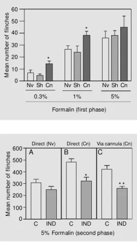
shows that Cn rats presented a higher fre-quency of flinches after stimulation with 0.3 and 1% of formalin when compared with Nv and Sh animals (P<0.05, Duncans test). An increase in the number of flinches in Cn animals was also observed in the second phase (data not shown). Dorsal injections of 5% formalin did not cause significant differences between the three groups of animals during the first phase but did in the second phase of the test (comparing con-trols of panel B with A and C with A of Figure 4).
Figure 4 also shows that the number of flinches observed during the second phase of the formalin test (5% formalin) was sig-nificantly inhibited when indomethacin was administered to chronically implanted ani-mals either directly into the intrathecal space or via the cannula (panels B and C, respec-tively; P<0.01 and P<0.001, unpaired Stu-dent t-test). Note that administration of in-domethacin (10 µg) by direct puncture did not significantly affect flinching in Nv ani-mals when compared with the control group (panel A; Tris-buffer).
In the third series of experiments, using thermal stimulation of the paw inflamed with increasing doses of carrageenin (31-125 µg), the Cn animals showed a significant sensiti-zation 1 h after carrageenin challenge (P<0.05, one-way ANOVA followed by Duncans test), compared with Nv or Sh animals (Figure 5). This differential sensiti-zation was no longer significant 3 h after carrageenin challenge, when paw edema reached maximal values (data not shown). Note that there were significant differences between the Cn controls and the Nv or Sh animals (C).
No inflammatory cells were found in sec-tions of spinal cords from Nv animals (N = 25). The presence of predominantly mono-nuclear inflammatory cell infiltration was observed in the spinal cords of 20 and 69% of Sh (N = 27) and Cn animals (N = 43), respectively.
Figure 3 - Differences in the in-tensity of response of chroni-cally cannulated (Cn), naive (Nv) and sham -operated (Sh) rats during phase 1 of the formalin test. Formalin w as administered at different doses of 0.3, 1, and 5% . Each bar is the mean ± SEM number of flinches of 6-12 animals, measured for 10 min after the nociceptive stimulus. Data w ere analyzed by one-w ay ANOVA follow ed by Duncan’s test. * P<0.05 compared to nor-mal or Sh aninor-mals.
M
e
a
n
n
u
m
b
e
r
o
f
fl
in
c
h
e
s 60
50
40
30
20
10
0
Nv Sh Cn Nv Sh Cn Nv Sh Cn
0.3% 1% 5%
Formalin (first phase)
Figure 4 - Inhibitory effect of indomethacin on the increased response of the second phase (10-60 min) of the 5% formalin test in cannulated (Cn) animals. Panels A and B show the ef-fects of direct injections of in-domethacin (IND, 10 µg/10 µl) adm inist ered 30 m in bef ore subcutaneous injection of for-malin to naive (Nv) and Cn ani-mals. Panel C show s the effect of IND administration through a cannula. The controls (C) re-ceived 10 µl of Tris-buffer. The bars represent the mean ± SEM number of flinches of the in-jected paw of 9-12 animals per group. The asterisks indicate significant differences (* P<0.01 and * * P<0.001, unpaired Stu-dent t-test).
M
e
a
n
n
u
m
b
e
r
o
f
fl
in
c
h
e
s 600
500
400
300
200
100
0
A B C
Direct (Nv) Direct (Cn) Via cannula (Cn)
*
* *
C IND C IND C IND
5% Formalin (second phase)
D iscussio n
In our first series of experiments with PGE2 and carrageenin, there was an increased
hyperalgesic response to paw pressure in Cn animals compared with Nv or Sh animals (Figure 1). This increased sensitivity of Cn animals could be noted either by an enhance-ment of the effect of lower doses of carra-geenin or by an early hyperalgesic plateau. Cn animals reached the plateau 1 h after intraplantar injection of carrageenin while 3 h were necessary for Nv or Sh animals (com-pare A and B in Figure 1). In Cn animals carrageenin caused an earlier plateau effect when compared with PGE2 (data not shown).
This might result from the fact that, in addi-tion to PGE2, carrageenin releases
prostacy-clin and this eicosanoid causes a much ear-lier response and plateau than that induced by PGE2 (21).
*
An increased response was also observed after chemical stimulation. Cn rats presented a higher frequency of flinches in response to 0.3 and 1% formalin during the first phase of the test when compared with Nv and Sh animals (Figure 3). The presence of the can-nula increased the number of flinches in response to 5% formalin in the second phase compared with control Nv animals (Figure 4).
The hypersensitivity to thermal stimula-tion in paws inflamed by increasing doses of carrageenin was significantly higher in Cn animals than in Nv or Sh animals (Figure 5). However, no significant differences were observed in the controls not sensitized with intraplantar administration of carrageenin.
Thus, the common denominator observed with different experimental methods of in-flammatory nociception was an increased responsiveness of the chronically cannulated animals to mechanical, chemical or thermal stimuli. It should be noted, however, that when mechanical (results not shown) and thermal (Figure 5) stimuli were used there was no difference between control paws of Nv and Cn animals. Thus, the presence of the cannula did not produce of itself a detect-able increase in nociception in tests using pressure or thermal stimuli. However, in Cn animals the inflammation occurring when hyperalgesia was induced by formalin (which produces a quick inflammatory response), carrageenin (which causes an inflammation
with a late plateau) or PGE2 (which mimics
inflammatory hyperalgesia) caused signifi-cantly higher nociceptive responses than observed in Sh or Nv animals. We observed no differences in the nociceptive response of the control Cn, Sh or Nv animals before the hyperalgesic treatment either in the paw pres-sure test (data not shown) or when the paw was thermally stimulated (Figure 5). Thus, it appears that sensitization of the primary sen-sory neuron is a condition necessary for detection of the hyperalgesic effect of the implanted cannula. A possible reason for the failure of the mechanical and thermal noci-ception test in detecting the presence of the cannula may be the low sensitivity of the test. Alternatively, and more plausibly, it may be that the lowering of the threshold of the primary sensory neurons induced by car-rageenin, PGE2 or formalin is a necessary
condition for detection of the supraspinal and/or spinal sensitization provoked by the presence of the spinal cannula. This inter-pretation explains why a blockade of spinal neuron sensitization by intrathecal adminis-tration of indomethacin inhibited in a dose-dependent manner the hyperalgesia induced by intraplantar injection of PGE2 in the
pres-sure test (Figure 2) or by carrageenin in the thermal test (Figure 5). The blockade of PGE2 paw hyperalgesia by intrathecally
ad-ministered indomethacin is noteworthy be-cause this effect is not blocked by systemic or intraplantar treatment with indomethacin (21).
Infection is not a usual complication in studies using the intrathecal cannulation pro-cedure. However, there are reports of altered responses to drug administration with an acute or chronic implantation of a catheter (16,22-24). Inflammatory changes along the catheter tract have been observed, as well as spinal cord compression in some cases (16). In fact, mononuclear cell infiltration was frequently found in cannulated animals, com-pared with absence of infiltration in Nv ani-mals. It should be pointed out that, as
previ-Figure 5 - Comparison of cannu-lated (Cn), naive (Nv) and sham (Sh) animals in terms of w ith-draw al latencies of carrageenin (Cg)-inflam ed rat paw s in re-sponse to thermal stimulation. The bars represent groups in-jected w ith different doses of carrageenin (intraplantar, ipl). C is the control group injected w ith saline. The results are the mean ± SEM percent ratio betw een carrageenin-injected and control paw s 1 h after injection of the stim ulus in 5-13 anim als per group. Data w ere analyzed by ANOVA follow ed by Duncan’s test. The asterisks indicate sig-nificant differences betw een Nv and Cn animals (P<0.05).
L
a
te
n
c
y
o
f
in
je
c
te
d
p
a
w
/c
o
n
tr
o
l
(%
)
120
100
80
60
*
*
Nv Sh
Cg (µg, ipl) *
Cn Nv Sh Cn Nv ShCn NvSh Cn
ously described, the presence of migrating cells in an injured tissue is not a determinant condition for induction of hyperalgesia (25). The presence of mononuclear cells in a num-ber of Sh animals (which was not observed in Nv animals) may indicate that mere punc-ture of the occipital membrane may induce a mild inflammatory reaction. Although the presence of cells per se does not indicate sensitization, in order to avoid this possibil-ity, we used Nv animals to replicate Malm-berg and Yakshs observation of the inhibi-tory effect of the non-steroid anti-inflamma-tory drug indomethacin on the second phase of the formalin test at the dose of 5% (13,14). Indomethacin administration either via a can-nula or directly by intrathecal puncture to chronically cannulated animals caused anti-nociception. On the other hand, direct intra-thecal injection of indomethacin had no sig-nificant antinociceptive effect on Nv ani-mals (Figure 4). Thus, the presence of the cannula permitted the detection of the anti-nociceptive effect of indomethacin in Cn but not in Nv animals.
The presence of a chronically implanted cannula (Cn group) also enhanced the sensi-tivity to otherwise ineffective intraplantar doses of PGE2 (10 ng/paw in Nv and Sh
animals) and in such instances, the adminis-tration of indomethacin (via cannula, di-rectly by spinal puncture or systemically) blocked this prostaglandin effect in a dose-dependent manner (Figure 2). In a pilot study with a higher dose of PGE2 (100 ng/paw),
administration of indomethacin either intra-peritoneally (5 mg/kg) or via an intrathecal cannula (50 µg) or directly into the spinal cord (50 µg) did not modify the magnitude of hyperalgesia (data not shown). It should be pointed out that this high PGE2 dose already
produces a maximal hyperalgesia in response to pressure when injected into Nv animals. Thus, the lack of effect of indomethacin might reflect the fact that abolition of spinal sensitization by the cannula does not modify the maximal effect of PGE2. The intrathecal
indomethacin blockade of nociceptive be-havior induced by mechanical or thermal stimulation of hyperalgesic paws may be due to the ongoing sensitization of the spinal sensory neurons caused by the presence of the cannula in the spinal cord and may there-fore reflect an inhibition of the formation of prostaglandins due to a mild inflammation (small presence of neutrophils) induced by the chronically implanted cannula. The sen-sory neurons sensitized by spinal cannula-tion are situated above the lumbar region, probably in the thoracic region (the distal tip of the cannula approaches the lumbar re-gion). Supporting this suggestion is the fact that the fluid of the direct intrathecal injec-tions bathes this region without reaching the cervical vertebrae (Ferreira SH, personal ob-servations).
We have previously reported that intrathe-cal administration of PGE2 (L4-L5) causes a
Re fe re nce s
1. Yaksh TL & Rudy TA (1976). Chronic cath-eterization of the spinal subarachnoid space. Physiology and Behavior, 17: 1357-1358.
2. Yaksh TL & Rudy TA (1976). Analgesia mediated by a direct spinal action of nar-cotics. Science, 192: 1357-1358. 3. M almberg AB & Yaksh TL (1993).
Phar-macology of the spinal action of ketorolac, morphine, ST-91, U50488H, and L-PIA on the formalin test and an isobolographic analysis of the NSAID interaction (see comments). Anesthesiology, 79: 270-281. 4. M almberg AB & Yaksh TL (1995). Cyclo-oxygenase inhibition and the spinal re-lease of prostaglandin E2 and amino acids evoked by paw formalin injection: a mi-crodialysis study in unanesthetized rats. Journal of Neuroscience, 15: 2768-2776. 5. M almberg AB & Yaksh TL (1995). The
effect of morphine on formalin-evoked behaviour and spinal release of excitatory amino acids and prostaglandin E2 using microdialysis in conscious rats. British Journal of Pharmacology, 114: 1069-1075. 6. Akerman B, Rosell S & Folkers K (1982). Intrathecal (D-Pro2, D-Trp7,9)-SP elicits hypoalgesia and motor blockade in the rat and antagonizes noxious responses in-duced by substance P. Acta Physiologica Scandinavica, 114: 631-633.
7. Yamamoto T & Yaksh TL (1993). Effects of intrathecal strychnine and bicuculline on nerve compression-induced thermal hyperalgesia and selective antagonism by M K-801. Pain, 54: 79-84.
8. Chaplan SR, Pogrel JW & Yaksh TL (1994). Role of voltage-dependent calcium chan-nel subtypes in experimental tactile allo-dynia. Journal of Pharmacology and Ex-perimental Therapeutics, 269: 1117-1123. 9. Ramw ell PW, Shaw JE & Jessup R (1966). Spontaneous and evoked release of pros-taglandins from frog spinal cord. Ameri-can Journal of Physiology, 211: 998-1004. 10. Ferreira SH (1983). Peripheral and central analgesia. In: Bonica JJ, Lindblom U &
Iggor A (Editors), Advances in Pain Re-search and Therapy. Raven Press, New York.
11. Taiw o YO & Levine JD (1986). Indometha-cin blocks central nociceptive effects of PGF2 alpha. Brain Research, 373: 81-84. 12. M inami T, Uda R, Horiguchi S, Ito S,
Hyodo M & Hayaishi O (1994). Allodynia evoked by intrathecal administration of prostaglandin E2 to conscious mice. Pain, 57: 217-223.
13. M almberg AB & Yaksh TL (1992). Antino-ciceptive actions of spinal nonsteroidal anti-inflammatory agents on the formalin test in the rat. Journal of Pharmacology and Experimental Therapeutics, 263: 136-146.
14. M almberg AB & Yaksh TL (1992). Hyper-algesia mediated by spinal glutamate or substance P receptor blocked by spinal cyclooxygenase inhibition. Science, 257: 1276-1279.
15. Ferreira SH, Lorenzetti BB & Correa FM (1978). Central and peripheral antialgesic action of aspirin-like drugs. European Jour-nal of Pharmacology, 53: 39-48. 16. Serpell M G, DeLeo JA, Coombs DW,
Colburn RW, Tw itchell BB, Willenbring S & Fromm C (1993). Intrathecal catheter-ization alone reduces autotomy after sci-atic cryoneurolysis in the rat. Life Sci-ences, 53: 1887-1892.
17. Papir-Kricheli D, Frey J, Laufer R, Gilon C, Chorev M , Selinger Z & Devor M (1987). Behavioural effects of receptor-specific substance P agonists. Pain, 31: 263-276. 18. M estre C, Pelissier T, Fialip J, Wilcox G &
Eschalier A (1994). A method to perform direct transcutaneous intrathecal injection in rats. Journal of Pharmacology and Toxi-cology M ethods, 32: 197-200.
19. Wheeler-Aceto H, Porreca F & Cow an A (1990). The rat paw formalin test: com-parison of noxious agents. Pain, 40: 229-238.
20. Hargreaves K, Dubner R, Brow n F, Flores C & Joris J (1988). A new and sensitive
method for measuring thermal nocicep-tion in cutaneous hyperalgesia. Pain, 32: 77-88.
21. Ferreira SH, Nakamura M & Abreu Castro M S (1978). The hyperalgesic effects of prostacyclin and prostaglandin E2. Prosta-glandins, 16: 31-37.
22. Long JB, M obley WC & Holaday JW (1988). Neurological dysfunction after in-trathecal injection of Dynorphin A (1-13) in the rat. I. Injection procedures modify pharmacological responses. Journal of Pharmacology and Experimental Thera-peutics, 246: 1158-1166.
23. St evens CW & Yaksh TL (1986). Dynorphin A and related peptides admin-istered intrathecally in the rat: A search for putative kappa opiate receptor activity. Journal of Pharmacology and Experimen-tal Therapeutics, 238: 833-838.
24. Bianchi M , Dib B & Panerai AE (1998). Interleukin-1 and nociception in the rat. Journal of Neuroscience Research, 53: 645-650.
25. Castro M SA & Ferreira SH (1979). Cell migration and hyperalgesia: A paradoxical effect of endotoxin. In: Weissmann G, Samuelsson B & Paoletti R (Editors), Ad-vances in Inflammation Research. Raven Press, New York.
26. Ferreira SH & Lorenzetti BB (1994). Gluta-mate spinal retrograde sensitization of pri-mary sensory neurons associated w ith nocicept ion. Neuropharm acology, 33: 1479-1485.
27. Ferreira SH & Lorenzetti BB (1996). In-trathecal administration of prostaglandin E2 causes sensitization of the primary af-ferent neuron via the spinal release of glutamate. Inflammation Research, 45: 499-502.