neuropathic pain complaints. The present review article provides relevant information on the understanding and recognition of NP, as well as evidence-based therapeutic approaches.
KEY WORDS: neuropathic pain, nociceptive pain, diagnosis, treatment.
o que os neurologistas gerais devem saber sobre dor neuropática?
resumo – A dor neuropática (DN) é definida como dor causada por lesão ou disfunção do sistema somatossensitivo, como resultado da ativação anormal da via nociceptiva (fibras de pequeno calibre e trato espinotalâmico). As principais causas desta síndrome são: diabetes, neuralgia pós-herpética, neuralgia trigeminal, acidente vascular encefálico, esclerose múltipla, trauma raquimedular, infecção por HIV, câncer. Nos últimos anos, a DN vem recebendo especial atenção por dois motivos principais: (1) refratariedade terapêutica de várias síndromes dolorosas com componentes neuropáticos predominantes e (2) desenvolvimento de ferramentas diagnósticas para o reconhecimento deste tipo de dor. O presente artigo de revisão fornece informações relevantes para o entendimento e reconhecimento da DN, bem como de abordagens terapêuticas baseadas em evidência.
PALAVRAS-CHAVE: dor neuropática, dor nociceptiva, diagnóstico, tratamento.
1MD, PhD, Department of Neurology, Hospital de Clínicas de Porto Alegre, Porto Alegre RS, Brazil; 2MD, PhD, Department of Neurology, Fluminense Federal University, Niterói RJ, Brazil.
Received 4 March 2009, received in inal form 9 May 2009. Accepted 11 June 2009.
Dr. Pedro Schestatsky – EMG Unit. Neurology Department / Hospital de Clínicas de Porto Alegre - Rua Ramiro Barcelos 2600 - 90035-003 Porto Alegre RS - Brasil. E-mail: [email protected]
Pain is deined as an unpleasant emotional experience related to potential or real tissue damage1, being
classi-ied as “nociceptive” or “neuropathic” pain types. Nocice-ptive pain is caused by physiological activation of pain re-ceptors and it is related to musculoskeletal tissue damage i.e., osteoarthritis, hand trauma and so forth2.
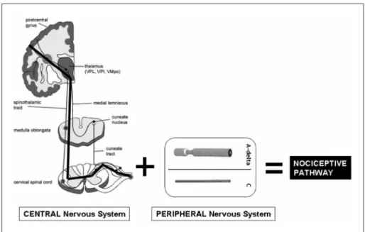
Neuropath-ic pain (NP), on its turn, is deined as pain initiated by a le-sion or dysfunction of the somatosensory system, resul-tant from abnormal activity of the nociceptive pathway3.
This pathway is formed by small ibers and spinothalam-ic tracts up to cerebral cortex4 (Fig 1). More recently,
be-cause of the possible coexistence of both types of pain, some authors recommend the use of the term
predom-inant neuropathic or predominant nociceptive types of pain, depending on the predominant clinical picture2.
epidemiology
A recent survey analysing 6.000 adults from the pri-mary health system of the United Kingdom, found a prev-alence of 8.2% of pain with predominant neuropathic characteristics5,6. Such prevalence represented
as-sociated with pain, such as cancer, AIDS and diabetes7,8.
Overall, the most common cause of NP is diabetes mellitus9,10. More recently, some authors reported a high
prevalence of this kind of pain in patients with glucose in-tolerance or prediabetes11. Table 1 shows the most
com-mon causes of peripheral and central NP.
physiopathology
There are more than 20 postulated theories trying to clarify the mechanisms underlying NP12. However, most of
them are based on complex neurochemical models9,13, that
are dificult to translate for clinical practice. The conse-quence of such complexity and lack of knowledge on NP mechanisms is the low eficacy of pharmacological drugs available for NP (only 30 to 50% of pain relief), observed in recent meta-analysis14. In the opinion of the present
au-thors and other colleagues7,15, another reason for the
per-sistent refractoriness of NP treatments is the excessive emphasis on the study of central sensitization
phenom-enon as the main cause of NP. Such theory was based on several studies with important methodological problems that have gained huge popularity7,15. However, according
to Occam’s law16 (“the simplest theory is to be preferred
over the complicated one”), the most plausible and con-vincing theory for NP comprehension is the ectopic gen-eration of impulses coming from C mechano-insensitive ibers which leads to positive neuropathic symptoms in-cluding pain17,18. Right after a nerve lesion, some patients
may develop changes in Na+ ion channels that leads to an axonal hyperexcitability and painful symptoms. Indeed, sodium channel dysfunction, seems to be the basis of ge-netic predisposition to chronic pain19. Apart from the
pri-mary lesion site, axonal hyperexcitability sometimes also occur far away from the nerve injury region, or even in healthy and distant nerves (ectopic nerve discharges), causing apparently unexplained pain syndromes7,10.
In-deed, NP may be relieved by antiepileptic agents, such as carmazepine or gabapentine, that block Na+ channels14.
Fig 1. The nociceptive pathway: small fiber and spinothalamic tracts up to cerebral cor-tex (Modiied from Treede4, 2003.)
Table 1. Most common causes of neuropathic pain (modiied from Bennett5, 2006).
Pain topography Structure involved Examples
Peripheral nervous system Nerve Diabetic neuropathy
Trigeminal neuralgia
Complex regional pain syndrome Neuropathy induced by tumoral invasion Chronic entrapment (i.e., carpal tunnel syndrome)
Dorsal root Post-herpetic neuralgia
Traumatic brachial plexus avulsion
Central nervous system Brain Post-stroke
Multiple sclerosis
Spinal cord Spinal cord injury
In fact, some neurologists considered NP to be “an ep-ilepsy of the nerve that responds satisfactorily to anti-convulsivants”.
diagnosis
Identifying NP in clinical practice is not an easy task. The painful sensation cannot be objectively measured and there is no universal consensus for the diagnosis of such condition. However, some authors proposed a prac-tical algorithm for NP8, in which three levels of
diagnos-tic certainty are considered: possible, probable or deini-tive diagnosis of NP (Fig 2). Nocicepdeini-tive pain and NP coex-ist in most of painful conditions and their identiication is crucial. Neuropathic pain demands speciic analgesic ap-proaches, quite different from the traditional approach to nociceptive pain type. An illustrative example is a diabet-ic patient with osteoarthrosis. In this case, the pain can be resultant from three possibilities: (A) small iber dysfunc-tion, (B) activation of pain receptors due to bone damage, or (C) both of them. A careful clinical evaluation can guide
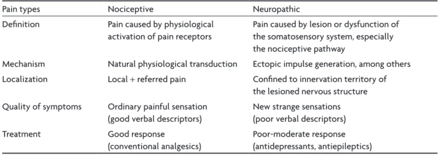
to a more rational and eficient therapeutic strategy for such kind of patient. Table 2 shows the main differences between neuropathic and nociceptive types of pain that could help to separating both entities7.
symptoms
Patients with NP can have multiple and complex sen-sory complaints. Differently from nociceptive pain, there are poor verbal descriptors for characterizing NP. Most patients report their symptoms using analogies (“ Doc-tor, my pain is like a…”). Such complaints are divided in spontaneous and evoked sensory symptoms. Spontane-ous pain, on its turn, can be subdivided into continuSpontane-ous or paroxistic ones. The latter is frequently, but not always, felt as if it is located at the supericial skin level and it is described in terms of dysaesthesias, such as burning, tin-gling, pricking and stabbing characteristics5.
signs
Abnormal and plausible indings on sensory exami-Fig 2. Levels of accuracy of the neuropathic pain diagnosis (Modiied from Treede8 et al.,
2007).
Table 2. Differences between nociceptive and neuropathic pain (modiied from Serra7, 2006).
Pain types Nociceptive Neuropathic
Deinition Pain caused by physiological
activation of pain receptors
Pain caused by lesion or dysfunction of the somatosensory system, especially the nociceptive pathway
Mechanism Natural physiological transduction Ectopic impulse generation, among others
Localization Local + referred pain Conined to innervation territory of
the lesioned nervous structure
Quality of symptoms Ordinary painful sensation
(good verbal descriptors)
New strange sensations (poor verbal descriptors)
Treatment Good response
(conventional analgesics)
nation in a patient with peculiar pain suggest the diag-nosis of NP10. Other neurological signs, such as hyper/
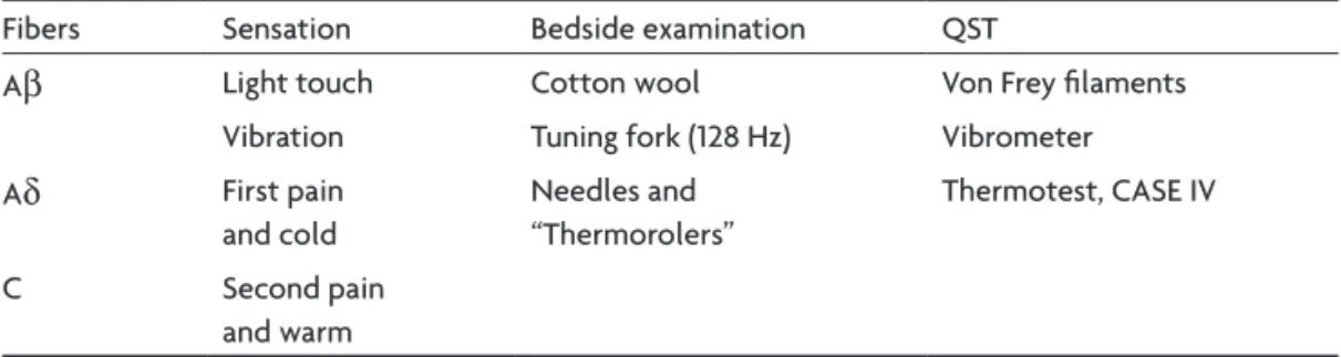
hypotony, focal paralysis and plantar cutaneous relex-es, among others, will also help to suggest the topogra-phy of pain (central vs. peripheral) in a patient with neu-ropathic symptoms. In order to improve the value of sen-sory indings in the diagnosis of NP, it is useful to classi-fy them in negative, positive and autonomic phenome-na5, as seen in Figure 3.
Negative phenomena result from loss of light touch, vi-bration and thermoalgesic senses. These sensations are me-diated by large myelinated Aβ (light touch and vibration), small poorly myelinated Aδ (cold and irst pain) and unmy-elinated type C ibers (warm and second pain). In order to assess the function of each type of iber, bedside maneu-vers and psychophysical tests (Table 3) can be employed3.
Positive phenomena of NP can be presented spon-taneously, evoked by sensory stimuli, or in combination. The most frequent described positive symptoms in clini-cal practice are the following5,7,10: (1) Allodynia: pain due to
stimulus which does not normally cause pain. Three types of allodynia are described, based on the precipitant stim-ulus: mechanical (or tactile), thermal (warm and cold) and kinetic (movement); (2) Hyperalgesia: painful sensation of
abnormal intensity in response to a nociceptive stimu-lus. Allodynia and hyperalgesia oftenly coexist in prac-tice and it can be dificult to differentiate the two. Both symptoms are considered essential features for NP, but they can also be present in the nociceptive type of pain; (3) Hyperpathia: painful reaction to repetitive nociceptive and non-nociceptive stimuli or prolonged pain sensation in response to nociceptive stimuli (aftersensation phe-nomenon); (4) Autonomic hyperactivity: abnormal blood low, cutaneous temperature and sweating can accompany painful states and contribute for its persistence. Trophic abnormalities can usually be developed in chronic pain states, such as seen in complex regional pain syndromes.
neuropathic pain scales
Verbal pain descriptor scales can provide important information to the assessment process alongside clinical history examination and investigation5. These scales also
provide standardized symptoms assessment for pain re-search and follow-up for NP treatments. Several scales have been published, with especial emphasis on NP in-tensity, such as the Neuropathic Pain Scale20, Neuropathic
Pain Questionnaire21 and Douleur Neuropatique 422. More
recently, the Leeds Assessment of Neuropathic Symptoms
Fig 3. Clinical picture of neuropathic pain (Bennett5, 2006).
Table 3. Bedside tests and quantitative sensory testing (QST) for different sensations conveyed by different types of sensory ibers (modiied from Cruccu et al.3, 2004)
Fibers Sensation Bedside examination QST
Aβ Light touch Cotton wool Von Frey ilaments
Vibration Tuning fork (128 Hz) Vibrometer
Aδ First pain
and cold
Needles and “Thermorolers”
Thermotest, CASE IV
C Second pain
and Signs Scale23 has been used as an index of
predomi-nant neuropathic vs. nociceptive pains. Such an instru-ment it is in validation process for Portuguese language in our center24.
most common neuropathic pain syndromes
Peripheral
Diabetic neuropathy – Burning feet sensation that gets
worse during the night is the typical clinical picture. The prevalence of NP in distal simetric polyneuropathy is 1–10%, depending on the degree of small iber involve-ment25. Rarely, pain occurs in the absence of large iber
signs and symptoms since small ibers are usually dysfunc-tional at early stages of the diabetic neuropathy.
Trigeminal neuralgia – Sudden, severe and usually
unilat-eral stabbing pain with V2 or V3 distribution of the V cra-nial nerve. Eighty percent of the cases are idiopathic, but 66% of these have evidence of vascular compression at the root entry zone26. Other causes include
demyelinia-tion secondary to multiple sclerosis, angioma, brainstem infarcts and tumors, such as acoustic neurinoma.
Post-herpetic neuralgia – Persistent pain over the skin
af-fected area that could last for more than 12 weeks after the healing of typical skin lesions, especially in elderly pa-tients. Pain may be disabling and recurs in months or even
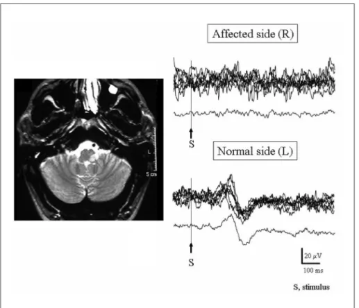
Fig 4. Absent laser-evoked potentials in a pa-tient with Wallenberg syndrome, supporting the diagnosis of neuropathic pain (From Hos-pital Clínic of Barcelona, 2005).
Table 4. Drugs for neuropathic pain (modiied from Finnerup14 et al., 2005 and Beniczky51 et al., 2005).
Neuropathic pain Drug NNT (CI 95%) Recommended doses
Peripheral Tricyclics 2.2 (1.9–2.6) Up to 150 mg/day
SNRI 6.8 (3.4–441) Up to 80 mg/day
Gabapentin 4.4 (3.4–6.2) 600 a 1200 mg 3×/day
Pregabalin 5.0 (3.5–8.6) 50 a 100mg 3×/day
Tramadol 3.9 (2.7–6.7) 200–400 mg/day
Oxicodone-CR 2.6 (1.9–4.1) 60–120 mg/day
Topical Lidocaine 4.4 (2.5–17.5) Patch or gel 5% (12 hs/day)
Carbamazepine 1.8 (1.4–2.7) Up to 1000 mg/day
Central Tricyclics 4.0 (2.6–8.5) Up to 75 mg/day
Lamotrigine 2.9 (1.3–5.0) Up to 200 mg/day
Carbamazepine 3.4 (1.7–105) Up to 1000 mg/day
years later. Both peripheral and central mechanisms con-tribute to the process27.
Cancer – Pain can be due to nerve lesion caused by
drect tumoral invasion, but also in case of secondary to i-brosis or myelopathy post-radiotherapy, chemotherapy or surgery, especially in cases of radical mastectomy or tho-racotomy5. Paraneoplastic painful neuropathies can
pre-cede tumor detection by months or years.
Drugs – Most of drug-induced neuropathies are of
ax-onal type, with a predilection for sensory nerves distally. The sensory injury can affect either large or small ibers. The most common drugs implicated in neuropathic pain are the following5: vincristine, zalcitabine and stavudine.
The treatment relies in durg interruption and the progno-sis is usually good.
Central
Any spinal-thalamic-cortical lesion can cause NP re-gardless its etiology (ischemic, inlamatory, infeccious and so on). Loss of pain descending inhibition is a less known physiopathological mechanism28 and can contribute for
NP maintenance in some patients with Parkinson’s dis-ease29. Stroke, multiple sclerosis and other myelopathies
are the most common causes of central NP5. This type of
pain is wrongly considered a rarity, since it occurs in 8% of patients with stroke30, 28% with multiple sclerosis, 75%
with syringomyelia31 and 70% with myelopathy30. Tactile
and cold allodynia are frequently seen in such patients. Approximately 70% of patients with post-stroke pain describe their pain in whole hemibody, ipsilateral to
mo-tor and sensory deficits31. There is no pathognomonic
characteristic of central NP. However, the pain is almost always dysaesthetic (“painful paresthesias”). Other aggra-vating factors are the psychiatric comorbities, commonly seen in patients with chronic pain, such as major depres-sion and anxiety that usually amplify pain perception10.
Central pain can initiate right after a structural damage, or it can take 2–3 years to arise after the insult. This may explain why central NP is so non-recognized in clinical practice. Figure 4 shows a patient with pain secondary to Wallenberg syndrome who had absent laser-evoked po-tentials contralateral to the neuropathic symptoms, indi-cating a lesion of the nociceptive pathway.
laboratory tests
Since NP results from lesion or dysfunction of small i-ber or spinothalamic tracts (nociceptive pathway), comple-mentary exams are used for direct or indirect demonstra-tion of lesion or dysfuncdemonstra-tion of the nociceptive pathway.
The quantitative sensory testing (QST) for temperature and pain helps to evaluate the nociceptive pathway as a whole, from the thermal receptor, through small ibers and spinothalamic tracts and up to patient’s verbal ex-pression of his/her perception. This is done using thresh-olds determination for different sensations32. By means
of a thermode with a temperature rising velocity of 1 to 4oC/s, placed over the skin affected by the pain, the
ateral area, for comparison purposes. Abnormal thermal thresholds (Fig 5B) signalize a lesion in any level of the no-ciceptive pathway, potentially causing NP symptoms.
Nerve conduction studies and electromyography – Although
this method does not assess small iber function, abnor-mal indings suggest a neuropathic process in a patient with pain10,33,34. For example: an altered test in a
diabet-ic patient with burning pain suggest a real NP since the small ibers usually are affected before the large ibers in the natural history of diabetic neuropathy35.
Microneurography – With a tungsten needle within the
nerve and using a speciic stimulation techniques it is pos-sible to identify and to record the activity of ive subtypes of C iber populations. By means of this method, Bostock and cols18 described the presence of double spikes from
mechano-insensitive C ibers in NP patients. This inding was considered to be a reliable marker of peripheral NP. Unfortunately, microneurography, a powerful tool for re-search, is too complex and time consuming to be used in clinical practice3.
Nociceptive RIII relex – By means of single or repetitive
electrical stimuli of the sural nerve it is possible to obtain electromyographic responses recorded at femoral biceps at latencies around 90 to 130 ms. The electrical threshold for its appearance and its maximal amplitude are measured7,34,36.
Because of its straight correlation with pain perception, the RIII responses have been used for monitoring the eficacy of pharmacological treatments for NP3.
Autonomic relexes – Besides carrying afferent signals of
pain and temperature, the C ibers are also involved with autonomic control (C efferent autonomic ibers) and its analysis can help in the etiological diagnosis of pain33,37. The
sympathetic skin response (SSR) is an autonomic relex me-diated by C efferent autonomic ibers29,34. Action potentials
are analyzed with respect to amplitudes and latencies. More recently, the analysis of SSR morphology and habituation
have been used for a better QST interpretation38 and for
the functional assessment of pain descending inhibition29.
Laser-evoked potentials (LEPs) – By means of scalp
elec-trodes, long latency brain potentials can be recorded in response to laser stimuli given on the skin, allowing the study of peripheral and central conduction of nocicep-tive ibers4. Abnormal LEPs (Fig 4) are seen in patients with
hemibody sensory syndromes, in which structural and asym-metrical lesions of the spinothalamic tract are found i.e., Wallenberg’s syndrome39 or syringomyelia40. According to
some authors, a lesion of the spinothalamic tract, demon-strable by abnormal LEPs is required for the establishment of NP diagnosis41,42. The only drawback of this method is
the undesirable burning of the skin after repetitive stimu-li in the same spot.
Contact-evoked heat potentials (CHEPS) – Differently from
the slow rising temperature thermode of QST, the ther-mode of this device is capable to increase the skin tem-perature with 70oC/s and, consequently, to generate long
latency brain potentials. One of the main advantages of the CHEPS in comparison with LEPs is the absence of cu-taneous lesions after several stimuli43. Recently, we
ob-served small amplitude CHEPS in patients with painful nerve entrapment of the thigh44, supporting the
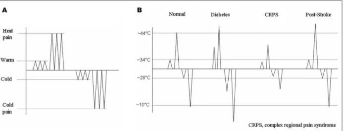
neuro-pathic character of pain complaints in meralgia paresthet-ica at latter stages (Fig 6A, 6B).
Functional neuroimaging – Functional magnetic
resso-nance image (fMRI) and positron emission tomography (PET) have been contributing for the mapping of cere-bral regions activated by nociceptive stimuli (“pain ma-trix”). These regions include the secondary somatosen-sory (SII), insular and cingulate cortex, as well as the up-per brainstem. Less consistently, the contralateral thala-mus and primary somatosensory are also considered pain matrix regions45. There is converging evidence that
spon-taneous NP is associated with less activity in contralat-Fig 6. Small amplitude contact-evoked heat potentials applied in the symptomatic side of a patient with meralgia
eral thalamus, whereas evoked pain is more associated with an increased activity in thalamic, insular and soma-tosensry regions3.
Skin punch biopsy – It allows the C iber quantiication
by measuring the intra-dermal C iber density. Loss of C ibers detected with the skin biopsy was seen in a large number of neuropathies46,47. The skin biopsy performed by
the punch technique is an easy and reliable method for the follow-up of NP patients, but it is unavailable in most medical centers throughout the world. Fortunately, there is a good correlation between intra-dermal C iber density and CHEPS’ amplitude48, a less invasive and more
accessi-ble method when compared o skin biopsy.
treatment
Recent studies have shown that most of patients treat-ed for NP receive drugs with non-provtreat-ed clinical eficacy or in inadequate dosages of appropriate medication49,50.
NP is usually refractory to ordinary analgesics. Tryciclic antidepressants and anticonvulsivants are the mainstay in the treatment of NP, regardless its topography (central vs. peripheral) and its etiology. Figure 7 shows a rational algorithm for the approach of patients with peripheral NP. Such an algorithm can also be used for patients with central NP14 with addition of few speciic drugs, such as
showed in Table 414,51.
Other treatment modalities, such as sensory stimula-tion (transcutaneous electrical nerve stimulastimula-tion or TENS, spinal cord stimulation, deep brain stimulation) and
sur-gery (thoracic sympathectomy, chordotomy, radicular neurolysis etc) are available in some centers, destinated to refractory patients52-54, but there is no strong enough
evidence for a systematical recommendation5,55. More
recently, transcranial magnetic stimulation of the motor cortex has been proposed in some forms of NP56, in order
to modulate painful symptoms through cortical reorga-nization. However, up to now, there are no deinitive con-clusions about the role of magnetic stimulation that could allow its wider utilization in clinical practice.
Final considerations
The three most important messages of this paper are: (1) NP is prevalent, frequently unrecognized and poorly treated; (2) A lesion in the somatosensory system - espe-cially the nociceptive pathway – is required for the diag-nosis of NP and (3) Neurologists have an important role in the approach of pain patients because of its capacity to detect subtle abnormalities in the neurological assess-ment that will support or not the diagnosis of NP.
reFerences
1. Merskey H, Bogduk N. Classiication of chronic pain. Seattle: IASP Press, 1994.
2. Bennett MI, Smith BH, Torrance N, Lee AJ. Can pain can be more or less neuropathic? Comparison of symptom assessment tools with rat
-ings of certainty by clinicians. Pain 2006;122:289-294.
3. Cruccu G, Anand P, Attal N, et al. EFNS guidelines on neuropathic pain assessment. Eur J Neurol 2004;11:153-162.
4. Treede RD. Neurophysiological studies of pain pathways in peripher
-al and centr-al nervous system disorders. J Neurol 2003;250:1152-1161.
Neurologist 2008;14:23-29.
12. Devor M. Neuropathic pain: what do we do with all these theories? Acta Anaesthesiol Scand 2001;45:1121-1127.
13. Woolf CJ, Salter MW. Neuronal plasticity: increasing the gain in pain. Science 2000;288:1765-1768.
14. Finnerup NB, Otto M, McQuay HJ, Jensen TS, Sindrup SH. Algorithm for neuropathic pain treatment: an evidence based proposal. Pain 2005;218:289-305.
15. Ochoa JL. Pain mechanisms in neuropathy. Curr Opin Neurol 1994;7: 407-414.
16. Jefferys WH, Berger JO. Ockham’s razor and Bayesian statistics. Amer Scientist 1991;80:64-72.
17. Serra J, Campero M, Bostock H, Ochoa J. Two types of C nociceptors in human skin and their behavior in areas of capsaicin-induced second
-ary hyperalgesia. J Neurophysiol 2004;91:2770-2781.
18. Bostock H, Campero M, Serra J, Ochoa JL. Temperature-dependent double spikes in C-nociceptors of neuropathic pain patients. Brain 2005;128:2154-2163.
19. Dib-Hajj SD, Yang Y, Waxman SG. Genetics and molecular pathophysi
-ology of Na(v)1.7-related pain syndromes. Adv Genet 2008;63:85-110. 20. Galer BS, Jensen MP. Development and preliminary validation of a pain
measure speciic to neuropathic pain: the Neuropathic Pain Scale. Neu
-rology 1997;48:332-338.
21. Krause SJ, Backonja MM. Development of a neuropathic pain question
-naire. Clin J Pain 2003;19:306-314.
22. Bouhassira D, Attal N, Alchaar H, et al. Comparison of pain syndromes associated with nervous or somatic lesions and development of a new neuropathic pain diagnostic questionnaire (DN4). Pain 2005;114:29-36. 23. Bennett M. The LANSS Pain Scale: the Leeds assessment of neuropath
-ic symptoms and signs. Pain 2001;92:147-157.
24. Schestatsky P, Felix-Torres V, Ehlers B, Camozzato AL, Nascimeno O, Chaves ML. Validação da Escala LANSS para a língua portuguesa. In process.
25. Little AA, Edwards JL, Feldman EL. Diabetic neuropathies. Pract Neu
-rol 2007;7:82-92.
26. Cruccu G, Biasiotta A, Galeotti F, Iannetti GD, Truini A, Gronseth G. Diagnostic accuracy of trigeminal relex testing in trigeminal neural
-gia Neurology 2006;66:139-141.
27. Truini A, Galeotti F, Haanpaa M. Pathophysiology of pain in postherpetic neuralgia: a clinical and neurophysiological study. Pain 2008;140:405-410. 28. Millan MJ. Descending control of pain. Prog Neurobiol 2002;66:355-474. 29. Schestatsky P, Kumru H, Valls-Solé J, et al. Neurophysiologic study of cen
-tral pain in patients with Parkinson disease. Neurology 2007;69:2162-2169. 30. Bonica JJ. Introduction: semantic, epidemiologic, and educational is
-sues. In: Casey KL (Ed) Pain and central nervous system disease: the central pain syndromes. New York: Raven Press 1991:13-29. 31. Boivie J. Central pain. In: Wall PD, Melzak R (Eds) Textbook of pain.
Edinburgh: Churchill Livingstone, 1999:879-914.
32. Fruhstorfer H, Lindblom U, Schmidt WC. Method for quantitative es-Fruhstorfer H, Lindblom U, Schmidt WC. Method for quantitative es-Method for quantitative es
-timation of thermal thresholds in patients. J Neurol Neurosurg Psychi-J Neurol Neurosurg Psychi
-atry 1976;39:1071-1075.
39. Hansen HC, Treede RD, Lorenz J, Kunze K, Bromm B. Recovery from brainstem lesions involving the nociceptive pathways: comparison of clinical indings with laser-evoked potentials. J Clin Neurophysiol 1996;13:330-338.
40. Veciana M, Valls-Sole J, Schestatsky P, Montero J, Casado V. Abnormal sudomotor skin responses to temperature and pain stimuli in syringo-syringo
-myelia.J Neurol 2007;254:638-645.
41. Casey KL, Beydoun A, Boivie J, et al. Laser-evoked cerebral potentials and sensory function in patients with central pain. Pain 1996;64:485-491. 42. Garcia-Larrea L, Convers P, Magnin M, et al. Laser-evoked potential ab-Laser-evoked potential ab
-normalities in central pain patients: the inluence of spontaneous and provoked pain. Brain 2002;125:2766-2781.
43. Chen AC, Niddam DM, Arendt-Nielsen L. Contact heat evoked poten
-tials as a valid means to study nociceptive pathways in human subjects. Neurosci Lett 2001;316:79-82.
44. Schestatsky P, Lladó-Carbó E, Casanova-Molla J, Alvarez-Blanco S, Valls-Solé J. Small ibre function in patients with meralgia parestheti
-ca. Pain 2008;139:342-348.
45. Peyron R, Laurent B, Garcia-Larrea L. Functional imaging of brain responses to pain. Areview and metaanalysis. Neurophysiol Clin 2000;30:263-288.
46. McCarthy BG, Hsieh ST, Stocks A, et al. Cutaneous innervation in senso-et al. Cutaneous innervation in senso-. Cutaneous innervation in senso
-ry neuropathies: evaluation by skin biopsy. Neurology 1995;45:1848-1855. 47. Holland NR, Stocks A, Hauer P, Cornblath DR, Grifin JW, McArthur
JC. Intraepidermal nerve iber density in patients with painful sensory neuropathy. Neurology 1997;48:708-711.
48. Atherton DD, Facer P, Roberts KM, Misra VP, Chizh BA, Bountra C. Use of the novel contact heat evoked potential stimulator (CHEPS) for the assessment of small ibre neuropathy: correlations with skin lare responses and intra-epidermal nerve ibre counts. BMC Neurol 2007;7:21.
49. Richeimer SH, Bajwa ZH, Kachramann SS, Ransil BJ, Warield CA. Uti
-lization patterns of tricyclic antidepressants in a multidisciplinary pain clinic: a survey. Clin J Pain 1997;13:324-329.
50. Finnerup NB, Johannesen IL, Sindrup SH, Bach FW, Jensen TS. Pain and dysaesthesia in patients with spinal cord injury: a postal survey. Spinal Cord 2001;39:256-262.
51. Beniczky S, Tajti J, Tímea Varga E, Vécsei L. Evidence-based pharma
-cological treatment of neuropathic pain syndromes. J Neural Transm 2005;112:735-749.
52. Meyerson BA. Neurosurgical approaches to pain treatment. Acta An
-aesthesiol Scand 2001;45:1108-1113.
53. Cheing GL, Luk ML. Transcutaneous electrical nerve stimulation for neuropathic pain. J Hand Surg 2005;30:50-55.
54. Cruccu G, Aziz TZ, Garcia-Larrea L, et al. EFNS guidelines on neuro
-stimulation therapy for neuropathic pain. Eur J Neurol 2007;14:952-970. 55. Pittler MH, Ernst E. Complementary therapies for neuropathic and neu
-ralgic pain: systematic review. Clin J Pain 2008;24:731-733.
56. Lefaucheur JP, Drouot X, Ménard-Lefaucheur I, Keravel Y, Nguyen JP. Motor cortex rTMS restores defective intracortical inhibition in chron