RBCCV 44205-1544 DOI: 10.5935/1678-9741.20140026
Improvement in cardioplegic perfusion technique
in single aortic clamping - initial results
Aperfeiçoamento em técnica de perfusão cardioplégica no pinçamento único de aorta - resultados iniciais
Marcelo Luiz Peixoto Sobral
1, PhD; Sérgio Francisco dos Santos Júnior
2; Juliano Cavalcante de
Sá
3;
Anderson da Silva Terrazas
4; Daniel Francisco de Mendonça Trompieri
1; Thierry Araújo Nunes
de Sousa
1; Gilmar Geraldo dos Santos
5, PhD; Noedir Antonio Groppo Stolf
6, MsC, PhD
1Real e Benemérita Associação Portuguesa de São Paulo, São Paulo, SP,
Brazil.
2Santa Casa de Itabuna (SCMI), Itabuna, BA, Brazil. 3Hospital Santa Izabel, Salvador, BA, Brazil.
4Hospital Universitário Francisca Mendes (UFAM), Manaus, AM, Brazil. 5Instituto do Coração da Faculdade de Medicina da Universidade de São
Paulo INCOR/FMUSP, São Paulo, SP, Brazil.
6Instituto do Coração da Faculdade de Medicina da Universidade Dde São
Paulo (FMUSP), São Paulo, SP, Brazil. Real e Benemérita Associação Por-tuguesa de São Paulo, São Paulo, SP, Brazil.
Correspondence address: Marcelo Luiz Peixoto Sobral
Real e Benemérita Associação Portuguesa de Beneicência de São Paulo Rua Maestro Cardim, 769, Bloco 5, 2º SS, Sala 30 – Paraíso - São Paulo, SP, Brazil - Zip code 01323-900
E-mail: [email protected]
This study was carried out at Real e Benemérita Associação Portuguesa de Beneicência de São Paulo, São Paulo, SP, Brazil.
No inancial support.
Article received on July 7th, 2013
Article accepted on November 17th, 2013 Abstract
Introduction: The most common method used for myocardial protection is administering cardioplegic solution in the coronary circulation. Nevertheless, protection may be achieved by inter-mittent perfusion of the coronary system with patient’s own blood. The intermittent perfusion may be performed by multiple sequences of clamping and opening of the aortic clamp or due single clamping and accessory cannulation of the aortic root as in the improved technique proposed in this study, reperfusion without the need for multiple clamping of the aorta.
Objective: To evaluate the clinical outcome and the occurrence of neurological events in in-hospital patients submitted to myo-cardial revascularization surgery with the “improved technique” of intermittent perfusion of the aortic root with single clamping.
Methods: This is a prospective, cross-sectional, observational study that describes a myocardial management technique that consists of intermittent perfusion of the aortic root with single clamping in which 50 patients (mean age 58.5±7.19 years old) have been submitted to the myocardial revasculrization surgery under the proposed technique. Clinical and laboratory variables, pre- and post-surgery, have been assessed.
Results: The mean peak level of post-surgery CKMB was 51.64±27.10 U/L in the second post-surgery and of troponin I was 3.35±4.39 ng/ml in the fourth post-surgery, within normal
limits. No deaths have occurred and one patient presented mild neurological disorder. Hemodynamic monitoring has not indi-cated any changes.
Conclusion: The myocardial revascularization surgery by perfusion with the improved technique with intermittent aortic root with single clamping proved to be safe, enabling satisfactory clinical results.
Descriptors: Anastomosis, surgical. Thoracic Surgery. Car-diopulmonary Bypass. Myocardial Reperfusion Injury.
Resumo
Introdução: O método mais comumente utilizado para a pro-teção miocárdica é o de administrar-se solução cardioplégica na circulação coronária. Entretanto, a proteção pode ser alcançada através da perfusão intermitente do sistema coronariano com sangue do próprio paciente, que é realizada por meio de múltiplas sequências de pinçamento e abertura do clamp aórtico ou por meio do pinçamento único e canulação acessória da raiz aórtica.
The aim of this study is to assess the clinical outcome and the occurrence of neurological events in hospital stay of patients undergoing CABG with improved technique of in-termittent perfusion of the aortic root with a single clamping.
METHODS
This is a cross-sectional observational study in which 50 patients with heart failure and indication of surgery for cor-onary artery bypass grafting with cardiopulmcor-onary bypass (CPB) underwent the procedure according to regular sched-ule and surgery by the same surgeon between August 2010 and June 2012.
Preoperative information (age, gender, diabetes mellitus II, hypertension, dyslipidemia, previous stroke, previous MI, history of smoking and alcohol consumption, COPD, CRF, ARF, LVEF and current medications) were compiled and an-alyzed. Intraoperative (duration of each distal anastomosis,
CPB low during each reperfusion, MAP during infusion of
the aortic root, time of each reperfusion and proximal anas-tomosis, number of distal and proximal anastomoses, CPB and anoxia, minimum temperature during CPB, using or not of the centrifugal pump during CPB or if the CPB
disconnec-tion was in sinus rhythm or ventricular ibrilladisconnec-tion, as well as
the HR, MAP, minimum and maximum temperature, diure-sis, inotropic support and IABP) and postoperative
in-hospi-tal period (neurological deicit, duration of ICU and hospiin-hospi-tal
stay, and perioperative as ECG changes, urea, creatinine, cre-atine kinase and troponin I).
INTRODUCTION
Coronary artery bypass graft (CABG) is a surgical pro-cedure well standardized in the world today, with survival rates at one year around 97%, remaining at 81% even after
ten years postoperatively[1]. The development of techniques
of myocardial protection during aortic clamping is largely responsible for these good results.
Myocardial protection can be achieved with the use of methods of transient ischemia, myocardial or atrial infusion of cardioplegic solution into the coronary circulation. The choice of method to be used generally depends on the
behav-ior of the service and experience of the surgeon[2].
In an adjuvant way, a current study reports that the technique of remote ischemic preconditioning reduced the amount of release of cardiac enzymes in patients undergo-ing myocardial revascularization, although no difference in length of hospital stay and creatinine values were reported.
This technique has its eficacy independent of myocardial
protection technique[3]. Hong et al.[4] conclude that the remote
ischemic preconditioning with remote ischemic postcondi-tioning of upper limb does not improve clinical outcomes in patients undergoing cardiovascular surgery.
However, performing multiple clamping of the ascending aorta during surgery with intermittent infusion may have the unintended effect of higher incidence of neurological events due to the manipulation of atheromatous plaques in this re-gion. A single aortic clamping should be preferred in order to reduce these events[5].
Abbreviations, acronyms & symbols
CVA Cerebrovascular accident AMI Acute Myocardial Infarction
ACE inhibitor Angiotensin Converting Enzyme CPB Cardiopulmonary bypass
CABG Coronary artery bypass grafting COPD Chronic Obstructive Pulmonary Disease ICU Intensive Care Unit
LVEF Left ventricular ejection fraction
Métodos: Descreve-se uma técnica de proteção miocárdica no uso do pinçamento único de aorta que consiste na canulação acessória da raiz aórtica com sistema aperfeiçoado para perfu-são coronária intermitente, foi realizado estudo observacional transversal prospectivo onde foram estudados 50 pacientes (idade
média 58,5±7.19 anos) submetidos à cirurgia de revascularização do miocárdio sob a técnica proposta. Foram avaliadas variáveis clínicas e laboratoriais pré e pós-operatórias.
Resultados: O nível médio de pico da CKMB pós-operatória foi de 51,64±27,10 U/L no segundo pós-operatório e da troponina I foi de 3,35±4,39 ng/ml no quarto pós-operatório, e estiveram dentro do limite da normalidade. Não foi observado nenhum óbito e um paciente evoluiu com alteração neurológica leve. A monitorização hemodinâmica não revelou alterações.
Conclusão: A cirurgia de revascularização do miocárdio com a técnica aperfeiçoada de perfusão intermitente da raiz da aorta com pinçamento único mostrou-se como método seguro, possibi-litando resultados clínicos satisfatórios nos pacientes estudados.
We also obtained radiographic, echocardiographic, and cardiac catheterization data. Patients were recruited only after signing Written Informed Consent (IC), in accordance with Resolution 196/96 of the National Health Council. The study was approved by the Research Ethics Committee at
Real e Benemérita Associação Portuguesa Beneicência de
São Paulo under the protocol number 659-10.
Inclusion criteria
The study included patients of both genders with indi-cation for CABG surgery performed with cardiopulmonary bypass, aged up to 70 years.
Exclusion criteria
The study excluded patients who had undergone surgery without cardiopulmonary bypass with moderate or severe aortic failure on echocardiography or aortography, with clin-ical evidence of previous cerebrovascular disease, clinclin-ical or ultrasound evidence of carotid system disease and concomi-tant surgery for aortic valve failure.
Surgical strategy
The approach to the heart was median longitudinal trans-sternal sternotomy , inverted “T” pericardiectomy, prepa-ration for CPB, full heparinization (3mg/kg), arterial can-nulation of the ascending aorta, cavoatrial cancan-nulation and number 14 catheter insertion in the ascending aorta for con-nection of airway suction and infusion, entry into cardiopul-monary perfusion (conventional membrane oxygenation), clamping of the ascending aorta, making of the distal and proximal anastomoses, in this sequence, separated by a peri-od of reperfusion of the aortic root performed by a catheter installed in the ascending aorta, the same used for aspiration of the aorta and left cavities being connected to the output side of the aortic perfusion cannula that supplied blood to reperfusion and additional via of the same side output
pro-vided low through the newly anastomosed graft.
When the distal anastomoses were performed, the coro-nary with more severe lesion or those which may revascular-ize greater myocardial injury territory, provided that it was not the anterior descending coronary artery so that there was risk of excessive traction of the graft of the left internal tho-racic artery (in the cases of other distal anastomoses in the left coronary).
Reperfusion blood of an average of three minutes was performed after each anastomosis. This method aims to
pro-vide distal blood low to coronary obstruction through the
newly anastomosed graft using cannula connection between the aorta and the graft itself.
Using standard monitoring equipment and surgical proce-dure, variables were assessed during surgery such as aspect of the ascending aorta for the presence of palpable plates cal-cium, ischemia time during performing the distal and
prox-imal anastomoses, mean arterial pressure of aortic root and
CPB low during periods of reperfusion of the aortic root,
time of each reperfusion, CPB time, anoxia and minimum temperature reached during surgery. After the allotted time the surgery was completed as usual.
Patients were monitored until discharge as the postoper-ative routine laboratory tests and evolution of clinical and hemodynamic parameters.
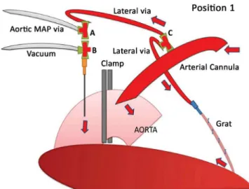
We propose a standard two basic positions, which begin after the clamping performed once proximally to the inser-tion of the arterial cannula in the ascending aorta. Posiinser-tion 1 for perfusion and reperfusion of the aortic root (Figure 1) where there is a direct connection between the arterial cannu-la and the intravascucannu-lar space existing between the ccannu-lamped region and the aortic valve apparatus that shall be competent, otherwise there would be excessive extravasation of blood
into the left ventricular cavity, reducing the inlow of blood to
the coronary ostia and distending the left ventricle.
In this position 1, the connections should still be posi-tioned so that the pressure is measured in the aortic root in order to ensure there is no method bias so that, during CPB with clamping and stopped heart, at this site there is no in-crease in pressure-induced vasopressors administered by an anesthesiologist or perfusionist, which could determine
mis-interpretations. It should also pay attention to the proper low
offered at CPB, which in this case requires no preference be-tween roller pump or centrifugal pump.
Position 2 is the one that should be used when the time of anoxia during anastomosis, both distal and proximal, at which point “B” is closed to the side via and open to the as-pirator used when necessary (Figure 2).
A third situation is still illustrated (Figure 3), which is initially important, of a cardioplegic solution backup because if we need to use it, it is ready requiring only release the car-dioplegia via and clamp and the route of the aspirator before “Y” bifurcation of the vias. In this situation the “B” point should be in the position of Figure 2.
Fig. 2 - It should be used during the anoxia time during anastomosis, both distal and proximal, in which the point “B” is closed to the side via and opened to the aspirator which is used according to the need
Table 1. Patient's characteristics.
Preoperative variables Age (years)
Gender (M/F) Diabetes Mellitus II
Systemic Arterial Hypertension Dyslipidemia
Previous Stroke Previous AMI Smoking COPD CRF ARF Alcoholism
Calcium in the aorta on X-ray
Left Ventricular Ejection fraction (LVEF) Good (>50%)
Regular (30-50%) Poor (<30%) Drugs
B-blocker ACEI Nitrate AAS Diuretic Clexane Clopidogrel Insulin Antiarrhythmic
Oral hypoglycemic Statins
Calcium channel blockers
N 58.5 ± 7.19
37/13 20 48 34 1 27 34 2 1 9 4
6 39
5
35
31 29 36 11 2 10
2 1 11 29 4 The “C” point is fundamental to the technique because it must provide at least two routes, because during reperfusion, one or more of them should be connected to the newly anas-tomosed grafts, which will ensure muscle myocardial
perfu-sion distally to the coronary leperfu-sion, since the antegrade low by ostia may be limited by signiicant injuries. It is important
to make sure that the “C” point is open during reperfusion. Thus, the technique of myocardial protection is describes when using the single aortic clamping accessory which con-sists of cannulation of the aortic root with improved support for intermittent coronary perfusion system.
Statistical Analysis
The data of the continuous and parametric variables were expressed as mean ± standard deviation.
Patients’ characteristics
Most of the patients had one or more co-morbidities such as diabetes mellitus, hypertension, dyslipidemia, smoking, COPD (Chronic Obstructive Pulmonary Disease), prior AMI (Acute Myocardial Infarction), among others (Table 1).
RESULTS
Of the 50 patients 37 were male and 13 were female with a mean age of 58.5 ± 19.7 years. LVEF was normal in 39
patients, good in six, and poor in ive of them. Regarding
the optimization of preoperative medical treatment there was great heterogeneity, where 36 patients were under use of aspirin (Salicilic Acid), 35 beta-blockers and statins 29. 31 were under use of ACE inhibitors (Inhibitor of Angiotensin Converting Enzyme) and only two were insulin dependent (Table 1).
In four patients plaques the ascending aorta were
iden-tiied on palpation. All patients underwent a single aortic
clamping during the entire procedure, having been required additional manipulation of the “clamp” for any reason. The average time for completion of each distal anastomosis was 9.20±1.15 minutes and during reperfusions of the aortic root
the mean CPB low was 3316.92±443.26 ml/min while the
average MAP during reperfusions was 67.85±10.86 mmHg. The time of each reperfusion averaged 3.56±0.62 min-utes. To perform the proximal anastomoses were required on average 5.56±1.44 minutes. The number of distal anas-tomoses was on average 2.56±0.57 per patient and prox-imal 1.44±0.50, and the mean CPB and anoxia times of 51.66±12.21 and 31.28±8.65 minutes respectively. The mini-mum temperature during surgery was on average 33.90±0.65 (Table 2).
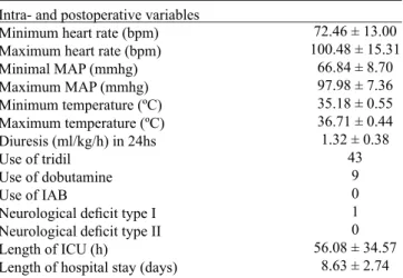
The electrocardiograms did not suffer changes different from those that may be present preoperatively and bleeding was within acceptable parameters (below 1ml/Kg/h in the early hours). There was no renal failure after CPB. The mean peak level of postoperative CK-MB was 51.64±27.10 UL in the second postoperative day and troponin I was 3.35±4.39 ng/ml on the fourth postoperative day (Table 3). Mild cog-nitive impairment was observed in one patient and hemody-namic monitoring revealed no changes during hospitaliza-tion, which was within the usual service standards (Table 4). No deaths were observed.
Table 4 - Intra- and postoperative variables
Intra- and postoperative variables Minimum heart rate (bpm) Maximum heart rate (bpm) Minimal MAP (mmhg) Maximum MAP (mmhg) Minimum temperature (ºC) Maximum temperature (ºC) Diuresis (ml/kg/h) in 24hs Use of tridil
Use of dobutamine Use of IAB
Neurological deicit type I Neurological deicit type II
Length of ICU (h)
Length of hospital stay (days)
72.46 ± 13.00 100.48 ± 15.31
66.84 ± 8.70 97.98 ± 7.36 35.18 ± 0.55 36.71 ± 0.44 1.32 ± 0.38
43 9 0 1 0 56.08 ± 34.57
8.63 ± 2.74
Table 3. Perioperative variables
Perioperative variables
ECG changes in the pre ► postoperative
Plaque of calcium in the ascending aorta Surgical complications
Preoperative urea Postoperative urea Preoperative creatinine Postoperative creatinine
Mean increase of creatinine in the pre ► postoperative
Peak of postoperative CK-MB (U/L) 2º PO Peak of postoperative troponin I (ng/ml) 4º PO
0 4 0 37.16 ± 12.47 50.50 ± 13.80 1.19 ± 0.24 1.32 ± 0.30 0.14 ± 0.25 51.64 ± 27.10
3.35 ± 4.39
DISCUSSION
The risk factor most important for intraoperative low cardiac output postoperatively is prolonged time of myo-cardial ischemia facing the effectiveness of the method used
for myocardial protection[6]. Many techniques for handling
myocardium during CABG are used and there is little safe evidence that a method is superior to another or that the same
method is suitable in all circumstances[7].
It is known that there may be a reduction of postoperative left ventricular function even without evidence of myocardial necrosis, in the case of myocardial protection is inappropri-ate. Conventionally, this situation is called “myocardial
stun-ning”, or that is, a stunned myocardium[8,9].
The arguments in favor of non-cardioplegic methods in-clude greater simplicity and, in the case of intermittent aor-tic cross-clamping, the proven ability of the myocardium to withstand short periods of ischemia, interspersed with periods of physiological reperfusion, without necrosis or loss of func-tion[10,11]. In our study we found that with single clamping, with
average short time of each anastomosis, either distal 9.20±1.15 minutes or proximal 5.56 ± 1.44 minutes, and always inter-spersed with blood reperfusion of the aorta 3.56±0.62 minutes.
Table 2. Intraoperative variables
Intraoperative variables
Time of each distal anastomosis (min.)
CPB low during each reperfusion (ml/min.)
MAP during root perfusion (mmHg) Time of each reperfusion (min.)
Time of each proximal anastomosis (min.) Number of distal anastomoses
Number of proximal anastomoses CPB time (min)
ANOXIA time (min)
Minimum temperature during CPB (°C) Use of centrifugal pump
Sinus CPB output PV CPB output
9.20 ± 1.15 3316 ± 443.26 67.85 ± 10.86 3.56 ± 0.62 5.56 ± 1.44 2.56 ± 0.57 1.44 ± 0.50 51.66 ± 12.21
31.28 ± 8.65 33.90 ± 0.65
It was found that brief periods of myocardial ischemia with perfusion intervals did not cause progressive depletion of high energy phosphates. This fact has a protective effect of myocardial preconditioning and proves that the injury pro-duced by repeated clamping is not cumulative, but rather, it seems to increase myocardial tolerance to subsequent periods
of ischemia[12-14]. This induction of tolerance to ischemia has
been termed ischemic preconditioning (IPC)[12]. These data
are conirmed in our study by myocardial markers CK-MB
and troponin I within normal limits, as well as the echocar-diographic control postoperatively.
The incidence of perioperative cerebrovascular accident (CVA) in the on-pump CABG ranges from 1.5% to 5.2% in
prospective studies and from 0.8% to 3.2% in retrospective[3]
vascular studies. The present study found only one case of mild cognitive impairment. The single most common cause of postoperative stroke is embolization of atherothrombotic debris from the aortic arch. Furthermore, only 45% of stroke
after CABG are identiied on the irst day after surgery, while
55% of them occur after the normal recovery from anesthesia
and are attributed to atrial ibrillation (AF), low cardiac out
-put or hypercoagulable states resulting from tissue injury[15].
Risk factors for intraoperative stroke are CPB time,
manip-ulation of the ascending aorta and arrhythmias[4]. The proposal
of the authors was to present method of intermittent infusion to minimize the risk of multiple aortic clamping, as in patients with presence of atheromatous plaques in the ascending aorta may be increased incidence of strokes. In our study, we did not observe any occurrence of adverse neurological event.
The incidence of perioperative myocardial infarction in
CABG ranges from 2 to 15%[16], and this variation can be
ex-plained by the absence of a gold standard test in the speciic
situation of infarction after coronary revascularization. In ad-dition, electrocardiographic changes and elevation of cardiac enzymes in the postoperative period, even in patients without
myocardial ischemia may occur[17,18].
International criteria for the diagnosis of MI after cardiac surgery is a new Q wave and CK-MB level > 3 times the reference value, new bundle branch block and level of CK-MB > 3 times the reference value or level of serum CK-CK-MB
> 8 times the reference value even in the absence of new Q[19]
wave. Several authors have reported different cutoff points
and different levels of sensitivity and speciicity for troponin
values as diagnostic of perioperative AMI.
Using the same enzymatic method Martinez et al.[20]
found a diagnostic cutoff for troponin I in values of > 12ng/
ml 10hrs after removal of the aortic clamp; Bonnefoy et al.[21]
values of> 10ng/ml; Alyanakian et al.[22] values of > 15ng/
mL; Gensini et al.[23] > 9.2 ng/ml after 12hrs and Sadony et
al.[24] > 11.6 ng/ml after 24 hours of removal of aortic clamp.
In the present investigation we found agreement with the studies described above, regarding the markers of myocardi-al ischemia and electrocardiographic changes.
REFERENCES
1. Sergeant P, Blackstone E, Meyns B. Validation and interdependence
with patient-variables of the inluence of procedural variables
on early and late survival after CABG. K. U. Leuven Coronary Surgery Program. Eur J Cardiothorac Surg. 1997;12(1):1-19. All patients in our study underwent surgery with cardio-pulmonary bypass. Various data available in the literature indicate advantage of CABG technique without
cardiopul-monary bypass in selected[25] cases, however, although
con-troversial in some points the “ROOBY” study showed no dif-ference between the results of neuropsychological outcomes
between CABG with and without CPB[26].
Intermittent perfusion of the aortic root is performed by some services as a method of myocardial protection and, al-though not standardized, it has been demonstrated reproduc-ible given the simplicity of the method that requires skill and dexterity to saving time during the primary surgical time and familiarity with connections alternating aortic perfusion, the graft and simultaneous measurement of pressure in the aortic root and ascending aortic aspiration during anoxia.
The intention to provide better nutrient supply to the myocardium during intermittent infusions by this method has
the beneits known and defended by adherents to the classic
“intermittent aortic cross-clamping” with the additional
ben-eit of minimal manipulation of the aorta, whereas only one
clamping and unclamping of the aorta occurs, the last at the end of the proximal anastomoses.
Ko at al.[27], in 15 years of follow-up, observed that the
overall mortality rate of patients undergoing CABG fell from 2.5% to 1.8%. In our study, despite the low number of pa-tients studied, there were no deaths.
CONCLUSION
Coronary artery bypass grafting with the technique of in-termittent perfusion of the aortic root with a single clamping proved to be safe, enabling satisfactory clinical and neurolog-ical outcomes in the patients studied.
MLPS Main Author
SFSJ Help in bibliographical survey and review of the study JCS Help in tabulating the data and monitoring of patients AST Help in the data tabulation
DFMT Help in the monitoring of patients in the perioperative period
TANS Help in perioperative monitoring of patients GGS Article review
NAGS Article review
2. Bonchek LI, Burlingame MW, Vazales BE, Lundy EF, Gassman CJ. Applicability of noncardioplegic coronary bypass to high-risk patients. Selection of patients, technique, and clinical experience in 3000 patients. J Thorac Cardiovasc Surg. 1992;103(2):230-7.
3. D’Ascenzo F, Cavallero E, Moretti C, Omedè P, Sciuto F, Rahman IA, et al. Remote ischaemic preconditioning in coronary artery bypass surgery:A Meta-analysis. Heart. 2012;98(17):1267-71.
4. Hong DM, Lee EH, Kim HJ, Min JJ, Chin JH, Choi DK, et al. Does remote ischaemic preconditioning with postconditioning improve clinical outcomes of patients undergoing cardiac surgery? Remote Ischaemic Preconditioning with Postconditioning Outcome Trial. Eur Heart J. 2014;35(3):176-83.
5. Djaiani G, Ali M, Borger MA, Woo A, Carroll J, Feindel C, et
al. Epiaortic scanning modiies planned intraoperative surgical
management but not cerebral embolic load during coronary artery bypass surgery. Anesth Analg. 2008;106(6):1611-8.
6. Francel TJ, Kouchoukos NT. Postoperative care. In: Kirklin/ Barratt-Boyes cardiac surgery. 3rd ed. Philadelphia: Churchill Livingstone; 2003.p.195-253.
7. Fontan F, Madonna F, Naftel DC, Kirklin JW, Blackstone EH, Digerness S. Modifying myocardial management in cardiac surgery: a randomized trial. Eur J Cardiothorac Surg. 1992;6(3):127-36.
8. Heyndricx GR, Millard RW, McRitchie RJ, Maroko PR, Vatner SF. Regional myocardial function and electrophysiological alterations after brief coronary artery occlusion in conscious dogs. J Clin Invest. 1975;56(4):978-85.
9. Braunwald E, Kloner RA. The stunned myocardium: prolonged, postischemic ventricular dysfunction. Circulation. 1986;66(6):1146-9.
10. Van der Veen FH, van de Vusse GJ, Reneman RS. Myocardial
blood low and oxygen consumption after aortic cross-clamping.
J Surg Res. 1989;47(4):319-24.
11. Fernandes PMP, Jatene FB, Gentil AF, Coelho FF, Kwasnicka K,
Stolf NAG, et al. Inluência do Pré-Condicionamento Isquêmico
na Proteção Miocárdica em Revascularização do Miocárdio com Pinçamento Intermitente da Aorta. Análise de Íons e Gasometria. Arq Bras Cardiol. 2001;77(4):311-7.
12. Murry CE, Jennings RB, Reimer KA. Preconditioning with ischemia: a delay of lethal cell injury in ischemic myocardium. Circulation. 1986;74(5):1124-36.
13. Yellon DM, Alkhulaii AM, Pugsley WB. Preconditioning the
human myocardium. Lancet. 1993;342(8866):276-7.
14. Jenkins DP, Pugsley WB, Alkhulaii AM, Kemp M, Hooper J, Yellon
DM. Ischemic preconditioning reduces troponin T release in patients undergoing coronary artery bypass surgery. Heart. 1997;77(4):314-8.
15. Task Force on Myocardial Revascularization of the European Society of Cardiology (ESC) and the European Association for
Cardio-Thoracic Surgery (EACTS); European Association for Percutaneous Cardiovascular Interventions (EAPCI), Wijns W, Kolh P, Danchin N, Di Mario C, Falk V, Folliguet T, et al. Guidelines on myocardial revascularization Eur Heart J. 2010;31(20):2501-55.
16. Sobral MLP, Santos GG, Santos LAS, Haddad VLS, Avelar Júnior SF, Stolf NAG. Estudo comparativo randomizado da evolução imediata dos pacientes com artéria radial anastomosada proximalmente na aorta ou como enxerto composto. Rev Bras Cir Cardiovasc. 2006;21(1):35-41.
17. Adam DH, Filsoui F, Antmann EM. Medical management of the patient
undergoing cardiac surgery. In: Zipes DP, Libby P, Bonow R, Braunwald E, eds. Braunwald’s heart disease: a textbook of cardiovascular medicine. 7th ed. Philadelphia: WB Saunders; 2004. p.1993-2019.
18. Leal JCF, Braile DM, Godoy MF, Purini Neto J, Paula Neto A, Ramin SL, et al. Avaliação imediata da troponina I cardíaca em pacientes submetidos à revascularização do miocárdio. Rev Bras Cir Cardiovasc. 1999;14(3):247-53.
19. Bojar RM. Cardiovascular management. In: Bojar RM. Manual of perioperative care in cardiac surgery. 4 ed. Malden: Blackwell Science; 2005. p.404-7.
20. de Castro Martínez J, Vázquez Rizaldos S, Velayos Amo C, Herranz Valera J, Almería Varela C, Iloro Mora MI.. Troponina I cardíaca en el infarto del miocárdio: perioperatorio trás cirurgía de revascularización coronária. Rev Esp Cardiol. 2002;55(3):245-50.
21. Bonnefoy E, Filley S, Guidollet J. Cardiac troponin I to diagnose perioperative myocardial infarction after bypass surgery. Eur Heart J. 1995;16 (Suppl): 325.
22. Alyanakian MA, Dehoux M, Chatel D, Seguret C, Desmonts JM, Durand G, et al. Cardiac troponin I in diagnosis of perioperative myocardial infarction after cardiac surgery. J Cardiothorac Vasc Anesth. 1998;12(3):288-94.
23. Gensini GF, Fusi C, Conti AA, Calamai GC, Montesi GF, Galanti G, et al. Cardiac troponin I and Q wave perioperative myocardial infarction after coronary artery bypass surgery. Crit Care Med. 1998;26:(12)1986-90.
24. Sadony V, Korber M, Albes G, Podtschaske V, Etgen T, Trösken T, et al. Cardiac troponin I plasma levels for diagnosis and quantitation of perioperative myocardial damage in patients undergoing coronary artery bypass surgery. Eur J Cardiothorac Surg. 1998;13(1):57-65.
25.Cantero MA, Almeida RMS, Galhardo R. Análise dos resultados imediatos da cirurgia de revascularização do miocárdio com e sem circulação extracorpórea. Rev Bras Cir Cardiovasc.2012;27(1):38-44.
26. Shroyer AL, Grover FL, Hattler B, Collins JF, McDonald GO, Kozora E, et al. On-pump versus off-pump coronary artery bypass surgery. N Engl J Med. 2009;361(19):1827-37.