113
SECONDARY METABOLITE PRODUCTION IN HYPERICUM PERFORATUM L.
CELL SUSPENSIONS UPON ELICITATION WITH FUNGAL MYCELIA
FROM ASPERGILLUS FLAVUS
SONJA GADZOVSKA-SIMIC1, O. TUSEVSKI1, S. ANTEVSKI1, NATALIJA ATANASOVA-PANCEVSKA2,
JASMINA PETRESKA3, MARINA STEFOVA3, D. KUNGULOVSKI2 and M. SPASENOSKI1
1 Laboratory for Plant Cell and Tissue Culture, Institute of Biology, Faculty of Natural Sciences and Mathematics, Uni-versity “Ss. Cyril and Methodius”, 1000 Skopje, Macedonia
2 Laboratory for Microbiology, Institute of Biology, Faculty of Natural Sciences and Mathematics, University “Ss. Cyril and Methodius”, 1000 Skopje, Macedonia.
3 Laboratory for Analytical Chemistry, Institute of Chemistry, Faculty of Natural Sciences and Mathematics, University “Ss. Cyril and Methodius”, 1000 Skopje, Macedonia.
Abstract – We investigated the production of phenylpropanoids (phenolic compounds, lavanols, lavonols and anthocya-nins) and naphtodianthrones (hypericins) in elicited Hypericum perforatum L. cell suspensions. To determine whether secondary metabolite production could be enhanced, Hypericum cell suspensions were exposed to mycelia extract from the fungus Aspergillus lavus. Elicited Hypericum cell suspension cultures displayed reduced growth and viability and a modiication of secondary metabolites production. Anthocyanins were only stimulated in fungal-elicited cell suspensions. Secondary metabolite production in elicited Hypericum cells revealed an antagonism between the lavonoid/naphtodian-throne and anthocyanin pathways. he data suggest a modiication of the channeling of the phenylpropanoid compounds. Together, these results represent useful data for monitoring the channeling in diferent secondary metabolite pathways during the scaled-up production of naphtodianthrones for medicinal uses.
Key words: Anthocyanins, Aspergillus lavus, elicitation, lavanols, lavonols, hypericin, Hypericum perforatum L., naphto-dianthrones, phenolics, phenylpropanoids.
INTRODUCTION
Hypericum perforatum L. is a well-known medicinal plant that has been in use for a decade. Studies of the pharmacological efects of the constitutive com-pounds are still in progress. Recently, the antiviral and antidepressant properties of Hypericum have become widely demonstrated (Di Carlo et al. 2001). Extracts from H. perforatum are known to contain compounds from six major natural product groups: naphtodianthrones, acylphloroglucinols, lavonol
1993). Little is known about the biosynthesis of hy-pericin other than it lies on the polyketide pathway (Zobayed et al., 2006). he gene hyp-1 encoding for a hypericin biosynthetic enzyme was cloned and char-acterized from H. perforatum cell cultures (Bais et al., 2003). However, the coordination of the produc-tion of naphtodianthrones with other secondary me-tabolites is still unknown. he levels of hypericin and pseudohypericin in Hypericum plants can vary from 0.03 to 0.3% dry weight under diferent environmen-tal parameters, but the precise environmenenvironmen-tal factors which inluence the biosynthesis of these naphtodi-anthrones and the partitioning with other secondary metabolites in Hypericum are not well understood.
Elicitation of secondary metabolite production in plant cell cultures could be induced either by biotic or abiotic molecules. Elicitors are now considered as signal molecules that activate the signal-transduction cascade and lead to the activation and expression of genes related with the biosynthesis of secondary me-tabolites (Zhao et al., 2005). Furthermore, elicitors stimulated the antioxidant defense systems of plant cells (De Gara et al., 2003). Treatments of cultured cells with fungal elicitors have also been shown to induce the phenylpropanoid/lavonoid biosynthetic pathways (Dixon et al., 2002; Tan et al., 2004). he fact that fungal elicitors stimulate strongly and rap-idly plant secondary metabolite accumulation has recently attracted considerable attention (Dixon et al., 2002; Zhao et al., 2005).
Cell suspension cultures from H. perforatum have already been established to study the overpro-duction of naphtodianthrones using phytohormones (Gadzovska et al., 2005) or various elicitors such as mannan, β-1,3-glucan, pectin (Kirakosyan et al., 2000), methyl jasmonate (Sirvent and Gibson, 2002), jasmonic acid (Walker et al., 2002; Gadzovska et al., 2007), salicylic acid (Sirvent and Gibson, 2002; Walk-er et al., 2002) or fungal elicitors from Colletotrichum gloeosporioides (Conceição et al., 2006) and Phytoph-thora cinnamomi (Walker et al., 2002). Hypericum in vitro culture systems have been shown to respond to elicitors by increasing naphtodianthrones, which have been considered to be the chemical defence
arsenal of plants against pathogens and herbivores (Zhao et al., 2005). Nevertheless, the coordination of the production of naphtodianthrone compounds with other secondary metabolites (lavonoids and phenolic compounds) remains unknown.
In this study, Hypericum cell suspensions were treated with mycelia extract from the pathogenic fungus Aspergillus lavus. A. lavus is a common plant pathogen fungus which causes diseases of agro-nomically important crops, such as corn and peanuts (St. Leger et al., 2000). A. lavus is responsible for the alatoxin contamination of crops prior to harvest or during storage (Yu et al., 2004). Investigations in this study focused on two areas: (1) the consequences of fungal elicitor treatments on cell biomass produc-tion, and (2) the partitioning between secondary metabolites as naphtodianthrones (hypericins) and phenylpropanoids (phenolic compounds, lavanols, lavonols and anthocyanins) in elicited cells. Activa-tion of the non-enzymatic antioxidant system was as-sessed by measuring the non-enzymatic antioxidant properties (NEAOP) in elicited cell suspensions.
MATERIALS AND METHODS
Plant material
H. perforatum seeds were collected from ield-grown plants on the Mt. Bistra (Mavrovo), Republic of Mac-edonia, at the altitude of 1200-1500 m. he seeds were washed overnight, air dried, surface sterilized with 1% NaOCl for 10 min, rinsed 3 times in ster-ile deionized water and cultured on MS macro and oligoelements (Murashige and Skoog, 1962), B5
vita-min solution (Gambrog et al., 1968), supplemented with 30 g⋅L-1 sucrose, and solidiied with 7 g⋅L-1
Dif-co Bacto agar. No growth regulator was added. he medium was adjusted to pH 5.6 before autoclaving (20 min at 120°C). he seeds were maintained in a growth chamber at 22°C under a 16 h photoperiod (700 µM/m2/s). he irst pair of leaves were excised
from 2 week-old in vitro plants and used as explants to establish callus cultures. hey were cultured in Petri dishes on MS/B5 medium supplemented with
(2,4-D), 0.5 mg⋅L-1 N6-benzyladenine (BA), 0.1 mg⋅L-1 α-naphthaleneacetic acid (NAA), 30 g⋅L-1 sucrose
and solidiied with 7 g⋅L-1 Difco Bacto agar. he
cal-lus cultures were initiated on the leaf surface ater 2 weeks. Subcultures were carried out every 14 days.
Cell suspension cultures were established from 28 day-old callus cultures. Green calli (2 g) were in-troduced in Erlenmeyer lasks containing 100 mL of MS/B5 medium, supplemented with 1.0 mg⋅L-1 2,4-D,
0.5 mg⋅L-1 BA, 0.1 mg⋅L-1 NAA and 30 g⋅L-1 sucrose.
he cultures were maintained on a rotary shaker at 100 rpm in the growth chamber at 25±1°C, under a 16 h photoperiod (700 µM/m2/s). Ater two weeks,
the cells released from the calli were transferred to 4 volumes of fresh liquid medium and subcultured, before treatment, every two weeks.
Fungal mycelium from Aspergillus lavus was grown in 250 ml lasks containing Sabouraud-Dex-trose broth (Oxoid). One-month-old mycelia were homogenized and centrifuged for 5 min. he fungal mycelia supernatant was re-suspended with sterile distilled water to a inal concentration of 50 mg⋅mL-1
and then autoclaved for 10 min at 120°C. he A. la-vus fungal mycelia extracts were stored at 4°C, until treatment.
Treatments of the cell suspensions with the fungal mycelia extract were performed 7 days ater subculture when the cells were in the log phase of growth. Cell suspensions cultivated on MS/B5
me-dium without elicitor were used as a control. Treated and control cell suspensions were then harvested by vacuum iltration on days 1, 4, 7, 14, and 21 post-elicitation, weighed for growth analysis, frozen in liquid nitrogen or lyophilized and stored at -80°C, until analysis.
Extraction and quantiication of secondary metabolites
Phenolic quantiication was performed as described by Gadzovska et al. (2007). Phenolic compounds were extracted from freeze-dried lyophilized and powdered plant material (0.2-0.5 g) with 80% (v/v)
methanol in an ultrasonic bath for 30 min at 4ºC. he total phenol content was determined when the methanolic extracts were mixed with the Folin-Cio-calteau reagent (Carlo Erba Reagenti, Rodano, Italy) and 0.7 M Na2CO3. Samples were incubated for 5 min
at 50ºC and then cooled for 5 min at room tempera-ture. Absorbance was measured spectrophotometri-cally at 765 nm. he concentration of total phenolic compounds was calculated using (+)-catechin (0-10 mg⋅ml-1) as a standard.
he lavanol contents were determined in the methanolic extracts with 4-(Dimethylamino)cin-namaldehyde (DMACA) reagent (Gadzovska et al., 2007). he DMACA reagent was added to the di-luted (1:10-1:100, v/v) extracts. he mixtures were incubated for 1 h at room temperature. Absorbance was measured at 637 nm. he content of lavanols was calculated using (+)-catechin (0-20 µg⋅ml-1) as
a standard.
he lavonol contents were determined in the methanolic extracts by the method described by Gadzovska et al. (2007). he lavonols were quanti-ied in the methanolic extracts iltered through Sep-pack C18 cartridges (Waters) to exclude chlorophyll
and carotenoid pigments (solid-phase extraction). Spectrophotometric measurements of the absorb-ance were made at 360 nm. Molar extinction coef-icient of quercetin (ε360=13.6 mM-1⋅cm-1) was used
to determine total lavonol contents.
Anthocyanin determination was performed as described by Gadzovska et al. (2007). Anthocyanins were extracted from freeze-dried lyophilized and powdered plant material (0.2-0.5 g) with 2 mL solu-tion of 1% HCl/CH3OH (15/85, v/v), ultrasonicated
for 60 min at 4°C and then centrifuged at 20,000 g for 30 min. he absorbance of supernatant was measured at 530 nm. he anthocyanin content was calculated using the molar extinction coeicient of cyanidin-3-glucoside (ε530=34300 M-1⋅cm-1) in acidic methanol.
were made at 590 nm. Standard solutions of hyper-icin (0-100 µg⋅ml-1) were prepared from pure
com-mercially available standard of hypericin (Sigma, Germany).
Non enzymatic antioxidant properties (NEAOP) assay
he antioxidant properties of soluble methanolic ex-tracts were estimated by the linoleic acid-β-carotene oxidation method adapted from Gadzovska-Simic, (2010). A linoleic acid-β-carotene emulsion was prepared by mixing 10 mg of linoleic acid with 750 µL of 0.2 mg⋅mL-1 chloroformic β-carotene solution
and 100 mg of Tween 40 (polyoxyethylene sorbitan monopalmitate). Chloroform was evaporated under nitrogen low for 10 min. he resulting mixture was adjusted to 25 mL with distilled water and shaken for 10 seconds. he reaction mixture was prepared as follows: 10 µL of extract were adjusted with 15 µL 80% (v/v) methanol and 225 µL of linoleic acid-β -carotene emulsion was added. he mixture was heat-ed to 50°C. he control consistheat-ed of 25 µL 80% (v/v) methanol and 225 µL of linoleic acid-β-carotene emulsion. Absorbance was measured at 470 nm eve-ry 15 min for 45 min. Results were computed as the ratio of β-carotene protection of the extract to the control (80 % methanol).
Statistical analyses
he experiments were independently repeated twice under the same conditions and all analyses
were performed in triplicate. Error bars of graphs show the standard error of mean value (±SE). he statistical analyses were performed with the SPSS statistical sotware program (SPSS version 11.0.1 PC, USA, IL). Means were expressed with their standard error and compared by one-way ANOVA (GML pro-cedure). All statistical tests were considered signii-cant at p ≤ 0.05.
RESULTS
Growth and viability of elicited H. perforatum L. cell suspensions
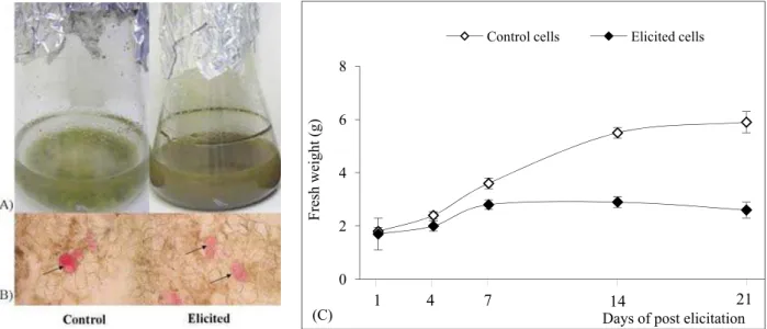
Optimization of cell proliferation was the irst step toward establishing elicited H. perforatum cell sus-pensions. he factors considered were the time of exposure to the treatment and cell biomass produc-tion. From 14 days ater elicitation, browning and cell aggregation appeared in the elicited cells (Fig. 1A). Outgoing results from this study showed that long-term treatment with the fungal elicitor caused a brown color and cell aggregation in the elicited cells. Hypericum-elicited cells tend to combine and form aggregates, resulting in heterogeneous populations of cell aggregates, ranging from a few cells to thou-sands of cells, and oten reaching a few centimeters in diameter. Localization of anthocyanins in the cell suspensions is shown in Fig. 1B (arrows). he fresh weight of the elicited cells was always below or equal to the control values throughout the time of culture maintenance (Fig. 1C). he efect of the fungal elici-tor was not signiicantly diferent compared to the Table 1. Correlations between secondary metabolite contents and non-enzymatic antioxidant properties (NEAOP) in H. perforatum cell suspensions elicited with fungal mycelia extract from Aspergillus lavus.
Pearson coeicient of correla-tion (r)
Phenolics Flavanols Flavonols Anthocyanins Hypericin
Flavanols 0.91 ***
Flavonols 0.92 *** 0.85 ***
Anthocyanins 0.12 ns 0.18 ns 0.31 ns
Hypericin 0.72 *** 0.67 ** 0.83 *** 0.46 ns
NEAOP 0.13 ns 0.20 ns 0.21 ns 0.54 ** 0.21 ns
control until day 7 of post-elicitation. From day 14, it was clear that biomass production in elicited cells was lower (2- to 3-fold in elicited cells) than in the control. Exogenously applied fungal elicitor medi-ates certain types of stress responses in Hypericum cells and its action results in a negative regulation of growth and development.
Secondary metabolite production in elicited H. perforatum cell suspensions
he inluence of the Aspergillus mycelium extract (50 mg·mL-1) on the secondary metabolite contents and
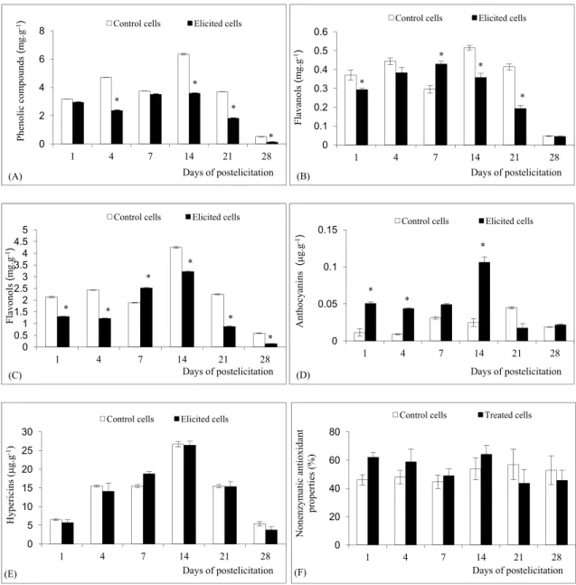
non-enzymatic activity (NEAOP) in the cell suspen-sions was tested. he fungal mycelia extract caused a 2-fold decrease of phenolic compounds ater 4 to 21 days of elicitation compared to the control (Fig. 2A). Hypericum-elicited cells had higher lavanol (Fig. 2B) and lavonol contents (Fig. 2C) at day 7: there-ater lavonoid accumulation decreased compared to the control cells. Anthocyanins were only stimulated in the elicited cell suspensions (Fig. 2D). Transient accumulations with maxima at day 14 of elicitation were found for anthocyanins. Namely, a 2- to 4-fold increase of anthocyanins from day 1 to day 14 was observed in elicited cells compared to the control
(Fig. 2D). Hypericins remained unchanged (Fig. 2E) in the corresponding samples of elicited cells com-pared. Induction of non-enzymatic antioxidant sys-tems was apprized by measuring the NEAOP in the elicited suspensions and an increase was monitored during the time of post-elicitation. NEAOP were higher (Fig. 2F) from day 1 to day 14 in elicited cells compared to the control.
Statistical analysis
Statistically analyses (Pearson’s correlation coeffi-cient) showed a positive correlation between the hypericin and phenylpropanoid contents in the cell suspensions upon elicitor treatment (Table 1). The production of phenolic compounds in the elicited cells was in positive correlations with the amounts of flavanols (r=0.91, p<0.001), flavonols (r=0.92, p<0.001) and hypericin (r=0.72, p<0.001). These results provide biochemical proof of a connec-tion between hypericin biosynthesis and flavonoid metabolism. NEAOP were only in positive corre-lation with anthocyanins (r=0.54, p<0.05). These results provide evidence that anthocyanins exhibit high antioxidant properties in Hypericum-elicited cells.
DISCUSSION
Efects of fungal elicitor on growth and viability in H. perforatum cell suspensions
Hypericum cell suspension cultures treated with the Aspergillus mycelia extract were negatively afected
for growth and turned brown in coloration. Such a result has already been reported in H. perforatum cell suspensions treated with jasmonic acid (Gadzovska et al., 2007), salicylic acid and fungal cell-wall elici-tors from Phytophthora cinnamomi (Walker et al., 2002). It has also been reported that the reduction in biomass might be due to membrane lipoxidation Fig. 2. Production of secondary metabolites of control and elicited H. perforatum cell suspensions. (A) Phenolic compounds; (B) la-vanols; (C) lavonols; (D) anthocyanins; (E) hypericins; (F) non-enzymatic antioxidant properties of plant extracts (NEAOP). *
induced by elicitor treatment (Radman et al., 2003). herefore, elicited plant cells show increased oxy-gen uptake that can manifest itself in the transient production of reactive oxygen species (ROS). he oxidative burst is a fast response of cells to elicitor treatment (Ortmann et al., 2004). Plant cells are usu-ally protected against the detrimental efects of oxy-gen species using non-enzymatic antioxidant and/or enzymatic scavenging systems (Blokhina et al., 2003; De Gara et al., 2003). In this study, exogenously ap-plied Aspergillus lavus mycelia extract stimulated the non-enzymatic antioxidant system in Hyperi-cum-elicited cells. hus, the NEAOP were found to be higher in elicited cells than in control ones. All these results indicated an eicient elicitation of Hy-pericum cell suspensions.
he relationships between subsequent suspension browning and the formation of cell aggregates with a strong induction of secondary metabolite pathways have already been reported (Zhao et al., 2005). hus, it has also been proposed that cells on the surface of cell aggregates may be strongly elicited, leading to the formation of a large number of apoptotic cells (Yuan et al., 2002). Accordingly, Hypericum cell sus-pensions elicited with fungal elicitor from Aspergil-lus turned brown, formed cell aggregates and accu-mulated the highest amounts of anthocyanins. his result is in agreement with relatively high NEAOP in Hypericum-elicited cells which positively correlated with the anthocyanins.
Efects of fungal elicitor on secondary metabolite production in H. perforatum cell suspensions
One of the problems encountered in the analysis of the metabolites produced by Hypericum is that anthocy-anins may have been misidentiied as hypericins; this probably occurred because both are red in color and because studies on hypericins do not take into con-sideration the presence of anthocyanins (Mulinacci et al., 2008). According to this study, hypericin only accumulates in the glandular structures of the leaf (Onelli et al., 2002), whereas anthocyanins accumu-late in the vacuole of parenchymatic cells of the leaf mesophyll (Pasqua et al., 2006). In contrast with this,
the results from our study suggested that one reason for the accumulation of both anthocyanins and hy-pericins in Hypericum-elicited cells might be the ex-istence of diferentiation and compartmentalization of cell aggregates in suspension cultures. Bais et al. (2003) reported that both cell growth and hypericin production were greatly afected by cell aggregate size and dark growth conditions in H.perforatum L. cell suspensions. According to these authors, the size of cell aggregates has major implications in secondary metabolite production, including hypericin content. A smaller size of cell aggregates has been preferred from the standpoint of process engineering, whereas a certain degree of cell-cell contact and cell difer-entiation was required for hypericin biosynthesis (Walker et al., 2002). hese authors reported that the production and localization of hypericin in cell sus-pensions is entirely diferent from intact plants. his could be because compounds like hypericins are ac-cumulated in specialized cells (dark glands) in plants (Zdunek and Alfermann, 1992).
Few studies have reported the efects of fungal elicitors on secondary metabolite production in Hy-pericum cells. Aspergillus mycelia extract strongly and usually early activated the biosynthesis of anthocy-anins analyzed in this study. he statistical analy-sis demonstrated positive correlations between the amounts of phenolics, naphtodianthrones and lavo-noids. However, an antagonism between lavonoid/ naphtodianthrone and anthocyanin pathways in Hy-pericum cells was shown. All these data suggest a par-titioning of the secondary compounds during elicita-tion. hus, the Aspergillus fungal elicitor was the most eicient in anthocyanin production. he efect of the biotic elicitor Colletotrichum gloeosporioides on sec-ondary metabolite production in H. perforatum cells has been examined (Conceição et al., 2006). In this study, elicited Hypericum cells showed a signiicant increase in xanthone accumulation.he fungal cell-wall elicitor from Phytophthora cinnamomi induced the production of hypericin in cell suspension cul-tures of H. perforatum (Walker et al., 2002). Little is known about the biosynthesis of naphtodianthrones; the unknown participating enzymes have to be char-acterized before enlarging the study to the efects of elicitation in Hypericum cell suspensions. It is still unclear whether distinct elicitors acted on the same or on distinct signaling pathways (Zhao et al., 2005). herefore, more work was needed to better under-stand the efects of various elicitors on the partition-ing between the secondary metabolites in Hypericum cell suspensions. Such a result could be explained by the distinct potential signaling pathways and could be a useful tool to monitor the scaled-up production of secondary metabolites for medicinal uses. hus, the mycelia extracts seem to be the most promising elicitors for such anapplication.
REFERENCES
Bais, H.P., Vepachedu, R., Lawrence, C. B., Stermitz, F. R., and
J. M. Vivanco (2003). Molecular and biochemical charac-terization of an enzyme responsible for the formation of hypericin in St. John’s Wort (Hypericum perforatum L.). J. Biol. Chem. 34, 32413-32422.
Blokhina, O., Virolainen, E., and K. V. Fagerstedt (2003). Anti-oxidants, oxidative damage and oxygen deprivation stress.
Ann. Bot. 91, 179-194.
Conceição, L., Ferrares, F., Tavares, R., and A. Dias (2006). In-duction of phenolic compounds in Hypericum perforatum
L. cells by Colletotrichum gloeosporioides elicitation. Phy-tochem. 67, 149–155.
De Gara, L., De Pinto, M., and F. Tommasi (2003). he anti-oxidant systems vis-à-vis reactive oxygen species during plant–pathogen interaction. Plant Physiol. Biochem. 41, 863–870.
Di Carlo, G., Borrelli, F., Ernst, E., and A. Izzo (2001). St. John’s wort: Prozac from plant kingdom. Trends Pharm. Sci. 22, 292-297.
Dixon, R. A., Achnine, L., Kota, P., Liu, C. J., Reddy, M. K. S.,
and L. Wang (2002). he phenylpropanoid pathway and plant defence - a genomics perspective. Mol. PlantPathol. 3, 371-390.
Falk, H. (1999). From the photosensitizer hypericin to the photo-receptor stentorin-he chemistry of phenanthroperylene quinines. Angew.Chem. Int. 38, 3116-3136.
Gadzovska, S., Maury, S., Ounnar, S., Righezza, M., Kascakova, S., Refregiers, M., Spasenoski, M., Joseph, C., and D. Hagège
(2005). Identiication and quantiication of hypericin and pseudohypericin in diferent Hypericum perforatum L. in vitro cultures. Plant Physiol. Biochem. 43, 591–601.
Gadzovska, S., Maury, S., Delaunay, A., Spasenoski, M., Joseph, C., and D. Hagège (2007). Jasmonic acid elicitation of Hy-pericum perforatum L. cell suspensions and efects on the production of phenylpropanoids and naphtodianthrones.
Plant Cell Tiss Organ Cult. 89, 1-13.
Gadzovska-Simic, S. (2010). Secondary metabolite production in cultures in vitro of Hypericum perforatum L. Efects of diferent exogenous factors. LAP Lambert Academic Pub-lishing, Saarbrücken, Germany, 1-192.
Gamborg, O. L., Miller, R. A., and K. Ojima (1968). Nutrient re-quirements of suspension cultures soybean root cells. Exp. Cell Res. 50, 148-151.
Katz, L., and S. Donadio (1993). Polyketide synthesis: Prospects for hybrid antibiotics. Ann. Rev. Microbiol. 47, 875-912.
Kirakosyan, A., Hayashi, H., Inoue, K., Charchoglyan, A., and H. Vardapetyan (2000). Stimulation of the production of hy-pericins by mannan in Hypericum perforatum shoot cul-tures. Phytochem. 53, 345-348.
Krikorian, A. D., and F. C. Steward (1969). Biochemical difer-entiation, the biosynthetic potentialities of growing and quiescent tissue, In: Plant Physiology, (Ed. F. C. Steward), Academic Press, London, 227-326.
shoots of Hypericum perforatum var. angustifolium (sin. Fröhlich) Borkh. Plant Physiol. Biochem. 46, 414-420.
Murashige T., and F. Skoog (1962). A revised medium for rapid growth and bioassays with tobacco tissue cultures. Physiol. Plant. 15, 473-497.
Nahrstedt, A., and V. Butterweck (1997). Biologically active and other chemical constituents of the herb of Hypericum per-foratum L. Pharmacopsych. 30, 129-134.
Onelli, E., Rivetta, A., Giorgi, A., Bignami, M., Cocucci, M., and
G. Patrignani (2002). Ultrastructural studies on the devel-oping secretory nodules of Hypericum perforatum. Flora, 197, 92-102.
Ortmann, I., Sumowski, G., Bauknecht, H., and B. M. Moersch-bacher (2004). Establishment of a reliable protocol for the quantiication of an oxidative burst in suspension-cultured wheat cells upon elicitation. Physiol. Mol. Plant Pathol. 64, 227-232.
Pasqua, G., Avato, P., Monacelli, B., Santamaria, A., and M. Ar-gentieri (2003). Metabolites in cell suspension cultures, calli, and in vitro regenerated organs of Hypericum perfo-ratum cv. Topas. Plant Sci. 165, 977-982.
Pasqua, G., Avato, P., and N. Mulinacci (2006). High-value me-tabolites from Hypericum perforatum: a comparison be-tween the plant and in vitro systems, In: Ornamental and Plant Biotechnology: Advances and Topical Tissue, (Ed. J. A. Teixeira da Silva), vol. II, Floriculture, Global Science Books, Japan, 507-513.
Radman, R., Saez, T., Bucke, C., and T. Keshavrz (2003). Elicita-tion of plants and microbial cell systems. Biotechnol. Appl. Biochem. 37, 91-102.
Sirvent, T., and D. Gibson (2002). Induction of hypericins and hyperforin in Hypericum perforatum L. in response to bi-otic and chemical elicitors. Physiol. Mol. Plant Pathol. 60, 311-320.
St. Leger, R.J., Screen, S.E., and B. Shams-Pirzadeh (2000). Lack of host specialization in Aspergillus lavus. App. Environ. Microb. 66, 320-324.
Tan, J., Bednarek, P., Liu, J., Schneider, B., Svatos, A., and K. Hahl-brock (2004). Universally occurring phenylpropanoid and species-speciic indolic metabolites in infected and unin-fected Arabidopsis thaliana roots and leaves. Phytochem. 21, 691–699.
Walker, S. T., Bais, H., and M. J. Vivanco (2002). Jasmonic acid - induced hypericin production in cell suspension cultures of Hypericum perforatum L (St. John’s wort). Phytochem. 60, 289-293.
Yu, J., Whitelaw, C. A., Nierman, W. C., Bhatnagar, D., and T. E. Cleveland (2004). Aspergillus lavus expressed sequence tags for identiication of genes with putative roles in ala-toxin contamination of crops. FEMS Microbiol Lett. 237, 333-340.
Yuan, Y. J., Li, J. C., Hu, Z. D., Wu, J. C., and A. P. Zeng (2002). Fungal elicitor-induced cell apoptosis in suspension cul-tures of Taxus chinensis var. mairei for taxol production.
Proc. Biochem. 38, 193-198.
Zdunek, K., and W. Alfermann (1992). Initiation of shoot organ cultures of Hypericum perforatum and formation of hy-pericin derivates. Planta Med. 58, 621-625.
Zenk, M. H. (1991). Chasing the enzymes of secondary metabo-lism: plant cultures as a pot of gold. Phytochem. 30, 3831-3863.
Zhao, J., Davis, L., and R. Verpoorte (2005). Elicitor signal trans-duction leading to protrans-duction of plant secondary metabo-lites. Biotechnol. Adv. 23, 283–333.