Rev Saúde Pública 2005;39(3) www.fsp.usp.br/rsp
INTRODUCTION
The importance of bioaerosols has been empha-sized in the last decades due to their relationship to human health, leading to the appearance of diseases that range from allergies to disseminated infections in susceptible patients.5 Determining the
composi-tion and concentracomposi-tion of airborne microorganisms in internal and/or external areas of critical hospital
Monitoring of airborne fungus and yeast
species in a hospital unit
José Nelson Martins-Diniz, Rosangela Aparecida Moraes da Silva, Elaine Toscano Miranda e Maria José Soares Mendes-Giannini
Departamento de Análises Clínicas. Faculdade de Ciências Farmacêuticas. Universidade Estadual Paulista. Araraquara, SP, Brasil
Correspondence to:
Maria José Soares Mendes Giannini Laboratório de Micologia Clínica - Unesp Rua Expedicionários do Brasil, 1621 Caixa Postal 502
14801-902 Araraquara, SP, Brasil E-mail: [email protected]
Financed by the Conselho Nacional de Desenvolvimento Científico e Tecnológico (CNPq - Grant n. 400141/1998-1). Study developed at the Laboratory of Clinical Mycology, Department of Clinical Analysis, Faculdade de Ciências Farmacêuticas of the Universidade Estadual Paulista Júlio de Mesquita Filho.
Part of the masters dissertation presented at the Faculdade de Ciências Farmacêuticas of the Universidade Estadual Paulista, Araraquara, SP, Brazil, 2003.
Presented at the XXII Congresso Brasileiro de Microbiologia, Florianópolis, SC, Brazil, 2003. Received on 22/12/2003. Reviewed on 15/10/2004. Approved on 2/2/2005.
Keywords
Fungi. Yeasts. Air conditioning. Cross infection, prevention and control. Aerosols. Infection control.
Abstract
Objective
To monitor and characterize airborne filamentous fungi and yeasts from abiotic and biotic sources within a hospital unit.
Methods
Collections were carried out on a monthly basis, at two different time periods, from the adult and pediatric intensive care units and surgical center of a hospital in Araraquara, Southeastern Brazil. Collection of airborne fungi was carried out using a simple-stage Andersen sample. The presence of yeasts was investigated in samples taken from the hands and oropharynx of staff members as well as from the surface of beds and doorknobs inside the critical areas.
Results
Thirty-two genera of airborne fungi and were recovered from the surgical center and 31 from the intensive care units. Genera most frequently isolated were Cladophialophora
spp., Fusarium spp., Penicillium spp., Chrysosporium spp. and Aspergillus spp. During the study period, a new unit was built in the hospital, which coincided with an increase in Cladophialophora spp., Aspergillus spp., and Fusarium spp. colony counts. Yeasts were found in 39.4% of samples obtained from healthcare staff (16.7% from interdigital spaces, 12.1% from nailbeds, and 10.6% from oropharynx) and in 44% of furniture samples, with a predominance of the Candida genus (C. albicans, C. guilliermondii, C. parapsilosis e C. lusitaniae), followed by Trichosporon spp.
Conclusions
We found a relatively high number of airborne fungi (potentially pathological) in special areas and expressive levels of yeasts in both biotic and abiotic samples. Microbiological and environmental monitoring should be conducted, especially in special areas which include immunocompromised patients, who are more susceptible to the exposure to environmental and staff-derived pathogens.
wards is considered as an extremely necessary meas-ure.1 Hospitals are environments that require great
Rev Saúde Pública 2005;39(3) www.fsp.usp.br/rsp Monitoring of fungus in a hospital unit
Martins-Diniz JN et al
nailbeds), and oral mucosa of 66 professionals di-rectly in contact with ICU and surgical-center pa-tients. Ninety-one swab samples were collected from doorknobs, beds, and telephones located inside the ICUs and surgical center.
Yeasts from biotic and abiotic sources were isolated on Sabouraud agar + chloramphenicol culture me-dium. Yeasts were identif ied previously on CHROMagar Candida medium. These results were associated to filamentation, germ-tube formation af-ter two hours, assimilation and fermentation of car-bon and nitrogen sources for confirmation. Identifi-cation was based on Kurtzman & Fell9 and
Mendes-Giannini & Melhem.12
Kruskal-Wallis, Mann-Whitney, Anova-Manova, and t-tests were performed in order to analyze vari-ables associated to collection time, location, and month of isolation, as well as the prevalence among the isolated genera. Statistical analyses were per-formed using Biostat 3.0 software.
RESULTS
Potentially pathogenic and toxigenic fungi were isolated from both environment and staff (Table). Thirty-one and 32 genera were isolated from surgi-cal center and ICUs, respectively. Quantitative analy-sis of colony counts at the two collection times was relatively similar in the ICUs, but not in the surgical center (p<0.05). Cladophialophora was the predomi-nant genus during morning (59.2%) and afternoon (74.4%), with significant differences (p<0.05) be-tween collection times and during certain months.
We obtained 33,317 CFU/m3 in the ICUs, with a
mean 317.1 CFU/m3 for internal environments and
454.6 CFU/m3 for environments outside special areas
but inside the building. From the surgical center, we isolated 34,301 CFU/m3, with a mean 332.2 CFU/m3
from internal environments and 482.8 CFU/m3 from
external ones. Forty-six samples were collected from locations outside the adult and pediatric ICUs and the surgical center, totaling 10,487 CFU/m3 and 4,401
CFU/m3,respectively. From the environment outside
the building, we isolated 5,388 CFU/m3 from eight
samples, with a mean 673.5 UFC/m3.
In the surgical center, Cladophialophora spp., Fusarium spp., Penicillium spp., and Aureobasidium
spp. were present in 10 of the internal points evalu-ated. Fusarium spp. were isolated from all rooms dur-ing the morndur-ing period and from nine of 10 points in the afternoon collection. Aspergillus spp. were found in four of 10 points during the morning period. render them powerful sources of dispersion of
bioaerosols.
Fungal infections of hospital origin are gaining in importance in recent years due to their progres-sive increase and to the high rates of morbidity and mortality with which they are associated.4,19 Many
of these infections are endogenous in nature, but others can be acquired by exogenous routes, through the hands of healthcare workers, contaminated infu-sion products and biomaterials, and abiotic envi-ronmental sources.19,25 Monitoring bioaerosols and
the microbiota of adjacent areas and hands of healthcare professionals may provide epidemiologi-cal information regarding the microorganisms in-volved in nosocomial infections.8,25
Thus, in the present study, we carried out the envi-ronmental monitoring of hospital areas, isolating and quantifying both fungal bioaerosols and yeasts from abiotic sources and healthcare workers.
METHODS
The present study was conducted in critical areas of a hospital in the city of Araraquara, Southeastern Brazil, with monthly collections between October 2001 and August 2002. Critical areas included adult and pediatric intensive care units (ICUs) and surgical center, all of which are climate-controlled by air-con-ditioning and/or ventilation, but which lack HEPA (high-efficiency particulate air) filters. Collection of air samples from the critical areas was carried out at two different times, one during the morning, immedi-ately following cleaning, and another during late af-ternoon, at the end of the regular shift.
Sampling totaled 196 air samples from critical ar-eas of the hospital (surgical center and adult and neonatal ICUs) collected using appropriate equip-ment, following the recommendations of Andersen.16
Samples from the access routes to the critical areas and from a spot outside the building were collected on the same days as the sampling of internal areas.
Following the results of a pilot-test, airborne fungi were isolated by seeding the contents of 80 liters of air on Sabouraud agar + chloramphenicol culture medium. The identification of airborne fungi was based on the association of macroscopic aspects and microscopic characteristics of primary cultures. Identifications were later confirmed by sporulation characteristics follow-ing microculture on Lactrimel medium.10,11
!
Rev Saúde Pública 2005;39(3) www.fsp.usp.br/rsp
Monitoring of fungus in a hospital unit Martins-Diniz JN et al
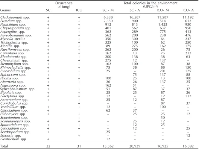
Fusarium spp. were prevalent (p<0.05) in the month of March during the morning; in May, Aspergillus
spp. were also isolated at similar levels to the dematious fungus, whereas, in the remaining months, there was a prevalence of Cladophialophora spp. This genus increased significantly (p<0.05) in the month of March during the afternoon, falling gradually in subsequent months (Figure 1).
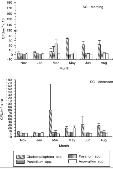
In the ICUs, over 60% of isolates from all rooms were of the Cladophialophora genus. There was a correlation between the number of colonies and the number of occupied hospital beds in both collection periods. Fusarium spp. were found in the five rooms, whereas Aspergillus spp. were found in four. In the adult ICU, during the morning period, Cladophialo-phora spp. remained stable during the months of Oc-tober, December, and February, began to increase in April, and peaked in July (p<0.05). The same occurred in the afternoon period, but counts were proportion-ally lower, and differences between the two periods were significant (p<0.05) in October, February, and July (Figure 2).
In the Neonatal ICU (Figura 3), Cladophialophora
spp. prevailed in July (p<0.05) and April (p<0.05), in the morning and afternoon periods, respectively, with a subsequent reduction in counts.
During renovation, there was a significant differ-ence (p<0.05) in airborne fungus counts in relation to the sampled months. Higher rates of the predomi-nant genus were found in external environments when compared to internal ones, especially during the af-ternoon (data not shown).
Another aspect of the survey focused on the healthcare staff of the adult and neonatal ICUs and of the surgical center. Yeasts were isolated from 39.4% of staff members. From nailbeds 12.1% of samples were isolated, 16.7% from interdigital spaces, and 10.6% from the oropharynx.
Of the positive samples obtained from staff mem-bers, Candida albicans accounted for 23%, C. parapsilosis for 19%, Candida spp. for 19%, C. guilliermondii for 8%, and Trichosporon spp. for 31%.
C. albicans was isolated mainly from the oropharynx and C. parapsilosis and Trichosporon spp, from interdigital spaces and nailbeds.
Table - Airborne fungi isolated from surgical center and intensive care units during different time periods.
Ocurrence Total colonies in the environment
of fungi (UFC/m3)
Genus SC ICU SC - M SC - A ICU- M ICU- A
Cladosporium spp. + + 6,338 16,587 11,587 11,192
Fusarium spp. + + 2,350 900 514 612
Penicillium spp. + + 912 813 1,425 950
Chrysosporium spp + + 401 562 637 950
Aspergillus spp. + + 362 289 775 413
Aureobasidium spp. + + 562 200 238 476
Mycelia sterilia + + 350 300 64 237
Trichoderma spp. + + 325 100 62 250
Monilia spp. + + 89 275 162 175
Paecilomyces spp. + + 262 200 26 75
Curvularia spp. + + 162 13 75 212
Rhodotorula spp. + + 200 138 38 63
Chaetomium spp. + + 275 12 137
Stemphylium spp. + + 162 100 87 38
Rhinocladiella spp. + + 75 38 88 150
Exserohilum spp. + + 25 201 125
Epicoccum spp. + + 75 137 88
Phoma spp. + + 100 25 13 100
Alternaria spp. + + 26 26 137 25
Nigrospora spp. + 162 51
Syncephalastrum spp. + + 51 87 37 37
Bipolaris spp. + + 25 25 87 26
Dactylaria spp. + + 12 12 112
Acremonium spp. + + 25 12 87
Conidiobulus spp. + 87 37
Verticillium spp. + + 12 100
Gliocladium spp. + 62 37
Pithomyces spp. + + 25 25 12
Sepedonium spp. + - 50
Scopulariopsis spp. + + 25 12
Sporotrichum spp. + + 12 25
Ulocladium spp. + + 12 25
Scedosporium spp. + 25
Emonsia spp. + 12
Geotrichum spp. + 12
Total32 31 13,362 20,939 16,925 16,392
" Rev Saúde Pública 2005;39(3) www.fsp.usp.br/rsp Monitoring of fungus in a hospital unit
Martins-Diniz JN et al
A total 91 samples was obtained from furniture sam-ples, of which 44% were positive for the presence of yeasts. Candida was the predominant genus (70%), followed by Trichosporon spp. Candidaguilliermondii
prevailed in the environment, especially on doorknobs (52%), where C. lusitaniae (5,0%), C. parapsilosis
(3,0%), Candida spp. (10%), and Trichosporon spp. (30%) were also isolated. Trichosporon spp. were iso-lated from all the environments investigates, prevail-ing on doorknobs and in the air inside the ICUs.
DISCUSSION
The incidence of fungi is widely variable, depend-ing on season, temperature, relative humidity, time of the day, speed and direction of winds, presence of human activity, and artificial climate control.6,11,13
The composition and concentration of airborne organisms in external and internal environments of
critical hospital areas have been little inves-tigated, but there are studies that emphasize their importance due to the appearance of these agents in nosocomial infections.1
In the present study, about 30 different gen-era were isolated from a hospital environment, with a predominance of Cladophialophora
spp., Fusarium spp., Penicillium spp., Chrysosporium spp., and Aspergillus spp. The number of studies addressing this subject in Brazil is limited, and data on the airborne microbiota in hospitals are sparse. Silva et al22
investigated the fungal microbiota of the air and floor of a hospital, and found mainly
Cladosporium spp. (65.0%), Aspergillus spp. (37.1%), Mycelia sterilia (26.9%), Fusarium
spp. (20.1%), Penicillium spp. (19.8%),
Aureobasidium spp.(18.4%), Curvularia spp. (16.2%), and Nigrospora spp.(15.3%). The prevalence of Cladophialophora has been reported in studies of aerosols from several Brazilian cities.6,13
During the study period, the hospital un-derwent extensive renovation for the imple-mentation of a new unit, and this coincided with an increase in propagule counts, espe-cially of Cladophialophora spp. In the pe-riod preceding renovation, there was a sig-nificant increase in Fusarium spp. fungi (p<0.05), which are potentially pathogenic and are currently being reported in cases of invasive disease.20Aspergillus spp. were
iso-lated from the special areas in both periods, and, at similar levels to Cladophialophora
spp., from inside the surgical center during the after-noon in the month of May, coinciding with renova-tion. In this period there was an increase in air con-tamination outside the building (data not shown), and a concomitant increase in the contamination of inter-nal air. The same genera of fungi were recovered from both environments, suggesting an influence of exter-nal factors on interexter-nal contamination levels, as has been described in the literature.2 There was also an
increase in levels of Cladophialophora spp. in all internal collection points when compared to the pre-vious months, and counts of this fungus were increased in the external environment. After the completion of the renovation process, there was a gradual decline, tending towards former levels, especially with respect to the Cladophialophora genus in the surgical center during the afternoon, indicating the importance of environmental monitoring to guide preventive meas-ures. Several studies suggest that the distribution of fungi, both in terms of concentration and of the
dif-Figure 1 - Major genera isolated from the surgical center, in CFU/m3
(geometric mean ± standard deviation), during morning and afternoon
collection periods in the months of November, January, March, May, June and August.
Nov Jan Mar May Jun Aug
-10 0 10 20 30 40 130 140 150 160 170 180
SC - Morning
CFU/m
3 x 10
Month
Nov Jan Mar May Jun Aug
-100 10 20 30 40 50 60 70 80 90 100 110 120 130 140 150 160 170 180
SC - Afternoon
CFU/m
3 x 10
Month
Cladophialophora spp.
Penicillium spp.
#
Rev Saúde Pública 2005;39(3) www.fsp.usp.br/rsp
Monitoring of fungus in a hospital unit Martins-Diniz JN et al
ferent genera encountered, vary among geo-graphical areas and is influenced by envi-ronmental and seasonal factors.6,11,13,17,24
The levels encountered are considered as high for such environments, especially if compared to the levels reported by Távora et al.24 A complicating factor is the lack of
ref-erence values for climate-controlled environ-ments considered as ‘special.’ Mean levels in the collection points outside the ICUs and surgical center were higher than those found in internal environments, but lower than those found outside the building. The two sectors investigated were climate-controlled, but did not use HEPA filters. However, cli-mate control in the ICUs was less regular, and natural ventilation was used on occa-sion. A difference in the total number of fungi isolated is evident in the surgical center, probably due to the different routines of the two sectors.
In a study lasting for over 30 weeks car-ried out during the partial renovation of a hospital in Pittsburgh, USA, Overberger et al17 (1995) isolated Penicillium spp.,
As-pergillus spp., and, to a lesser extent, Alter-naria spp., commonly isolated fungi, and
Cladosporium spp. were the fungi most fre-quently isolated from the air. The authors reported an increase in levels of viable spores during renovation, and a subsequent decrease to former levels after renovation was con-cluded, results similar to those of the present
study. Likewise, Bouza et al2 (2002) found that the
demolition of a hospital building was associated with increased colony counts in external and non-HEPA protected internal air, and that counts returned to former levels after 11 days. Aspergillus fumigatus was the fungus most frequently isolated, followed by Peni-cillium spp., Aspergillus niger, Aspergillus flavus, As-pergillus spp., and Fusarium spp., among other spe-cies. On the other hand, Aspergillus spp. were the f ilamentous fungus most frequently isolated by Panagopoulou et al18 (2002) in a multicenter
hospi-tal study, corresponding to 70,5% of all isolates. Mousa et al14 (1999), while investigating the
correla-tion between fungi isolated from burn wounds and environmental surfaces of a burn care unit, found a greater proportion of Aspergillus niger in both pa-tients and environments. However, Ulocladium spp. were the most common isolates in the control group (cultures obtained from outside the burn care unit), suggesting that patients may have acquired fungal infections while in the burn care unit.
In the present study, we also monitored the area around the beds and the healthcare professionals that worked inside the surgical center and ICUs. C. albi-cans, described in the literature as the major agent of nosocomial infections,4 was the prevailing species
among the samples obtained from the healthcare staff. The presence of yeasts on the hands of these profes-sionals is a source of concern for hospital-infection control teams, since this may be a potential source of contamination and dissemination of microorganisms. In the present survey, yeasts were found in samples obtained from 39.4% of the healthcare staff. Data on this subject in the literature show great disparity, with rates ranging between 17% and 80%.8,23
Candidiasis and invasive aspergillosis have in-creased in recent years, especially among immuno-compromised patients.1,25C. albicans is still the
ma-jor species involved in fungemia, although
non-albicans species have increased in frequency, and are often refractory to conventional treatment.3,4 On
Figure 2 - Major genera isolated from the adult ICU, in CFU/m3
(geometric mean ± standard deviation), during morning and afternoon
collection periods in the months of October, December, February, April, May and July.
Oct Dec Feb Apr May Jul
-10 0 10 20 30 40 50
60 Adult ICU - Morning
CFU/m
3 x 10
Month
Oct Dec Feb Apr May Jul
-10 0 10 20 30 40 50 60
Adult ICU - Afternoon
CFU
/m
3 x 10
Month
Cladophialophora spp. Penicillium spp.
$ Rev Saúde Pública 2005;39(3) www.fsp.usp.br/rsp Monitoring of fungus in a hospital unit
Martins-Diniz JN et al
REFERENCES
1. Alberti C, Bouakline A, Ribauad P, Lacroix C, Rousselot P, Leblanc T et al. Relationship between environmental fungal contamination and the incidence of invasive aspergillosis in haematology patients. J Hosp Infect 2001;48:198-206.
2. Bouza E, Peláez T, Pérez-Molina J, Marín M, Alcalá L, Padilla P et al. Demolition of a hospital building by controlled explosion: the impacto on filamentous fungal load in internal and external air. J Hosp Infect 2002;52:234-42.
the other hand, infections by resistant microorgan-isms are increasing, resulting in higher mortality rates, increased hospitalization periods, and consequent higher cost to hospital services.21
In the present study, yeasts of the Trichosporon
genus, considered as emergent agents of hospital infections,15 were also isolated at
substantial levels, both from the healthcare staff and from all environmental surfaces in-vestigated.
Of the samples obtained from the surfaces of furniture, two Candida lusitaniae isolates are worthy of note, since this yeast has mecha-nisms to develop resistance to Amphotericin B (certain strains being primarily resistant).7
One of these samples was isolated from a doorknob in the neonatal ICU, and the other in the following week from a doorknob in the adult ICU, which are physically close to each other. Although a genotypic characteri-zation was not carried out, such a result may suggest that yeasts are carried from one place to the next within critical areas. Although
Candida spp. were the yeasts most frequently isolated from healthcare staff and environ-ment, the isolation of Trichosporon spp. was important due to its potential to resist anti-fungal therapy.
The characterization of environmental fungi from internal environments in critical hospital areas and of the fungal microbiota found on the hands of healthcare profession-als is recognized worldwide as an important measure aimed at substantially reducing morbidity, mortality, and high hospital costs.
Environmental sources must hence be moni-tored, especially in special rooms, which ac-commodate immunosuppressed patients, susceptible to environmental pathogens exposure. Only then, it will be possible to design appropriate measures for controlling such pathogens and to choose the adequate patients therapies to be instituted.
Figure 3 - Major genera isolated from the neonatal ICU, in CFU/m3
(geometric mean ± standard deviation), during morning and afternoon
collection periods in the months of October, December, February, April, May, and July.
Oct Dec Feb Apr May Jul
0 10 20 30 40 50 60 70 80 90 100 110 120
Neonatal ICU - Morning
CFU/m
3 x 10
Month
Oct Dec Feb Apr May Jul
-10 0 10 20 30 40 50 60 70 80 90 100 110
120 Neonatal ICU - Afertnoon
CFU/m
3 x 10
Mont
Cladophialophora spp. Penicillium spp.
Fusarium spp. Aspergillus spp.
3. Colombo AL, Nucci M, Salomão R, Branchini ML, Richtmann R, Derossi A et al. High rate of non-albicans candidemia in Brazilian tertiary care hospitals. Diagn Microbiol Infect Dis 1999;34:281-6.
4. Colombo AL. Epidemiology and treatment of hematogenous candidiasis: a Brazilian perspective.
%
Rev Saúde Pública 2005;39(3) www.fsp.usp.br/rsp
Monitoring of fungus in a hospital unit Martins-Diniz JN et al
5. Cooley JD, Wong WC, Jumper CA, Straus DC. Correlation between the prevalence of fungi and sick building syndrome. Ocupp Environ Med 1998;55:579-84.
6. Gambale W, Purchio A. Influência de fatores abióticos na dispersão aérea de fungos na cidade de São Paulo. Rev Microbiol 1983;14:204-14.
7. Hawkins JL, Baddour LM. Candida lusitaniae infections in the era of fluconazole availability. Clin
Infect Dis 2003;36:14-8.
8. Horn WA, Larson EL, Mcginley KJ, Leyden JJ. Microbial flora on the hands of health care personnel: differences in composition and antibacterial
resistance. Infect Control Hosp Epidemiol 1988;(9):189-93.
9. Kurtzman CP, Fell JW. The Yeasts: a taxonomic study. 4ª ed. New York: Elsevier; 1998. p. 605.
10. Lacaz CS, Porto E, Heins-Vaccari E. Guia para identificação: fungos, actinomicetos e algas de interesse médico. São Paulo: Sarvier; 1998.
11. Lacaz CS, Porto E, Martins JEC, Heins-Vaccari EM, Melo NT. Tratado de Micologia Médica Lacaz. São Paulo: Sarvier; 2002. p. 1104.
12. Mendes-Giannini MJS, Melhem MSC. Fungos. In: Ferreira AW, Ávila SLM, coordenadores. Diagnóstico laboratorial das principais doenças infecciosas e auto-imunes. Rio de Janeiro: Guanabara-Koogam; 2001. p. 333-403.
13. Mezzari A, Perin C, Santos Jr AS, Bernd LAG, Gesu GD. Fungos anemófilos e sensibilização em indivíduos atópicos em Porto Alegre, RS. Rev Assoc
Med Bras 2003;49(3):270-3.
14. Mousa HAL, Al-Bader SM, Hassan DA. Correlation between fungi isolated from burn wounds and burn care units. Burns 1999;25:145-7.
15. Moretti-Branchini M, Fukushima K, Schreiber AZ, Nishimura K, Papaiordanou PMO, Trabasso P et al. Trichosporon species infection in bone marrow transplanted patients. Diagn Microbiol Infect Dis 2001;39:161-4.
16. Nesa D, Lortholary J, Bouakline A, Bordes M, Chandenier J, Derouin F et al. Comparative performance of impactor air samples for
quantification of fungal contamination. J Hosp Infect 2001;47:149-55.
17. Overberger PA, Wadowsky RM, Schaper MM. Evaluation of airborne particulates and fungi during hospital renovation. Am Ind Hyg Assoc 1995;56:706-12.
18. Panagopolou P, Filioti J, Petrikkos G, Giakouppi P, Anatoliotaki M, Farmaki E et al. Enviromental surveillance of filamentous fungi in three tertiary care hospitals in Greece. J Hosp Infect 2002;52:185-91.
19. Pfaller MA. Nosocomial candidiasis: emerging species, reservoirs, and modes of transmission. Clin
Infect Dis 1996;22(Suppl 2):89-94.
20. Raad I, Tarrand J, Hanna H, Albitar M, Janssen E, Boktour M et al. Epidemiology, molecular mycology, and environmental sources of Fusarium infection in patients with cancer. Infect Control Hosp Epidem 2002;23:532-7.
21. Rentz AM, Halpern MT, Bowden R. The impact of candidemia on length of hospital stay, outcome and overall cost of illness. Clin Infect Dis 1998;27:781-8.
22. Silva MG, Moreira YK, Cisalpino IO. Flora fúngica do ar e do piso no Hospital das Clínicas da Universidade de Minas Gerais, Belo Horizonte, Brasil. Rev
Microbiol 1983;14:215-22.
23. Strausbaugh LJ, Sewell DL, Ward TT, Pfaller MA, Tjoelker RC, Heitzman T. High frequency of yeast carriage on hands of hospital personnel. J Clin
Microbiol 1994;32:2299-300.
24. Távora LGF, Gambale W, Heins-Vacari EM, Arriagada GLH, Lacaz CS, Santos CR et al. Comparative performance of two air samples for monitoring airborne fungal propagules. Braz J Med
Biol Res 2003;36:613-6.