with or without Dental Caries
Pernilla Lif Holgerson1
*, Carina Öhman1,2, Agneta Rönnlund1,2, Ingegerd Johansson2
1Department of Odontology/section of Pedodontics, UmeåUniversity, Umeå, Sweden,2Department of Odontology/section of Cariology, UmeåUniversity, Umeå, Sweden
Abstract
Background
The aim of this longitudinal study was to evaluate the oral microbiota in children from age 3 months to 3 years, and to determine the association of the presence of caries at 3 years of age.
Methods and findings
Oral biofilms and saliva were sampled from children at 3 months (n = 207) and 3 years (n = 155) of age, and dental caries was scored at 3 years of age. Oral microbiota was assessed by culturing of total lactobacilli and mutans streptococci, PCR detection ofStreptococcus
mutansandStreptococcus sobrinus, 454 pyrosequencing and HOMIM (Human Oral
Mi-crobe Identification Microarray) microarray detection of more then 300 species/ phylotypes. Species richness and taxa diversity significantly increased from 3 months to 3 years. Three bacterial genera, present in all the 3-month-old infants, persisted at 3 years of age, whereas three other genera had disappeared by this age. A large number of new taxa were also ob-served in the 3-year-olds. The microbiota at 3 months of age, except for lactobacilli, was un-related to caries development at a later age. In contrast, several taxa in the oral biofilms of the 3-year-olds were linked with the presence or absence of caries. The main species/phy-lotypes associated with caries in 3-year-olds belonged to theActinobaculum,Atopobium,
Aggregatibacter, andStreptococcusgenera, whereas those influencing the absence of car-ies belonged to theActinomyces,Bergeyella,Campylobacter,Granulicatella,Kingella, Lep-totrichia, andStreptococcusgenera.
Conclusions
Thus, during the first years of life, species richness and taxa diversity in the mouth increase significantly. Besides the more prevalent colonization of lactobacilli, the composition of the overall microbiota at 3 months of age was unrelated to caries development at a later age. Several taxa within the oral biofilms of the 3-year-olds could be linked to the presence or ab-sence of caries.
OPEN ACCESS
Citation:Lif Holgerson P, Öhman C, Rönnlund A, Johansson I (2015) Maturation of Oral Microbiota in Children with or without Dental Caries. PLoS ONE 10(5): e0128534. doi:10.1371/journal.pone.0128534
Academic Editor:Zezhang Wen, LSU Health Sciences Center School of Dentistry, UNITED STATES
Received:March 11, 2015
Accepted:April 28, 2015
Published:May 28, 2015
Copyright:© 2015 Lif Holgerson et al. This is an open access article distributed under the terms of the Creative Commons Attribution License, which permits unrestricted use, distribution, and reproduction in any medium, provided the original author and source are credited.
Data Availability Statement:All original sequence data are available athttp://dx.doi.org/10.6084/m9. figshare.1384833All other relevant data are within the paper and its Supporting Information files.
Funding:This work was supported by the Swedish patent revenue foundation (PLH) (http://www.pmf.se), and the Västerbottens Läns landsting (PLH) (http:// www.fou.nu/is/vll). The funders had no role in study design, data collection and analysis, decision to publish, or preparation of the manuscript.
Introduction
The microbiota of the oral cavity and other parts of the gastro-intestinal (GI) tract develops from virtual sterility at birth into one of the most heavily colonized parts of the human body [1,2,3,4], but with distinct bacterial communities at the various anatomical niches [5]. In the mouth alone, more than 700 taxa have been identified, with approximately two thirds belong-ing to cultivable species (named or unnamed), and one-third belongbelong-ing to the uncultivable phylotypes (www.homd.org) [6].
Microbial variations during the first few years of life lead to the establishment of a stable bacterial ecosystem in the GI tract, including the mouth [4]. Generally, facultative anaerobic genera, such asStreptococciandActinomyces, are the initial colonizers, succeeded by more an-aerobic genera, such asBifidobacteriain the gut, andVeillonellaeandFusobacteriain the mouth [1,7]. Molecular based methods such as PCR, cloning and Sanger sequencing, microar-rays, and multiplex pyro- or Illumina sequencing, have provided insight into both cultivable and uncultivable bacterial species in health and disease. For example, several studies suggest that changes in microbial flora during the early variation period, and in the final composition of the gut flora, are related to negative health outcomes, such as allergy, obesity, and intestinal diseases during childhood [4,8,9], and obesity and myocardial infarction in adults [10,11]. There are few studies examining the impact of early microbial colonization in the mouth, but early acquisitions of the cariogenic species,Streptococcus mutansandStreptococcus sanguinis, have been associated with increased and decreased risks of dental caries, respectively [12,13]. Microarray and multiplex sequencing are less labor intense when compared to single species PCR and sequencing, after cloning, for in-depth characterization of the microbiota. Although microarrays provide taxonomic resolution at the species level or of a group of similar species of predetermined bacteria, multiplex sequencing determines the whole array of bacteria. The limi-tation of the latter method relates to sequencing errors and the length of the obtained sequence, which affects the taxonomic resolution. These limitations have been met by quality filtering, and the obtained sequence length has increased over time to approximately 450 bp with the 454 FLX+ pyrosequencing methods. The most commonly used gene in multiplex sequencing, and for the taxonomic assignment of oral microbiota, is 16S rRNA, as in the HOMD (Human Oral Microbiome Database) curated database on common oral bacteria (www.HOMD.org) [14].
Dental caries is a highly prevalent polymicrobial infectious disease characterized by demin-eralization of tooth tissues, and dysbiosis of the tooth colonizing microbiota [15,16,17,18]. Several studies have applied newer multiplex sequencing methods such as 454 FLX pyrose-quencing; in order to identify caries associated microbiota [19,20,21]. However, the results ob-tained from these studies, conducted mostly in young children with early childhood caries (ECC), lack uniformity. The microbial species reported to be associated with ECC are, Strepto-coccus,Veillonella,Lactobacillus(especially in dentin caries),Olsenella,Actinomyces,Prevotella,
Granulicatella,Leptotrichia,Propionibacterium,Megasphaera, andScardovia[18,22]. In con-trast, several studies have found species such asCapnocytophaga,Fusobacteria,Tannerella,
Phorpyromonas,Abiotrophia, andStreptococcus, on healthy tooth surfaces [21,22].
Material and Methods
Ethics statements
The study was approved by the Regional Ethical Review Board in Umeå, Umeå University (http://www.epn.se), Sweden (Dnr 07-100M), and was conducted according to the principles described in the Declaration of Helsinki. Written informed consent was obtained from all caregivers.
Subjects and study design
This is a longitudinal cohort study (acronym, MamBa, Mamma-Barn, Swedish for Mother-Child) of infants from the age of 3 months to 3 years. Information on the association between mode of delivery and feeding in relation to microarray-assessed microbiota has been described previously [23,24]. Briefly, all mothers with babies born between September 2007 and January 2009 in a small inland town or a coastal university city in Northern Sweden, and with available contact details, were eligible for the study. However, only those parents, who were likely to stay in the area, were invited to provide consent for their child’s participation in the study (Fig 1). Children with a severe disease, and those associated with complicated pregnancies or deliveries, were excluded. A total of 207 (52%) mothers consented to participate in the study. At 3 months of age, the child visited the hospital along with his/her mother, who was then asked to complete a questionnaire regarding the feeding mode (exclusive or partial breast feeding, or exclusive formula feeding), mode of delivery, gestational age at birth, infant health (allergy, infections, stomach discomfort), use of antibiotics or products containing probiotic bacteria, use of a paci-fier, and presence of teeth. Samples were collected from the infant at this stage. At 3 years of age, the child attended a regular oral examination at the nearest Public Dental clinic, with his/ her mother/father, as part of the free annual examinations running from 3 to19 years of age, in Sweden. During this visit, an oral examination was performed and samples were collected by a specialist in pediatric dentistry (PLH). The caregiver was asked to complete another question-naire covering the same topics as that at 3 months of age. Information regarding infant body weight and length, at birth, at 3 months, and at 3 years of age, was obtained from child health care and medical records.
Caries scoring
Caries was registered using a mirror and probe, under good lighting conditions, and bitewing radiographs were used when approximal surfaces were unavailable for visual inspection. The criteria for caries detection were as described by the World Health Orginization [25]. Non-cav-itated lesions (initial caries lesions) were identified when the surfaces presented a“ chalky-white”appearance. Decayed (d), missing (m), and filled (f) surfaces (s) were recorded, and the sum (dmfs score), including carious lesions in enamel and dentine, were calculated.
Samplings at 3 months and 3 years of age
At 3 month of age, the buccal mucosa, tongue, and alveolar ridges were swabbed carefully using sterile cotton swabs (Applimed SA, Chatel-St-Denis, Switzerland). At 3 years of age, this procedure was repeated, and the teeth were scraped with a sterile wooden toothpick, for the presence of dental plaque, which, along with the mucosa-adherent biofilm, was immediately pooled into Eppendorf tubes (Sarstedt, Nümbrecht, Germany) with 200μl TE-buffer (10 mM
previously [23]. All samples were obtained between 1–3 hours (mean 2 hours) after the latest meal, and stored at−80°C.
Bacteria cultivation
Colony-forming units (CFU) of mutans streptococci per mL of saliva, were estimated by culti-vation on mitis salivarius sucrose agar supplemented with 0.2 U bacitracin at 37°C in 5% CO2.
Lactobacilli CFUs were estimated by culture on Rogosa agar (Merck, Darmstadt, Germany)
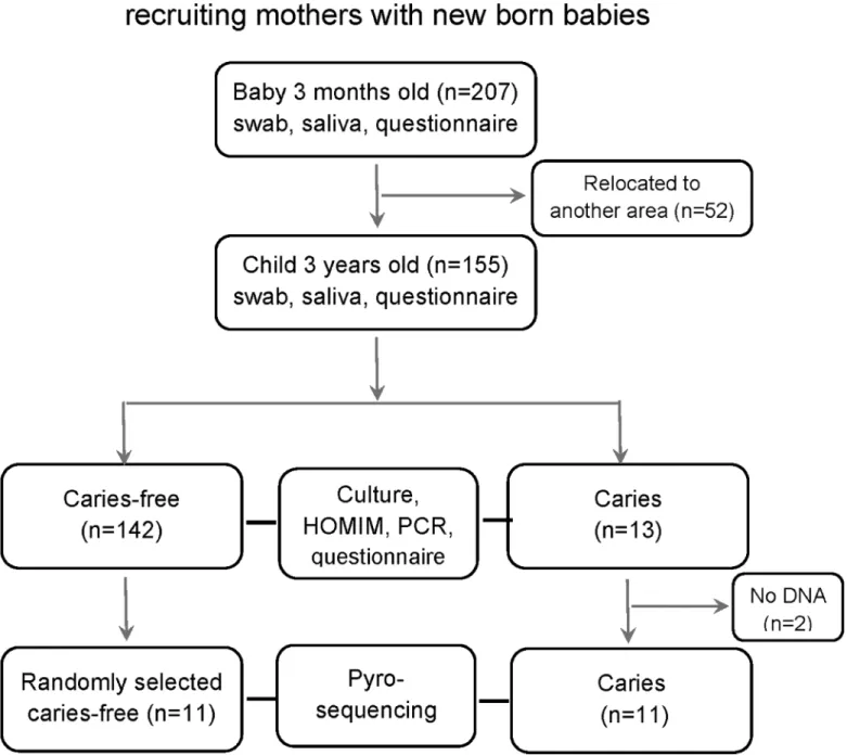
Fig 1. Flow chart diagram.Description of number of study participants and dropouts, data collection and analyses and caries status at 3 years of age.
anaerobically incubated at 37°C for 48–72 h. Total viable counts were cultured on blood agar (Columbia base agar, supplemented with 5% horse blood), and anaerobically incubated at 37°C for 48–72 h. This was done for all children at 3 months and 3 years of age.
DNA extraction
Bacterial DNA was purified from all swab and swab/tooth samples, using the GenElute Bacteri-al Genomic DNA kit (Sigma Aldrich, St. Louis, MO), according to the manufacturer’s instruc-tions, and as described previously [23]. The final quality and quantity of the DNA was evaluated using a Nanodrop 1000 spectrophotometer (Thermo Scientific, Wilmington, DE). Polymerase chain reaction (PCR) ofStreptococcus mutansandStreptococcus sobrinuswas per-formed using HotStarTaq Mastermix (Qiagen, Hamburg, Germany) with specific primers de-scribed in Yano et al [26]. ForS.mutansthe forward primer was 5’AGCCATGCGCAATC AACAGGTT 3’and the reverse 5’CGCAACGCGAACATCTTGATCAG 3’, and forS.sobrinus
the forward primer was 5’GAAACCAACCCAACTTTAGCTTGGAT 3’and the reverse 5’ATGGAGTGATTTTCCATCGGTACTTG 3’with the thermal cycling conditions: 15 min-utes at 95°C; 30 cycles of 30 seconds at 95°C, 30 seconds at 60°C, 1 minmin-utes at 72°C; followed by 5 minutes at 72°C [26].
Pyrosequencing and data processing
Oral samples, from all children with caries at 3 years of age, and an equal number of randomly selected children who were free of clinical signs of caries, were selected for microbiota charac-terization by 454 FLX+ pyrosequencing. The V3-V4 hypervariable region of the 16S rRNA gene was amplified by PCR, using the universal forward primer, 347F, and the reverse primer, 803R [27]. For sample identification, fusion primers with unique barcode sequences according to the“Guidelines for Amplicon Experimental Design (www.454.com), by Roche Diagnostics, were created. DNA was amplified under the following running conditions: initial denaturation at 94°C for 3 min, followed by 30 cycles at 94°C for 15 s, 58°C for 15 s, and 72°C for 30 s, with a final extension at 72°C for 8 min.
Amplicon processing and 454 sequencing were conducted at the Lund University Sequenc-ing Facility (Faculty of Science, Lund, Sweden), usSequenc-ing the Lib-A chemistry supported by the FLX+ platform. Complementary runs were performed on a 454 Roche Junior apparatus at Umeå University (Department of Immunology). After cleaning (Agencourt AMPure XP, Beckman Coulter, Brea, CA) and inspection (DNA 1000 kit on a 2100 Bioanalyzer, Agilent Technologies, Palo Alto, CA), the amplicons were quantified (Quant-iT ds DNA assay kit, Invi-trogen, Carlsbad, CA, and a Quantifluor fluorometer, Promega, Madison, WI), and diluted to obtain a total of 1×10⁷copies/uL. Titration and library production (aiming at 10
–15% enrich-ment) were performed using emulsion PCR, and the Lib-A kit (Bi-directional sequencing, Roche Diagnostics, Branford, CT). DNA positive beads were enriched, counted (Innovatis CASY particle counter, Roche Innovatis, Bielefeld, Germany), processed (XLR70 sequencing kit, Roche Diagnostics), and loaded onto a picotiter plate for pyrosequencing on a 454 Life Sci-ences Genome Sequencer FLX+ machine (Roche Applied Science, Penzberg, Germany). In Umeå, Roche’s bench-top machine, GC Junior sequencer (Roche Applied Science, Penzberg, Germany), was used according to the manufacturer’s guidelines.
sequences starting with the reverse primer were reverse complemented. The sequences were clustered into Operational Taxonomic Units (OTUs) at a sequence similarity of 97% in the 16S rRNA chimera checked Human Oral Microbiome Database [14], using USEARCH [28]. OTUs with a single sequence were removed, and representative sequences for each of the remaining OTU clusters were taxonomically assigned against the Human Oral Microbiome Database (www.HOMD.org) [14]. Sequences with98.5% identity were taxonomically assigned to the
species (named or unnamed species, or uncultivable species) level, while those between 97.0 and<98.5% were assigned to the genus level.
Microarray analysis
Microarray identification of microbes was done in all children, at 3 months and 3 years of age, using the HOMIM facility at the Forsyth Research Institute, Boston, MA, (http://mim.forsyth. org/homim.html) as described previously [23,29]. The HOMIM (Human Oral Microbe Identi-fication Microarray) microarray holds 422 oligonucleotide probes, targeting more than 300 bacterial taxa. Hybridization signals are read on a scale of six (0–5) with a lower limit of detec-tion of 104cells.
Data analyses
IBM SPSS Statistics (version 22.0; IBM Corporation, Armonk, NY, USA) was used for descrip-tive analyses and univariate testing of differences and associations. Normally distributed vari-ables are presented as means with 95% confidence interval (CI), and differences between group means were tested using parametric testing (unpaired t-test). Non-normally distributed vari-ables (including all sequence varivari-ables) are presented as medians with range, and non-paramet-ric testing (Mann-Whitney U test) were used to test if the subjects in the two age groups and in the caries-affected and caries-free groups, respectively, were samples from the same or different populations. Chi-square test was used to test differences in-group frequency distributions. For taxa comparisons, a p-value of0.008 was considered statistically significant after accounting
for multiple testing by the false discovery rate. Univariate comparisons were not applied for single taxa due to the combination of small groups and a high number of repeated tests. For other variables a p-value of<0.05 was considered statistically significant.
Rarefaction curves were calculated to compare microbial richness among the clinical sam-ples, and principal coordinate analysis (PCoA) to compare the phylogenetic diversity (β diver-sity) of these samples, using QIIME.
Multivariate principal component (PCA) was used to identify clustering of subjects, and partial least square (PLS) (SIMCA P+, version 12.0, Umetrics AB, Umeå, Sweden) to identify taxa associated with caries status [18,30,31]. Models with pyrosequencing data only, and those with the addition of lactobacilli and streptococci by culture and PCR, were tested. Vari-ables were autoscaled to unit variance, and cross-validated predictions of Y were calculated. Clustering of subjects is displayed in a score-loading plot, and the importance of each x-vari-able is displayed in a loading plot. Varix-vari-ables with a 95% confidence interval for the PLS correla-tion coefficient that did not include zero, were considered statistically significant. The Q2value
gives the capacity of the x-variables to predict the outcome (caries).
Results
At 3 years of age, 155 of the original 207 participating children had an oral examination. The remaining 52 did not visit the clinic mainly due to relocation to another area. The number of erupted teeth, among the 3-year-old children who participated in the study, ranged from 16–
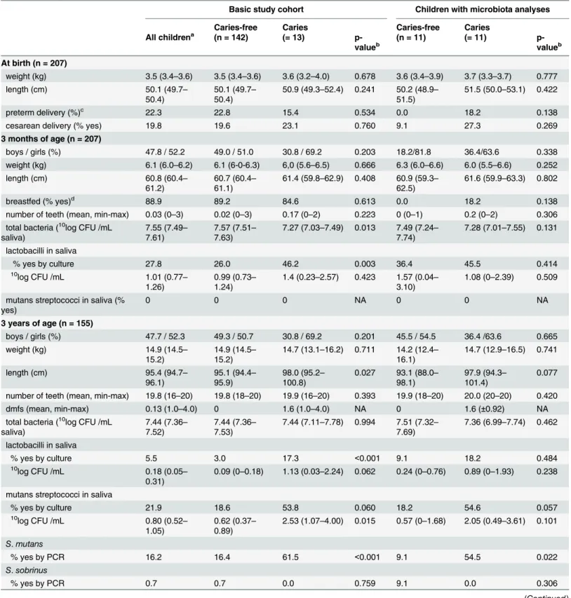
Table 1. Study group characteristics at birth, 3 months and 3 years of age.
Basic study cohort Children with microbiota analyses
Caries-free Caries Caries-free Caries
All childrena (n = 142) (= 13)
p-valueb (n = 11) (= 11) p-valueb
At birth (n = 207)
weight (kg) 3.5 (3.4–3.6) 3.5 (3.4–3.6) 3.6 (3.2–4.0) 0.678 3.6 (3.4–3.9) 3.7 (3.3–3.7) 0.777
length (cm) 50.1 (49.7–
50.4)
50.1 (49.7– 50.4)
50.9 (49.3–52.4) 0.241 50.2 (48.9– 51.5)
51.5 (50.0–53.1) 0.422
preterm delivery (%)c 22.3 22.8 15.4 0.534 0.0 18.2 0.138
cesarean delivery (% yes) 19.8 19.6 23.1 0.760 9.1 27.3 0.269
3 months of age (n = 207)
boys / girls (%) 47.8 / 52.2 49.0 / 51.0 30.8 / 69.2 0.203 18.2/81.8 36.4/63.6 0.338
weight (kg) 6.1 (6.0–6.2) 6.1 (6-0-6.3) 6,0 (5.6–6.5) 0.666 6.3 (6.0–6.6) 6.0 (5.5–6.6) 0.252
length (cm) 60.8 (60.4–
61.2)
60.7 (60.4– 61.1)
61.4 (59.8–62.9) 0.408 60.9 (59.3– 62.5)
61.6 (59.9–63.3) 0.802
breastfed (% yes)d 88.9 89.2 84.6 0.613 0.0 18.2 0.138
number of teeth (mean, min-max) 0.03 (0–3) 0.02 (0–3) 0.17 (0–2) 0.223 0 (0–1) 0.2 (0–2) 0.306 total bacteria (10log CFU /mL
saliva)
7.55 (7.49– 7.61)
7.57 (7.51– 7.63)
7.27 (7.03–7.49) 0.013 7.49 (7.24– 7.74)
7.28 (7.01–7.55) 0.131
lactobacilli in saliva
% yes by culture 27.8 26.0 46.2 0.003 36.4 45.5 0.414
10log CFU /mL 1.01 (0.77
– 1.26)
0.99 (0.73– 1.24)
1.4 (0.23–2.57) 0.423 1.57 (0.04– 3.10)
1.08 (0–2.39) 0.509
mutans streptococci in saliva (% yes)
0 0 0 NA 0 0 NA
3 years of age (n = 155)
boys / girls (%) 47.7 / 52.3 49.3 / 50.7 30.8 / 69.2 0.201 45.5 / 54.5 36.4 /63.6 0.665
weight (kg) 14.9 (14.5–
15.2)
14.9 (14.5– 15.2)
14.7 (13.1–16.2) 0.711 14.2 (12.4– 16.1)
14.7 (12.9–16.5) 0.741
length (cm) 95.4 (94.7–
96.1)
95.1 (94.4– 95.9)
98.0 (95.2– 100.8)
0.027 93.1 (88.0– 98.1)
97.9 (94.3– 101.4)
0.077
number of teeth (mean, min-max) 19.8 (16–20) 19.8 (18–20) 19.9 (16–20) 0.393 19.9 (18–20) 20.0 (20–20) 0.420
dmfs (mean, min-max) 0.13 (1.0–4.0) 0 1.6 (1.0–4.0) NA 0 1.6 (±0.92) NA
total bacteria (10log CFU /mL
saliva)
7.44 (7.36– 7.52)
7.44 (7.36– 7.53)
7.44 (7.11–7.78) 0.994 7.51 (7.32– 7.69)
7.36 (6.99–7.74) 0.462
lactobacilli in saliva
% yes by culture 5.5 3.0 17.3 <0.001 9.1 18.2 0.484
10
log CFU /mL 0.18 (0.05–
0.31)
0.09 (0–0.18) 1.13 (0.03–2.24) 0.062 0.24 (0–0.76) 0.89 (0–1.93) 0.238
mutans streptococci in saliva
% yes by culture 21.9 18.6 53.8 0.060 18.2 54.6 0.057
10log CFU /mL 0.80 (0.52
– 1.05)
0.62 (0.37– 0.89)
2.53 (1.07–4.00) 0.015 0.57 (0–1.68) 2.05 (0.49–3.61) 0.101
S.mutans
% yes by PCR 16.2 16.4 61.5 <0.001 9.1 54.5 0.022
S.sobrinus
% yes by PCR 0.7 0.7 0.0 0.759 9.1 0.0 0.306
Mean dmfs (sum of decayed missing filled surfaces) among the 13 children with caries was 1.6 (1.0–4.0), with 38% of the caries lesions located in the enamel only, whereas 62% had extended into the dentin. A majority (90%) of the caries lesions were in the tooth fissures, and 10% on the smooth buccal surfaces, whereas the lingual or approximal surfaces of the teeth remained unaffected. The mean body weight at birth, 3 months and 3 years of age, as well as the propor-tion of boys versus girls, breastfed versus not, and those delivered by Cesarean secpropor-tion did not differ between children who developed caries and those who did not (Table 1). However, at 3 years of age, children who developed caries were taller than those who did not (Table 1).
DNA from two children with caries, ended during the 3-month analyses with HOMIM and PCR (Fig 1), and these children could therefore not be represented in the pyrosequencing anal-yses, resulting in a total of 11 children in the caries group and 11 in the caries-free group. How-ever, all 13 children with caries were included in the other analyses performed in this study (culture, PCR, and HOMIM microarray).
Culture, 16S rRNA Gene Sequencing and Microarray detection
The mean bacterial count estimated from culture on the anaerobically incubated blood agar plates was 3.2x10⁷with a range from 3.8x10³-1.6x10⁸. From the 44 samples analyzed by
pyrose-quencing, 932,510 sequences passed quality filtering, with a mean number of 20,514 (95% CI 17,744–23,284) sequences per sample, and a variation of 5,119 to 44,625 sequences. The origi-nal sequence data is available athttp://dx.doi.org/10.6084/m9.figshare.1384833. All 932,510 se-quences could be clustered into 376 OTUs at 97% identity against the HOMD database. Eight of these OTUs could not be assigned to the bacterial kingdom, while another 26 presented with a single sequence. The remaining 342 OTUs represented 7 phyla, or divisions (Actinobacteria,
Bacteriodetes,Firmicutes,Fusobacteria,Proteobacteria,SR1,TM7), and 72 genera (S1 Table). Fifty-eight OTUs had a sequence identity of<98.5%, and could only be assigned to the genus
level, while 284 sequences had an identity of98.5%. The latter 284 sequences represented 196 unique HOT numbers in the HOMD database; named (n = 115) or unnamed (n = 43) spe-cies, and uncultivable phylotypes (n = 38). The HOMIM microarray gave a positive signal (score1) for 151 unique species/phylotypes.
Table 1. (Continued)
Basic study cohort Children with microbiota analyses
Caries-free Caries Caries-free Caries
All childrena (n = 142) (= 13)
p-valueb (n = 11) (= 11) p-valueb
Intake of sweet fruit soupe 19.7 16.8 54.5 0.031 27.2 44.4 0.705
Data are presented as means (95% CI) or proportions (%). Differences between groups were tested with Student´st-test or a Chi2 test, respectively. a) numbers varies by age as displayed
b) p-value<0.05 statistical significant. Tested between caries diseased and caries-free groups
c) gestational week 38 or earlier d) exclusively breast fed infants
e) intake of sweet fruit soup. Measured with a questionnaire, frequency of intake per week. Proportion presented is based on an intake more than 3 times a week.
Microbiota at 3 months versus 3 years of age
Cultivation and PCR. The total number of bacteria (CFU/mL saliva on blood agar) did not differ between samples collected at 3 months and 3 years of age (Table 1). Mutans strepto-cocci were not detected by culture, pyrosequencing, or microarray at 3 months of age, but at 3 years of age 16% had become colonized withS.mutansand 1 child withS.sobrinus.Notably, girls were more frequently colonized with mutans streptococci (by cultivation) than boys, at 3 years of age (28.8 versus 13.9%, p = 0.029).
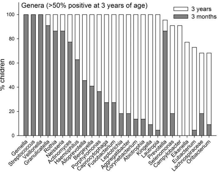
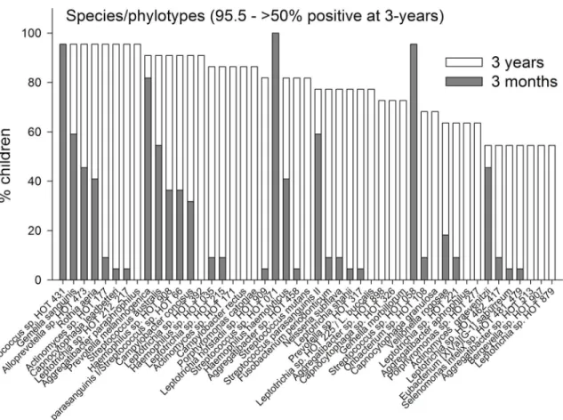
Pyrosequencing and Microarray. The prevalences of the 7 identified phyla are compared for the ages 3-months and 3-years inFig 2. InFig 3the prevalences of genera are present for genera that could be detected in>50% of the 3-year olds. Data for all detected genera are
shown inS1 Table. All 196 unique HOT species/phylotypes identified by pyrosequencing were present among the 3-year-old children, but only 176 were observed among the 3 months old infants. The mean (min–max) number of unique species/phylotypes was 31 (14–59) among the 3-month-old infants and 89 (67–128) in the 3-year-old children. In addition, the 3 month and 3-year-old children had 16 (3–28) and 31 (24–42) OTUs, respectively, defined at the genus level. Comparisons of the prevalence of species/phylotypes found in all children at 3-years of
Fig 2. Phyla prevalence.Prevalence at 3-months and 3-years of age of detected phyla.
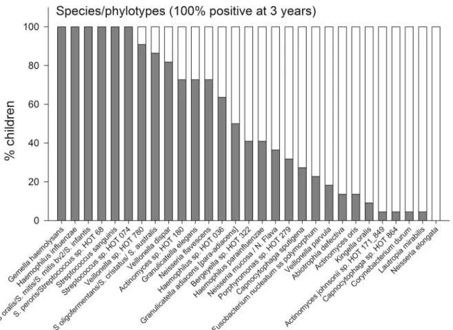
age are shown inFig 4, and for species/phylotypes found in less than 100% but more than 50% are shown inFig 5. Data for all detected species/phylotypes are shown inS2 Table. A full de-scription of the core microbiome at age 3 months and 3 years are shown inS3 Table.
Significantly lower species richness was confirmed in 3 months compared to 3 year olds in rarefaction analyses (Fig 6). The HOMIM microarray identified 56 unique species/phylotypes among the 3-month-old infants, and 94 unique species/phylotypes among the 3-year-old children.
PCoA modeling (weighted and unweighted) of pyrosequencing taxa revealed distinct clus-tering of the 3-month-old children from the 3-year olds (Fig 7). Similarly, PLS multivariate modeling with age as the dependent variable, and pyrosequencing, or HOMIM identified spe-cies/phylotypes as independent variables, revealed 2 significant components with explanatory capacities (R2) of 87.6% and 97.2%, and predictive capacities (Q2) of 77.5% and 89.8%,
respec-tively (S1 Table). Three genera (Streptococcus,Veillonella, andGemella) were present in all chil-dren at both 3 months, and 3 years of age, whereas another 3 genera (Escherichia(E.coli),
Staphylococcus(S.epidermidis), andPseudomonas(no species with>98.5 identity) were
Fig 3. Genera prevalence.Prevalence at 3-months and 3-years of age of genera detected in>50% of the children at 3 years of age.
significantly more prevalent (p<0.008) at 3 months compared with 3 years of age. In addition,
2 lactobacillus species (L.crispatusandL.gasseri), and one Streptococcus phylotype ( Strepto-coccus sp.HOT 058) were more prevalent, a borderline significance, in 3-months old infants than 3-years old children by pyrosequencing (S2 Table). Totally, 23 genera were significantly less prevalent (p<0.008) at 3 months compared to 3 years of age (S1,S2andS3Tables).
Microbiota in children with or without caries at 3 years of age
Cultivation and PCR. Children who had caries at 3 years of age had significantly lower CFU/mL saliva of total bacteria during infancy (3 months) than those who did not (Table 1). The proportion of children with lactobacilli (culture) in saliva was higher both at 3 months and 3 years of age, in the children who had caries at 3 years of age compared to those who did not (Table 1). Further, at 3 years of age, the CFU/mL saliva of total bacteria and the proportion of children colonized with mutans streptococci were higher in children with caries when com-pared to those who were caries-free (Table 1). Thus, slightly more than half of the children in the former group had mutans streptococci (culture), mainlyStreptococcus mutans(PCR) com-pared to less than every 5thchild in the latter group (Table 1). Only one child with caries was positive forStreptococcus sobrinusby PCR analysis (Table 1).
Pyrosequencing and Microarray. Species richness, determined by pyrosequencing of samples collected at 3 months and 3 years of age, did not differ between children with or with-out caries at the age of 3 months (Fig 7). Further, unweighted or weighted PCoA did not
Fig 4. Species/phylotype prevalence.Prevalence at 3-months and 3-years of age of species/phylotypes detected in all (100%) of the children at 3 years of age.
Fig 5. Species/phylotype prevalence.Prevalence at 3-months and 3-years of age of species/phylotypes detected in>50% but<100% of the children at 3
years of age.
doi:10.1371/journal.pone.0128534.g005
Fig 6. Rarefaction curves.Curves from children at 3 months and 3 years of age, stratified by caries development at 3 years of age. Comparisons include 11 children with caries, and 11 caries-free children.
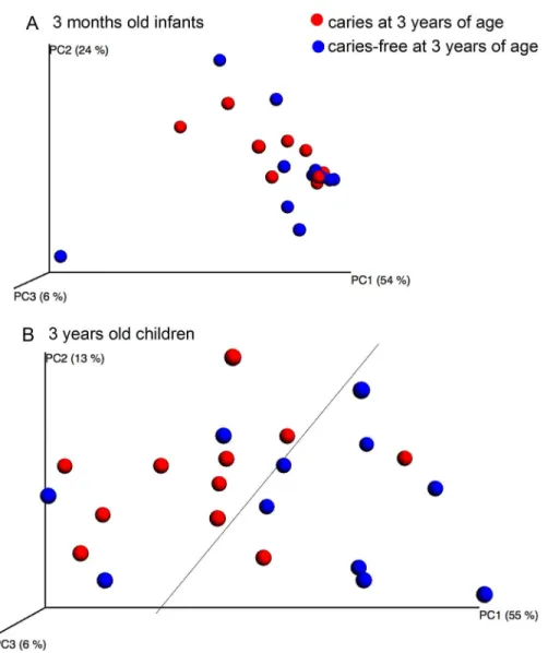
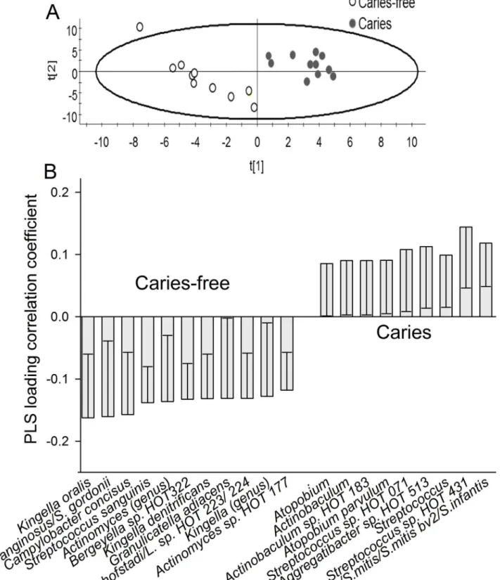
display any cluster formation based on the pyrosequencing determined microbiota at 3 months of age (Fig 8A), and no significant component was found by PLS when pyrosequencing and HOMIM microarray determined microbiota were used as independent variables, and caries status as the dependent variable. However, PCoA tended to separate the 3-year-olds with caries from those without caries (Fig 8B), and two significant components were identified by PLS, when pyrosequencing and HOMIM microarray taxa were used as independent variables, and caries status as the dependent variable. The predictive power of this model was 21.0%. The most influential variables in the 3-year-old caries-free children were (in alphabetical order): Ac-tinomycesgenus,Actinomyces sp. HOT 177, Bergeyella sp. HOT 322,Campylobacter concisus,
Granulicatella adiacens,Kingella genus,Kingella dentifricans,Kingella oralis,Leptrotrichia hof-stadii/Leptricihia sp. HOT 223 or 234,Streptococcus anginosus/S.gordonii, andStreptococcus sanguinis, and those in the children having caries were:Actinobaculumsp. HOT 183, Atopo-biumgenus,Atopobium parvulum,Aggregatibacter sp. HOT 513,Streptococcusgenus, Strepto-coccus sp. HOT 431,Streptococcus oralis, andS.mitis/Smitis bv2/ S.infantis(Fig 9A and 9B).
Discussion
The present study, monitored the oral microbiota, in the same children, at 3 months and 3 years of age, and, using DNA based methods, demonstrated that the overall species richness and taxa diversity in the mouth increases significantly during the first years of life. Most bacte-ria that colonize the oral cavity at 3 months of age persist at 3 years of age, but a few disappear, and a large number of different species are introduced during this time period. The major, and novel, finding, in this study was that, besides a more prevalent colonization with lactobacilli, the overall microbiota composition at 3 months of age was unrelated to caries development at a later age, whereas, several bacterial taxa in the oral biofilms of the 3 year olds could be linked to having caries or not at the same age.
The strengths of the present longitudinal study are that a specialist in pediatric dentistry, single-handedly, performed all clinical assessments, and that approximately 75% of the origi-nally included infants could be seen at 3 years of age. The reason for the comparably high fol-low-up percentage is that, parents who were likely to leave the area, within the coming years, were not invited to participate in the study. Consequently, despite the low overall inclusion rate, the long-term compliance for those who consented to participate in the present study was high. This concept was chosen because one of the two towns where the participants were resi-dent is a university town, comprising a large proportion of sturesi-dents of childbearing ages, who
Fig 7. PCoA clustering analysis by age.Red dots indicate samples from the 3-month-old infants, and blue dots indicate samples from the 3-year-olds.
knew they would leave the area in the coming years. This may pose as a potential risk of selec-tion bias in the present study cohort; however, a comparison between age, social status, and lifestyle factors, in a recent study involving early pregnant women living in the same area did not support the aforementioned notion [32].
The number of caries-affected children was small. This reflects that Sweden is a country with skewed caries prevalence in children and adolescents,i.e. a large portion being caries-free. The highest incidence is seen in children living in families with low socioeconomic status, and some immigrant groups. The incidence of caries in 3-year-olds in Sweden is reported to be 3 percent. The presently found prevalence is slightly higher compared to national data, but in ac-cordance with other studies performed in the region [33,34]. A potential limitation for caries detection was that the oral status did not justify a complementary X-ray in any child (see line 127). X-ray might have identified single children with initial approximal caries, but this was not doable due to tightly regulated criteria for X-ray in children by Swedish legislation. Based
Fig 8. PCoA clustering analysis by caries status.The upper figure (A) shows the microbiota variation between 3-month-old infants who developed caries at 3 years of age (red dots) and those who did not (blue dots). The lower figure (B) shows the microbiota variation between 3-year-old children who had caries (red dots) and those who did not (blue dots).
on that the children were included from the general population and the caries prevalence was as in the region combined with that an analysis method that is specially suited to find hidden structures was used, the present result should represent the situation in similar populations.
The sources of bacterial transmission to the infant’s mouth during the first few years of life include the mother’s vaginal, gut, and oral microbiota, the skin of the caretakers and siblings, breast milk, and other foods [33,35]. In the present study group, taxa belonging to the Escheri-chia,Staphylococcus, andPseudomonasgenera were significantly more prevalent at 3 months of age than at 3 years of age, which may be a result of transmission through one or more of the aforementioned routes. Only 3 taxa (Streptococcus,Veillonella, andGemella)in the core micro-biome of the 3-month-old infants (present in all infants) persisted in all of the 3-year-olds. The
StreptococcusandVeillonellagenera produce lactate, and have been associated with ECC [22], whereas,Gemella, and otherStreptococcusspecies such asStreptococcus sanguinis, have been found to be more prevalent in caries-free children [21]. The alterations in oral biofilm micro-biota, between the 3-month-olds and the 3-year-olds in the present study, largely conform to that reported in the literature [36].
The proportion of children with lactobacilli in the mouth was 46% at 3 months, and 17% at 3 years of age and the speciesL.crispatusandL.gasseritended to be more prevalent. We had previously reported the presence of lactobacilli in the mouths of infants who were exclusively breastfed, withLactobacillus gasseribeing the most dominant of the species [24]. Furthermore,
Lactobacillus gasseriisolated from the infants was found to have antibacterial traits, suggesting that the early microbiota in the mouth may program for later healthy conditions. However, the present study does not support the hypothesis that the presence ofLactobacillus gasseri, or any other lactobacilli species at 3 to 4 months of age, has a major impact on the composition of the mature oral microbiota, or the possibility of remaining caries-free. On the contrary, the pro-portion of lactobacilli at 3 months of age was significantly higher in children who had caries at 3 years of age. This is contradictory to what has been reported for the gut microbiota, where the presence of lactobacilli has been reported to influence immune responses, nutrition, and overall wellbeing [37], but, is in line with the finding that supplementation with the probiotic
Lactobacillus paracaseistrain, F19, during early infancy, did not protect against caries develop-ment at a later age [38].
Mutans streptococci, especiallyS.mutans, were found to be more prevalent in children who had caries, in the present study. This is in agreement with several studies, which have reported the association between infections with mutans streptococci, mostlyS.mutans, during the tooth eruption period, and early childhood caries (ECC) [39,40]. However, similar to previous reports, not all children who developed caries were colonized with mutans streptococci (cul-ture) orS.mutans(PCR), in the present study [13,19].
generally well nourished, and the present study included only those children who were healthy. Therefore, we presume that the accelerated growth in the 3 year olds with caries in this study is purely coincidental; however, we also believe that it should be followed-up in a future,
larger study.
The present study identified taxa fromActinomyces,Bergeyella,Campylobacter, Granulica-tella,Kingella,Leptrotrichia,and Streptococcus genera, to be associated with healthy teeth at 3 years of age. Several of these genera have previously been associated with healthy teeth (caries-free) in studies focusing on the whole microbiota. For example,Actinomyces,Campylobacter,
Leptotrichia,and Streptococcusgenera have been reported in Swedish adolescents living in the same area as those in the present study (Johanssonet al., personal communication). Further-more,Campylobacter[43],Granulicatella[44],Kingella[43],Leptotrichia[21],and Streptococ-cus(particularlyStreptococcus sanguinis)[13,39,44,45] have been associated with caries-free teeth in pre-school, and schoolchildren. However, comparisons with other studies is hampered by the fact that they are performed in different socio-economic conditions, different stages of the disease and different ages. It should also be kept in mind that to prove a causal or protective trait, identified species need to be tested in various types of experimental studies.
In contrast, the following taxa were associated with caries at 3 years of age:Actinobaculum
sp. HOT 183,Atopobiumgenus,Atopobium parvulum,Aggregatibacter sp. HOT 513, Strepto-coccusgenus,Streptococcus sp. HOT 431,Streptococcus oralis, andS.mitis/Smitis bv2/ S. infan-tis. Previous studies have linked the genera,Atopobium[45,46], andStreptococcus[12,21,40,
45,47,48], with enamel or dentin caries in pre-school children, schoolchildren, and adoles-cents. Notably, the bacteria that were found to be overrepresented in caries may represent the causally related species, or those favored by the caries associated environment, such as, the spe-cies utilizing lactate for metabolism.
Based on the results in the present study, we conclude that both species richness and taxa di-versity are significantly increased in the mouth, during the first years of life. Nevertheless, some taxa disappear with age. Furthermore, besides the more prevalent colonization of lactoba-cilli, the overall microbiota composition at 3 months of age was unrelated to caries develop-ment at a higher age. However, several taxa in the oral biofilms of the 3-year-olds could be associated with the presence or absence of caries at that age.
Supporting Information
S1 Table. Phyla and genera in 3 month and 3-year-old children.Proportion (%) children with detectable sequences in various phyla and genera, and mean prevalence of sequence pro-portions in these phyla and genera. Differences between age groups and groups with or without caries as 3 years of age were tested with Mann-Whitney test. P-values p0.008 are considered statistically significant.
(DOCX)
S2 Table. Species/phylotypes in 3 month and 3-year-old children.Proportion (%) children with detectable sequences in various phyla and genera, and mean prevalence of sequence pro-portions in these phyla and genera. Differences between age groups were tested with Mann-Whitney test, whereas no univariate statistical analyses were performed between children with or without caries at 3 years of age (seemethodssection). P-values p0.008 are considered statistically significant.
(DOCX)
HOMIM microarray.The oral microbiota was determined by (A) 454 FLX+ pyrosequencing and (B) the HOMIM microarray in samples from 3-months old infants and repeated when the child was 3-years of age. Core microbiota refers to bacteria detected in all children. Age charac-teristic species/phylotypes from (A) pyrosequencing and (B) HOMIM analyses were identified by PLS modeling with species/phylotypes as the independent block and age as the dependent variable. Species/phylotypes where the 95% CI did not include zero, i.e. statistically significant, are listed in alphabetical order.
(DOCX)
Acknowledgments
We would like to thank Tomas Johansson for his valuable support in pyrosequencing analysis, and Rolf Claesson for technical support.
Author Contributions
Conceived and designed the experiments: PLH IJ. Performed the experiments: CÖ AR. Ana-lyzed the data: PLH CÖ IJ. Contributed reagents/materials/analysis tools: PLH CÖ AR IJ. Wrote the paper: PLH CÖ AR IJ.
References
1. Könönen E. Development of oral bacterial flora in young children. Ann Med. 2000; 32: 107–112. PMID: 10766401
2. Könönen E. Anaerobes in the upper respiratory tract in infancy. Anaerobe. 2005; 11:131–136. PMID: 16701543
3. Palmer C, Bik EM, Digiulio DB, Relman DA, Brown PO. Development of the human infant intestinal microbiota. PLoS Biol. 2007; 5:e177. doi:10.1371/journal.pbio.0050177PMID:17594176
4. Arrieta MC, Stiemsma LT, Amenyogbe N, Brown EM, Finlay B. The intestinal microbiome in early life: health and disease. Front Immunol. 2014; 5:427. doi:10.3389/fimmu.2014.00427PMID:25250028
5. Segata N, Haake SK, Mannon P, Lemon KP, Waldron L, Gevers D, et al. Composition of the adult di-gestive tract bacterial microbiome based on seven mouth surfaces, tonsils, throat and stool samples. Genome Biol. 2012; 13:R42. doi:10.1186/gb-2012-13-6-r42PMID:22698087
6. Dewhirst FE, Chen T, Izard J, Paster BJ, Tanner AC, Yu WH, et al. The human oral microbiome. J Bac-teriol. 2010; 192:5002–5017. doi:10.1128/JB.00542-10PMID:20656903
7. Koenig JE, Spor A, Scalfone N, Fricker AD, Stombaugh J, Knight R, et al. Succession of microbial con-sortia in the developing infant gut microbiome. Proc Natl Acad Sci 2010; 108: 4578–4585. doi:10.1073/ pnas.1000081107PMID:20668239
8. West CE. Gut microbiota and allergic disease: new findings. Curr Opin Clin Nutr Metab Care. 2014; 17:261–6. doi:10.1097/MCO.0000000000000044PMID:24535216
9. Luoto R, Collado MC, Salminen S, Isolauri E. Reshaping the gut microbiota at an early age: functional impact on obesity risk? Ann Nutr Metab. 2013; 63:17–26. doi:10.1159/000354896PMID:24217033
10. Flint HJ. Obesity and the gut microbiota. J Clin Gastroenterol. 2011; 45:128–132. doi:10.1097/MCG. 0b013e31821f44c4
11. Ettinger R, MacDonald K, Reid G, Burton JP. The influence of the human microbiome and probiotics on cardiovascular health. Gut Microbes. 2014 Dec 20:0.
12. Jiang W, Jiang Y, Li C, Liang J. Investigation of supragingival plaque microbiota in different caries sta-tus of Chinese preschool children by denaturing gradient gel electrophoresis. Microb Ecol. 2011; 61: 342–352. doi:10.1007/s00248-010-9753-zPMID:20927511
13. Kanasi E, Dewhirst FE, Chalmers NI, Kent R Jr, Moore A, Hughes CV, et al. Clonal analysis of the microbiota of severe early childhood caries. Caries Res. 2010; 44:485–497. doi:10.1159/000320158 PMID:20861633
15. Takahashi N, Nyvad B. The role of bacteria in the caries process: ecological perspectives. J Dent Res. 2011; 90:294–303. doi:10.1177/0022034510379602PMID:20924061
16. de Soet JJ, Nyvad B, Kilian M. Strain-related acid production by oralstreptococci. Caries Res. 2000; 34:486–490. PMID:11093023
17. Mantzourani M, Gilbert SC, Sulong HN, Sheehy EC, Tank S, Fenlon M, et al. The isolation of bifidobac-teria from occlusal carious lesions in children and adults. Caries Res. 2009; 43:308–313. doi:10.1159/ 000222659PMID:19494490
18. Tanner AC, Kent RL Jr, Holgerson PL, Hughes CV, Loo CY, Kanasi E, et al. Microbiota of severe early childhood caries before and after therapy. J Dent Res. 2011; 90:1298–1305. doi:10.1177/
0022034511421201PMID:21868693
19. Crielaard W, Zaura E, Schuller AA, Huse SM, Montijn RC, Keijser BJ. Exploring the oral microbiota of children at various developmental stages of their dentition in the relation to their oral health. BMC Med Genomics. 2011; 4:22. doi:10.1186/1755-8794-4-22PMID:21371338
20. Ling Z, Kong J, Jia P, Wei C, Wang Y, Pan Z, et al. Analysis of oral microbiota in children with dental caries by PCR-DGGE and barcoded pyrosequencing. Microb Ecol. 2010; 60:677–690. doi:10.1007/ s00248-010-9712-8PMID:20614117
21. Jiang W, Zhang J, Chen H. Pyrosequencing analysis of oral microbiota in children with severe early childhood dental caries. Curr Microbiol. 2013; 67:537–542. doi:10.1007/s00284-013-0393-7PMID: 23743597
22. Xu H, Hao W, Zhou Q, Wang W, Xia Z, Liu C, et al. Plaque bacterial microbiome diversity in children younger than 30 months with or without caries prior to eruption of second primary molars. PLoS One. 2014; 9:e89269. doi:10.1371/journal.pone.0089269PMID:24586647
23. Lif Holgerson P, Harnevik L, Hernell O, Tanner AC, Johansson I. Mode of birth delivery affects oral microbiota in infants. J Dent Res. 2011; 90:1183–1188. doi:10.1177/0022034511418973PMID: 21828355
24. Holgerson PL, Vestman NR, Claesson R, Ohman C, Domellof M, Tanner AC, et al. Oral Microbial Pro-file Discriminates Breast-fed From Formula-fed Infants. J Pediatr Gastroenterol Nutr 2013; 56:127– 136. doi:10.1097/MPG.0b013e31826f2bc6PMID:22955450
25. World Health Organization. Oral health surveys: basic methods. 4th edition. 1997. World Health Orga-nization, Geneva.
26. Yano A, Kaneko N, Ida H, Yamaguchi T, Hanada N. Real-time PCR for quantification of Streptococcus mutans. FEMS Microbiol Lett. 2002; 19:23–30.
27. Nossa CW, Oberdorf WE, Yang L, Aas JA, Paster BJ, Desantis TZ, et al. Design of 16S rRNA gene primers for 454 pyrosequencing of the human foregut microbiome. World J Gastroenterol. 2010; 16: 4135–4144. PMID:20806429
28. Edgar RC. Search and clustering orders of magnitude faster than BLAST. Bioinformatics 2010; 26: 2460–2461. doi:10.1093/bioinformatics/btq461PMID:20709691
29. Colombo AP, Boches SK, Cotton SL, Goodson JM, Kent R, Haffajee AD, et al. Comparisons of subgin-gival microbial profiles of refractory periodontitis, severe periodontitis, and periodontal health using the human oral microbe identification microarray. J Periodontol. 2009; 80:1421–1432. doi:10.1902/jop. 2009.090185PMID:19722792
30. Sjöström M, Wold S, Söderström B. PLS discriminant plots; Elsevier: Amsterdam. 1986. pp. 461–470.
31. Haenlein M. A Beginner’s Guide to Partial Least Squares Analysis. Understanding statistics. 2004; 3:283–297.
32. Lundqvist A, Johansson I, Wennberg A, Hultdin J, Högberg U, Hamberg K, et al. Reported dietary in-take in early pregnant compared to non-pregnant women—a cross-sectional study. BMC Pregnancy Childbirth. 2014; 14:373. doi:10.1186/s12884-014-0373-3PMID:25361589
33. Isolauri E. Development of healthy gut microbiota early in life. J Paediatr Child Health. 2012; 48 Suppl 3:1–6. doi:10.1111/j.1440-1754.2012.02489.xPMID:22681492
34. Stecksén-Blicks C, Hasslöf P, Kieri C, Widman K. Caries and background factors in Swedish 4-year-old children with special reference to immigrant status. Acta Odontol Scand. 2014; 72:852–858. doi:10. 3109/00016357.2014.914569PMID:24823934
35. Munyaka PM, Khafipour E, Ghia JE. External influence of early childhood establishment of gut micro-biota and subsequent health implications. Front Pediatr. 2014; 2:109. doi:10.3389/fped.2014.00109 PMID:25346925
37. Preidis GA, Saulnier DM, Blutt SE, Mistretta TA, Riehle KP, Major AM. Probiotics stimulate enterocyte migration and microbial diversity in the neonatal mouse intestine. FASEB J. 2012; 26:1960–1969. doi: 10.1096/fj.10-177980PMID:22267340
38. Hasslöf P, West CE, Videhult FK, Brandelius C, Stecksén-Blicks C. Early intervention with probiotic Lactobacillus paracasei F19 has no long-term effect on caries experience. Caries Res. 2013; 47: 559– 565. doi:10.1159/000350524PMID:23838478
39. Parisotto TM, Steiner-Oliveira C, Silva CM, Rodrigues LK, Nobre-dos-Santos M. Early childhood caries and mutans streptococci: a systematic review. Oral Health Prev Dent. 2010; 8:59–70. PMID:20480056
40. Hughes CV, Dahlan M, Papadopolou E, Loo CY, Pradhan NS, Lu SC, et al. Aciduric microbiota and mutans streptococci in severe and recurrent severe early childhood caries. Pediatr Dent. 2012; 34:16– 23.
41. Kaul SS, Pathak RK, Santosh A. Emergence of deciduous teeth in Punjabi children, north India. Z Mor-phol Anthropol. 1992; 79:25–34. PMID:1441721
42. Leroy R, Bogaerts K, Lesaffre E, Declerck D. The emergence of permanent teeth in Flemish children. Commun Dent Oral Epidemiol. 2003; 31:30–39. PMID:12542430
43. Torlakovic L, Klepac-Ceraj V, Ogaard B, Cotton SL, Paster BJ, Olsen I. Microbial community succes-sion on developing lesucces-sions on human enamel. J Oral Microbiol. 2012; 4. doi:10.3402/jom.v4i0.16125 PMID:23248741
44. Luo AH, Yang DQ, Xin BC, Paster BJ, Qin J. Microbial profiles in saliva from children with and without caries in mixed dentition. Oral Dis. 2012; 18:595–601. doi:10.1111/j.1601-0825.2012.01915.xPMID: 22458262
45. Gross EL, Leys EJ, Gasparovich SR, Firestone ND, Schwartzbaum JA, Janies DA, et al. Bacterial 16S sequence analysis of severe caries in young permanent teeth. J Clin Microbiol. 2010; 48:4121–4128. doi:10.1128/JCM.01232-10PMID:20826648
46. Obata J, Takeshita T, Shibata Y, Yamanaka W, Unemori M, Akamine A, et al. Identification of the micro-biota in carious dentin lesions using 16S rRNA gene sequencing. PLoS One. 2014; 9:e103712. doi:10. 1371/journal.pone.0103712PMID:25083880
47. Kianoush N, Adler CJ, Nguyen KA, Browne GV, Simonian M, Hunter N. Bacterial profile of dentine car-ies and the impact of pH on bacterial population diversity. PLoS One. 2014; 9:e92940. doi:10.1371/ journal.pone.0092940PMID:24675997