Morphology and function of the ovipositor mechanism in Ceraphronoidea... 25
Morphology and function of the ovipositor mechanism
in Ceraphronoidea (Hymenoptera, Apocrita)
Andrew F. Ernst1, István Mikó1,2, Andrew R. Deans1,2
1 North Carolina State University, Department of Entomology, Raleigh, NC 27695-7613 USA 2 Pennsylva-nia State University, Department of Entomology, 501 ASI Building, University Park, PA 16802 USA
Corresponding author:István Mikó ([email protected])
Academic editor:M. Yoder | Received 25 March 2013 | Accepted 4 July 2013 | Published 1 August 2013
Citation: Ernst AF, István Mikó I, Deans AR (2013) Morphology and function of the ovipositor mechanism in Ceraphronoidea (Hymenoptera, Apocrita). Journal of Hymenoptera Research 33: 25–61. doi: 10.3897/JHR.33.5204
Abstract
he ovipositor of apocritan Hymenoptera is an invaluable source of phylogenetically relevant characters, and our understanding of its functional morphology stands to enlighten us about parasitoid life history strategies. Although Ceraphronoidea is one of the most commonly collected Hymenoptera taxa with considerable economic importance, our knowledge about their natural history and phylogenetic relation-ships, both to other apocritan lineages and within the superfamily itself, is limited. As a irst step towards revealing ceraphronoid natural diversity we describe the skeletomuscular system of the ceraphronoid ovi-positor for the irst time. Dissections and Confocal Laser Scanning Microscopy 3D media iles were used to visualize the ovipositor complex and to develop character concepts. Morphological structures were described in natural language and then translated into a character-character state format, whose terminol-ogy was linked to phenotype-relevant ontologies. Four unique anatomical phenotypes were revealed: 1. he irst valvifer (gonangulum) of the genus Trassedia is composed of two articulating sclerites, a condi-tion present only in a few basal insect taxa. he biparticondi-tion of the irst valvifer in Trassedia is most likely secondary and might allow more rapid oviposition. 2. Ceraphronoids, unlike other Hymenoptera, lack the retractor muscle of the terebra; instead the egg laying device is retracted by the seventh sternite. 3. Also unlike other Hymenoptera, the cordate apodeme and the anterior lange of the second valvifer are fused and compose one ridge that serves as the site of attachment for the dorsal and ventral T9-second valvifer muscles. Overall, the ceraphronoid ovipositor system is highly variable and can be described by discrete, distinguishable character states. However, these diferences, despite their discrete nature, do not relect the present classiication of the superfamily and might represent parallelisms driven by host biology.
JHR 33: 25–61 (2013) doi: 10.3897/JHR.33.5204 www.pensoft.net/journals/jhr
Copyright Andrew F. Ernst et al. This is an open access article distributed under the terms of the Creative Commons Attribution License 3.0 (CC-BY), which permits unrestricted use, distribution, and reproduction in any medium, provided the original author and source are credited.
Skeletomusculature, second valvula, ovipositor sheath, Aphanogmus, Ceraphron, Conostigmus, Megaspi-lus, Dendrocerus, Lagynodes, Hymenoptera Anatomy Ontology, Phenotypic Quality Ontology, Spatial Ontology
introduction
Ceraphronoidea has, until recently, been one of the most neglected groups of parasitic Hymenoptera, despite their ubiquity in the environment and fascinating life history strategies, which span from primary to quaternary parasitism (hyper-hyper-hyperpara-sitism) (Haviland 1920). Since the establishment of Ceraphronoidea as a superfamily (Masner 1956, Masner and Dessart 1967) signiicant revisionary work has addressed Lagynodinae (Dessart 1977, 1987), species of Conostigmus Dahlbom (Dessart 1997) and species groups of Dendrocerus Ratzeburg (Dessart 1972, Fergusson 1980, Dessart 1985, Dessart 1995a, Dessart 1999, Dessart 2001). Records of parasitism by ceraphro-noids cover a wide range of hosts, from at least eight insect orders: Hemiptera, hy-sanoptera, Hymenoptera, Neuroptera, Coleoptera, Trichoptera, Diptera and Mecop-tera (Austin 1984, Chiu et al. 1981, Cooper and Dessart 1975, Dessart 1967, 1992, Dessart and Bournier 1971, Evans et al. 2005, Fergusson 1980, Ghesquiere 1960, Ishii 1937, Luhman et al. 1999, Muesebeck 1979, Priesner 1936, Sinacori et al. 1992). Two species, Dendrocerus carpenteri (Curtis), and Dendrocerus aphidum (Rondani), serve as models for research concerning resource use and allocation by parasitoids (Araj et al. 2006), mate location (Schwörer et al. 1999), development and host interactions (Marris et al. 2000), sex determination of ofspring (Chow and Mackauer 1996), host discrimination (Chow and Mackauer 1999), and behavioral evolution in bio-control systems (Müller et al. 1997).
he phylogenetic placement of Ceraphronoidea remains uncertain despite the re-cent eforts to resolve the Hymenoptera tree of life (Heraty et al. 2011, Sharkey et al. 2011). Using expressed sequence tag (EST) data and limited exemplars, Sharanowski et al. (2010) placed Ceraphronoidea as sister to Evanioidea, but with low support. Heraty et al. (2011), using data from four genetic loci, placed Ceraphronoidea as sister to Stephanidae or Stephanidae + Orussidae, depending on the analysis. Vilhelmsen et al. (2010) found Ceraphronoidea to be sister to Megalyroidea within Evaniomorpha, based on morphological data. Sharkey et al. (2011) using molecular data and morpho-logical data - based largely on Vilhelmsen et al. (2010) - also suggested a sister relation-ship between Ceraphronoidea and Megalyroidea.
Morphology and function of the ovipositor mechanism in Ceraphronoidea... 27
c, 2001) and with the discovery of the irst ceraphronid wasp with well-developed pterostigma even the family level classiication of the superfamily has been challenged recently (Mikó and Deans 2009). Our recent observations on the genus Trassedia pro-vide additional epro-vidence that the current classiication of the superfamily needs to be revised (Mikó et al. in press). he genus was erected by Cancemi (1996), who classiied the type species within Megaspilinae and considered it to be closely related to Con-ostigmus. he original classiication is supported by two distinct character states: the presence of a fore wing pterostigma (which has subsequently been found in Ceraphro-nidae; Mikó and Deans 2009) and the presence of 11 antennomeres (Ceraphronidae have 10). Our observations of multiple specimens of Trassedia luapi reveal numerous morphological character states that are speciic to Ceraphronidae (Mikó and Deans 2009, Mikó et al. in press): absence of mesotibial apical spur, absence of narrow sclerite anterior to synsternum, presence of Waterston’s evaporatorium, and numerous male genitalia characters. Based on these diferences the genus has recently been transferred to Ceraphronidae (Mikó et al. in press), a classiication we follow in this study.
It is evident that a comprehensive study using both morphological and molecular characters is necessary for the reevaluation of Ceraphronoidea systematics. Since tra-ditionally used morphological characters have been phylogenetically inconsistent the utilization of unexplored character systems, such as the male and female terminalia might ofer additional data relevant to more robustly estimate the phylogenetic history of this group.
Despite the extensive descriptive work done on comparative morphology of the Hymenoptera female terminalia (Oeser 1961, Smith 1970, Le Ralec et al. 1996, Quicke et al. 1992, Quicke et al. 1994, Quicke et al. 1999, Vilhelmsen 2000, Vil-helmsen et al. 2001), the available morphological data on the ceraphronoid female terminalia are restricted to the distal region of the terebra (Quicke et al. 1994, Le Ralec et al. 1996) and the accessory glands (Höller et al. 1993). he skeletomuscular system of ceraphronoid ovipositor remained, until now, relatively unexplored. he main aim of the present study is to describe morphological diversity of the female terminalia in Ceraphronoidea and compare its anatomical structures with that of other Hymenop-tera taxa giving special emphasize on the skeletomuscular system. With this study, we establish a baseline for further phylogenetic analyses of the superfamily using oviposi-tor characters.
Materials and methods
techniques, imaging method and collecting locality (verbatim collecting data are stored at ighsare.com.).
Taxon Specimen
identiier
Imaging techniques
Locality DOI of CLSM media iles stored at http://igshare.com
Aphanogmus sp. 1 NCSU 0055648 brightield Hungary Aphanogmus sp. 1 NCSU 0002419 CLSM 40×
water imer-sion
Hungary doi: 10.6084/m9.igshare.156446 doi: 10.6084/m9.igshare.156439 Aphanogmus sp. 2 PSUCIM_5009 brightield Madagascar
Ceraphron sp. NCSU 0071198 brightield Madagascar Ceraphron sp. NCSU 0071197 brightield,
CLSM 10x
Madagascar doi: 10.6084/m9.igshare.156445 Ceraphron sp. PSUCIM_5005,
5006 CLSM Madagascar doi: 10.6084/m9.igshare.156470 doi: 10.6084/m9.igshare.156469 doi: 10.6084/m9.igshare.156447 Conostigmus
abdominalis (Boheman)
NCSU 0056302 brightield Sweden
Conostigmus abdominalis (Boheman)
NCSU 0056301 brightield Sweden
Conostigmus abdominalis (Boheman)
NCSU 0055647 brightield,
CLSM 20× Sweden doi: 10.6084/m9.igshare.156448 doi: 10.6084/m9.igshare.156433 Dendrocerus
spis-sicornis Hellén PSUCIM_5001, 5002 CLSM Sweden doi: 10.6084/m9.igshare.156452 doi: 10.6084/m9.igshare.156451 doi: 10.6084/m9.igshare.156450 doi: 10.6084/m9.igshare.156449 Dendrocerus
spis-sicornis Hellén PSUCIM_5010 CLSM Sweden doi: 10.6084/m9.igshare.156459 doi: 10.6084/m9.igshare.156453 Lagynodes sp. NCSU 0055643 brightield USA
Lagynodes sp. NCSU 0056306 CLSM 20×, 40× water immersion
USA doi: 10.6084/m9.igshare.156454 doi: 10.6084/m9.igshare.156443 Lagynodes
crassi-cornis
Megaspilus
arma-tus (Say) NCSU 0071199 brightield USA Megaspilus
arma-tus (Say) NCSU 0055645 brightield, CLSM 20× USA doi: 10.6084/m9.igshare.156442 doi: 10.6084/m9.igshare.156434 Megaspilus
arma-tus (Say)
NCSU 0056307 CLSM 40× water imer-sion
USA doi: 10.6084/m9.igshare.156437
Megaspilus ar-matus
PSUCIM_5003 CLSM 20X Canada doi: 10.6084/m9.igshare.156458 doi: 10.6084/m9.igshare.156457 doi: 10.6084/m9.igshare.156456 doi: 10.6084/m9.igshare.156455 Trassedia luapi
Cancemi NCSU 0056318 brightield, CLSM 10× Madagascar doi: 10.6084/m9.igshare.156440 doi: 10.6084/m9.igshare.156438 doi: 10.6084/m9.igshare.156436 Trassedia luapi
Morphology and function of the ovipositor mechanism in Ceraphronoidea... 29
Specimens used in the present study were stored in 95% ethanol. Some specimens were critical point dried and dissected on Blue-Tack (Blue Tack, Bostik inc.) medium. his method is mostly used to reveal the spatial relationships between muscles. Other specimens were dissected in glycerin on a concave microscope slide or were macerated in KOH to visualize the skeletal structures. Dissections and observations were made using an Olympus SZX16 stereomicroscope and an Olympus CX41 compound mi-croscope.
Bright ield images were taken using an Olympus CX41 compound microscope, equipped with an Olympus DP71 digital camera. Image stacks were combined using CombineZP (Hadley 2010) “do stack” command. SEM micrographs were made using a Hitachi S-3200 Scanning Electron Microscope (wd=23.5, av=5kV). Specimens were critical point dried and coated with palladium prior to examination. CLSM images were made on glycerin-stored specimens between 1.5 mm thick, 24×50 mm cover glasses with a Leica LSM 710 and Olympus Fluoview 1000 confocal laser scanning microscopes (CLSM) using the 488 nm laser to excite the sample. We collected the autoluorescence of insect anatomical structures between 500 and 700 nm with two channels (500–580; pseudocolor green and 580–700 pseudocolor red) using 106 and 206 Plan Achromat objectives. Volume rendered images and media iles were gener-ated by Imaris Bitplane (Bitplane, Zürich, Switzerland) and ImageJ (Schneider et al. 2012) software. Bright ield and CLSM micrographs were edited with Adobe Pho-toshop CS4 (Adobe) changing “Gamma Correction” value, resize images to 7.3 cm width at 400 dpi resolution, standardize style and width of scale bars and create image annotations. Media iles, SEM micrographs and bright ield images are available from igshare.com (Table 1).
To verify the relationships between anatomical structures, serial transverse section-ing was carried out on a Lagynodes specimen (Table 1). he specimen was embedded in Araldit®, cut at 1 µm with a Microm microtome (HM 360), and stained with toluidine blue.
Anatomical terms used in the descriptions are linked to concepts in the Hymenop-tera Anatomy Ontology (HAO; HymenopHymenop-tera Anatomy Portal (http://portal.hymao. org), Yoder et al. 2010), the Phenotypic Quality Ontology 1.2 (PATO; Gkoutos et al. 2004) and the Biospatial Ontology (Mungall 2013) via a table of uniform resource identiiers (URIs) (Appendix) following Seltmann et al. 2012.
Towards semantic statements
a more accessible format. Deans et al. (2012a) proposed a new description model for taxonomists applying semantic statements. hese statements are written in a logic and queryable format and are linked to the concepts of biomedical ontologies. Semantic descriptions are therefore not only transparent for researchers unfamiliar with speciic morphological jargon, but can be executed via an automated reasoning mechanism (Balhof et al. in press, Mullins et al. 2012).
To meet the grand challenge of describing phenotypes in a semantic way, using Web Ontology Language (OWL; http://www.w3.org/TR/owl-features/) for example, one must be familiar with tools of the Semantic Web (e.g., Protégé, http://protege. stanford.edu/ and Manchester Syntax, http://www.w3.org/TR/owl2-manchester-syn-tax/). Perhaps more importantly, one also has to provide a character/character state description with terms explicitly linked to ontologies.
We provide here an example of the transformation of our natural language descrip-tions to character/character state format and to link the terminology to relevant pheno-type ontologies. Our goal is to make this product more accessible to future reasoning applications. During the “ontologization” procedure the describer is forced to provide strict, structure-based deinitions for each anatomical concept, which itself enhances the readability, objectivity, consistency and comparability of the research product.
Results i: Natural language descriptions of anatomical structures in the ceraphronoid ovipositor assembly and s7
Integument
he irst valvifer is dorsoventrally elongated in lateral view (ch1: 0; 1vf: Figs 2E, 5A, E,
6A, D) with convex anterior (ch2:0) and straight (ch3:0; p1v: Figs 2D, 4A, 6A) or
con-cave posterior margins (ch3:1; p1v: Fig. 3D) in all taxa except Dendrocerus where the posterior margin of the irst valvifer is angled at the tergo-valviferal articulation (ch3:2;
p1v: Fig. 4F). he irst valvifer is not subdivided (ch4:1; 1vf: Figs 2E, 5A, E, 6A, D) except in Trassedia, where the transvalvifer conjunctiva (ch4:0; tvc: Fig. 3E) separates
the elongate dorsal sclerite (ch5:0; d1vf: Fig. 3E) from the triangular ventral sclerite
of the irst valvifer (ch6:0; v1vf: Fig. 3E). he sclerites articulate with one another at the intravalvifer articulation (ch7:0; iava: Figs 3C, E), which is located anteriorly on the border between the two sclerites (ch8:0). he ventral margin of the dorsal sclerite (ch9:0) and the anterodorsal margin of the ventral sclerite are thickened relative to surrounding regions (ch10:0). he anterior lange of the irst valvifer (af1: Fig. 1F)
overlapping the second valvifer is present (ch11:0) in Conostigmus, Dendrocerus and Megaspilus, and Lagynodes but absent from Ceraphronidae (ch11:1). he irst valvifer articulates with the second valvifer on its posteroventral corner (ch12:0; intervalvifer articulation, iva: Figs 1B, 2C, 3A–C, E, 4A, F, 5A, B, E, 6A, C, D) and with T9 on its
Morphology and function of the ovipositor mechanism in Ceraphronoidea... 31
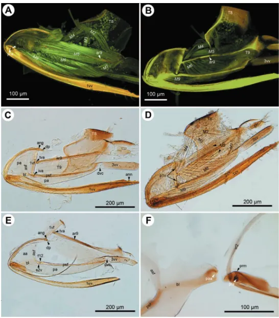
Figure 1. CLSM micrographs and bright ield image showing the female terminalia of Megaspilus armatus
margin of the irst valvifer in Conostigmus and Lagynodes (ch14:0; tva: Figs 2C, 4A), it
is on the ventral half of the margin (ch14:1; tva: Figs 3B, C, E, 5A, B, E, 6D) in
Cer-aphron, Aphanogmus sp. 2 and Trassedia, where it is located ventrally on the posterior margin of the dorsal sclerite of the irst valvifer (ch15:0; tva: Fig. 3B, C, E), it is in the upper half of the margin in Megaspilus and Dendrocerus (ch14:2; tva: Figs 1B, 4F) and adjacent to the anterior angle in Aphanogmus sp. 1 (ch14:3; tva: Figs 6A, C).
he anterior area of the second valvifer is expanded dorsally into a broad, lat, laterally directed surface (ch16:0; aa: Figs 1A, 2E). he basal line of the second valvi-fer (ch17:0) is a sharply deined ridge in Ceraphron, Trassedia, and Aphanogmus sp. 2 (ch18:0; bl: Figs 3B–D, 5A, E, 6D) but a thickening with difuse margins in Aphanog-mus sp.1 and Megaspilidae (ch18:1; bl: Figs 1D, 2C, E, 4A, F). he dorsal projection
of the second valvifer (ch19:0) is longer than the length of the anterior area of the second valvifer in Ceraphron and Trassedia, and Aphanogmus sp. 2 (ch20:1; dp: Figs 3C, E ) whereas the projection is shorter than the length of the anterior area in other taxa examined (ch20:2. dp: Figs 2E, 4A). he anterior section of the dorsal lange of the second valvifer is sharply deined ridge in Ceraphron, Aphanogmus sp. 2, and Tras-sedia (ch21:0; asf: Figs 3B, C, D, 5A–E, 6D) but a thickening with difuse margins in
Megaspilidae and Aphanogmus sp. 1 (ch21:1; asf: Figs 2C, E, F, 4A, F). he posterior section of the dorsal lange of the second valvifer is sharply deined (ch22:0; psf: Figs 2C, E, 4A, D, 5A, E). he area of the second valvifer posterior to the intervalviofer articulation is elongate (ch23:0 pa: Figs 2C, E). he ventral margin of the second
valvifer curves mediodorsally (ch24:0) encircling the posterior second valvifer-second valvula muscle (ch25:0. 2vf, M9: Fig. 1C). he genital membrane of the second valv-ifers accommodates the terebra when it is retracted (ch26:0; gm: Fig. 1C). he venom gland reservoir is surrounded by the second valvifer is present in Ceraphronidae and Lagynodes (ch27:0; res: Figs 3A–E). he reservoir was not observed in other
Megaspili-dae (ch27:1). he content of the reservoir is yellowish, transparent and hard, resin-like (ch28:0; res: Fig. 5F) in critical point dried specimens.
he irst valvula tapers distally in lateral view in Ceraphron, Aphanogmus sp. 2 and Trassedia (ch29:0; 1vv: Figs 3B, 5A, F, 6D), whereas it is spatulate in Megaspilidae and Aphanogmus sp. 1 (ch29:1; 1vv: Figs 1A, D, 2A, C, D, E, 4A, B, 6B, C). he banding
pattern and annuli are missing from the irst valvula (ch30:0; ch31:0).
he second valvulae are expanded proximally into the bulb (ch37:0; bulb: Figs 1A, D) and fused distal to the bulb (ch35:0). he dorsal valve tapers distally in dorsal view (ch36:0) and the anterior margin of the bulb is curved dorsally. he processus articula-ris is located laterally (ch32:0; pra: Figs 2F, 5D), the anterior notch of the dorsal valve
anteriorly (ch33:0) and the processus muscularis anterodorsally on the bulb (ch34:0;
Morphology and function of the ovipositor mechanism in Ceraphronoidea... 33
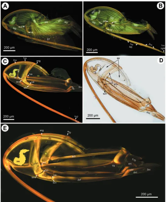
Figure 2. CLSM micrographs and bright ield image showing the female terminalia of Conostigmus ab-dominalis (Boheman). A CLSM, medial view (doi: 10.6084/m9.igshare.156448) B CLSM, lateral view (doi: 10.6084/m9.igshare.156433) C bright ield, medial view, muscles removed D bright ield, medial view e bright ield, medial view, irst valvifer and T9 separated from second valvifer, muscles removed; F, bright ield, second valvifer removed from second valvulae, muscles removed. All anterior to the left.
number of annuli is four (ch41:0) in Conostigmus and Lagynodes and six (ch41:1) in Megaspilus.
(ch45:0; 3vv: Figs 1B, 2B–E, 3C–E, 4D, F, 5E, F, 6A, B, F), its lateral wall is convex (ch46:0) and sclerotized (ch47:0) whereas its medial wall is concave (ch48:0; 3vv: Fig.
3E) and membranous (ch49: 0; 3vv: Fig. 3F).
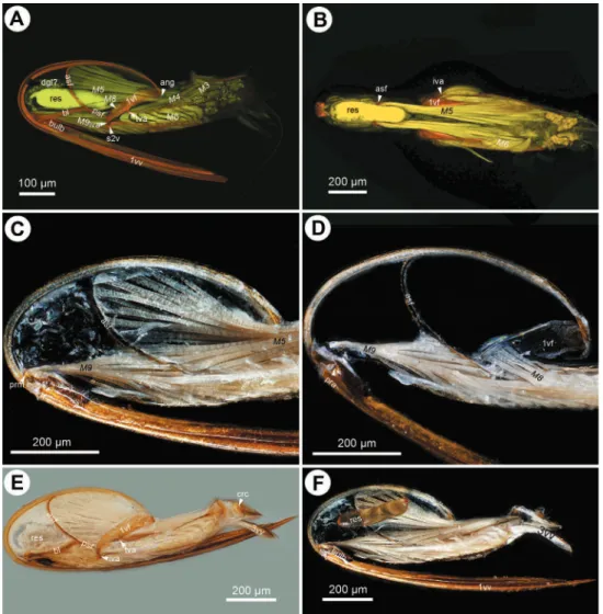
Figure 3. CLSM micrographs and bright ield image showing the female terminalia of Trassedia luapi
Morphology and function of the ovipositor mechanism in Ceraphronoidea... 35
T9 is quadrangular in lateral view (ch50:0; T9: Figs 2B, C) and wider than long dorsomedially (ch51:0; T9: Figs 3D, E). he cordate apodeme and the anterior lange are continuous, composing the anterior ridge of T9 (ch52:0; ar9: Figs 2A–C, 3E, 4B), which is adjacent anteriorly with and separated posteriorly from the anterior margin of T9 (ch53:0.); the distance between the anterior margin of T9 and the ridge increases gradually posteriorly (ch54:0).
Muscles (numbered according to Vilhelmsen 2000)
S7-irst valvula muscle (M1: Figs 1B, D, 3A) arises anteriorly from S7 (ch55:0) and
inserts on the irst valvula at the dorsal margin of the ovipositor adjacent to the bor-der of the irst valvula and the dorsal end of the irst valvifer (ch56:0). he muscle is oriented posterodorsally (ch57:0). he dorsal T8-T9 muscle (M2: Figs 2A, 3A, 4A, B) arises from along the anterior margin of T8 (ch58:0) and inserts dorsally on the anterior margin of T9 (ch59:0). he lateral T8-T9 muscle (M3: Figs 1B, 2A, B, D,
Morphology and function of the ovipositor mechanism in Ceraphronoidea... 37
3A, 4A, 5A) arises from T8 posterodorsally of the dorsal T8-T9 muscle (ch60:0), and inserts on the anterior ridge of T9 (ch61:0). he T8-irst valvifer muscle (M4: Figs 1B, 2A, 2B, 3A, 5A) arises from T8 dorsally of the site of attachment of the lateral T8-T9 muscle (ch62:0) and inserts on the irst valvifer adjacent to the ninth tergal condyle of the irst valvifer (ch63:0). he muscle inserts on the ventral sclerite of the irst valvifer adjacent to the intravalvifer articulation (ch64:0; M4: Fig. 3A)
D, 3B, 4A, B, D, E, F, 5A–C, 6A) arises dorsally and ventrally from the anterior ridge of T9 with sites of origins extending ventrally and dorsally on the wall of the tergite (ch65:0) and inserts along the posterior margin of the anterior area of the second valvifer (ch66:0). he ventral T9-second valvifer muscle (M6: Figs 1A, B, D, E, 2A, B, D, 3B, 4A, B, D, F, 5A, B, 6A) is not subdivided (ch67:0) and arises from the medial side of the posterior area of the second valvifer (ch68:0). he site of insertion of the muscle extends along the anterior part of the anterior ridge of T9 in most taxa examined (ch69:0) except Megaspilus, and Dendrocerus where the muscle seemingly inserts partly on the interarticular ridge of the irst valvifer (ch70:0; M6: Figs 1B, D, 4F). he posterior T9-second valvifer muscle (M7: Figs 1A, 2A, D, 4E) arises from the strap-like dorsal area of T9 (ch71:0) and inserts on the median bridge (ch72:0). he fan shaped irst valvifer-genital membrane muscle (ch73:0; M8: Figs 1A, D, E, F, 3B, 5A, D) arises from the medial surface of the irst valvifer near the intervalvifer articulation (ch74:0.). he site of insertion of the muscle extends along the genital membrane (ch75:0). In Trassedia the muscle arises from the medial surface of the dorsal sclerite of the irst valvifer (ch76:1; M8:
Fig. 3B). he posterior second valvifer-second valvula muscle (M9: Figs 1A–F, 2A,
B, D, 3A, B, 4B, D–F, 5A, C, D) arises from the medial side of the posterior area of the second valvifer (ch77:0), and inserts on the processus musculares (ch78:0). In Ceraphron and Trassedia some bands of the muscle arise from the anterior area of the second valvifer (ch.77:1; M9: Figs 3A, B, 5A, C, D). T9-genital membrane
muscle (ch79:0), lateral T9-second valvifer muscle (ch80:0), second valvifer-genital membrane muscle (ch81:0) and the anterior second valvifer-second valvula muscles (ch82:0) are absent from Ceraphronoidea.
Results ii: semantically enhanced characters and character states for ceraphronoid female terminalia.
Integument
First valvifer: shape
(0) elongated in lateral view Anterior margin of irst valvifer: shape
(0) convex in lateral view
Posterior margin of irst valvifer: shape (0) straight in lateral view
(1) concave in lateral view
(2) bent at tergo-valvifer articulation Transvalvifer conjunctiva: count
Morphology and function of the ovipositor mechanism in Ceraphronoidea... 39
Dorsal sclerite of the irst valvifer: shape (0) elongated in lateral view
Ventral sclerite of the irst valvifer: shape (0) triangular in lateral view
Intravalvifer articulation: count (0) present
(1) absent
Intravalvifer articulation: position (0) anterior region of irst valvifer
Ventral margin of dorsal sclerite of the irst valvifer: thickness (0) thickened
Anterodorsal margin of ventral sclerite of the irst valvifer: thickness (0) thickened
Anterior lange of the irst valvifer: count (0) present
(1) absent
Second valviferal condyle of the irst valvifer: position (0) at the posteroventral corner of the irst valvifer Ninth tergal condyle of the irst valvifer: position
(0) posterior margin of the irst valvifer
Distance between tergo-valvifer articulation and intravalvifer articulation (D1): pro-portion to the distance between tergo-valvifer articulation and anterior angle of irst valvifer (D2).
(0) D1=D2 (1) D1<D2 (2) D1>D2 (3) D2=0
Ninth tergal condyle of the irst valvifer: position (0) on dorsal sclerite of the irst valvifer
Anterior area of the second valvifer: height vs. height of posterior area of second valvifer
(0) 3≤:1
Basal line of the second valvifer: count (0) present
Basal line of the second valvifer: sharpness (0) sharp
(1) blunt
Dorsal projection of the second valvifer: count (0) present
Length of dorsal projection of second valvifer in lateral view (L1) vs. length of ante-rior area of second valvifer in lateral view (L2)
(0) sharp (1) blunt
Posterior section of dorsal lange of second valvifer: sharpness (0) sharp
Posterior area of second valvifer: shape (0) elongated
Ventral margin of the second valvifer: curvature
(0) curved medially and curved dorsally in lateral view Ventral margin of the second valvifer: position
(0) surrounds posterior second valvifer-second valvula muscle Genital membrane: position
(0) surrounds terebra
Venom gland reservoir of the second valvifer: count (0) present
(1) absent
Venom gland reservoir of the second valvifer: physical quality (0) resinous
Distal region of irst valvula: shape (0) tapered
(1) spatulate
Banding pattern on irst valvula: count (0) absent
Annuli on irst valvula: count (0) absent
Processus articularis: position
(0) anterolateral region of the bulb Anterior notch of the dorsal valve: position
(0) anterior region of the bulb Processus musculares: position
(0) anterodorsal region of the bulb Distal notch of the dorsal valve: count
(0) absent
Distal region of the dorsal valve: shape (0) tapered in dorsal view
Anterior region of dorsal margin of the bulb: curvature (0) curved dorsally in lateral view
Bulb: height in lateral view vs. height of anterior area of the second valvifer in lateral view
(0) =0.5 (1) <0.5
Morphology and function of the ovipositor mechanism in Ceraphronoidea... 41
Annuli on dorsal valve: count (0) present
(1) absent
Annuli on dorsal valve: count value (0) four
(1) six
Median bridge: count (0) present
Distal vertical conjunctiva of the second valvifer-third valvifer complex: count (0) present
Distal vertical conjunctiva of the second valvifer-third valvifer complex: overlap rela-tionship with dorsal margin of second valvifer-third valvula complex
(0) does not overlap
Distal region of the third valvula: shape (0) tapered in lateral view
Lateral region of the third valvula: shape (0) convex in posterior view
Lateral region of the third valvula: entity (0) is_a sclerite
Medial region of the third valvula: shape (0) concave in posterior view
Median region of the third valvula: entity (0) is_a conjunctiva
T9: shape
(0) quadrangular in lateral view
Dorso-median region of T9: width in dorsal view vs. length in dorsal view (0) width > length
Anterior ridge of T9: count (0) present
Anterolateral region of anterior ridge of T9: position (0) adjacent to anterior margin of T9
Distance between Anterior ridge of T9 and anterior margin of T9: anterior-posterior gradient
(0) increasing
Muscles
Attachment site of S7-irst valvula muscle (M1) on S7: position (0) adjacent to the anterior margin of S7
(0) anterior to attachment site of S7-irst valvula muscle on S7 Attachment site of dorsal T8-T9 muscle on T8: position
(0) adjacent to the anterior margin of T8
Attachment site of dorsal T8-T9 muscle on T9: position
(0) adjacent to anterior margin of the dorsomedial region of T9 Attachment site of lateral T8-T9 muscle on T8: position
(0) posterodorsal to the attachment site of dorsal T8-T9 muscle on T8 Attachment site of lateral T8-T9 muscle on T9: position
(0) adjacent to anterior ridge of T9
Attachment site of T8-irst valvifer muscle on T8: position
(0) dorsal to the attachment site of lateral T8-T9 muscle on T8 Attachment site of T8-irst valvifer muscle on irst valvifer: position
(0) adjacent to the ninth tergal condyle of the irst valvifer Attachment site of T8-irst valvifer muscle on irst valvifer: position
(0) on ventral sclerite of the irst valvifer adjacent to the intravalvifer articulation Attachment site of dorsal T9-second valvifer muscle on T9: position
(0) adjacent to the anterior ridge of T9 and the region of T9 dorsal to and ventral to the anterior ridge of T9
Attachment site of dorsal T9-second valvifer muscle on second valvifer: position (0) adjacent to the anterior section of the dorsal lange of the second valvifer Ventral T9-second valvifer muscle: count value
(0) 1
Attachment site of ventral T9-second valvifer muscle on second valvifer: position (0) posterior area of the second valvifer
Attachment site of ventral T9-second valvifer muscle on T9: position (0) adjacent to anterior region of anterior ridge of T9
First valvifer-second valvifer muscle: count (0) present
(1) absent
Attachment site of posterior T9-second valvifer muscle on T9: position (0) on the dorsal region of T9
Attachment site of posterior T9-second valvifer muscle on second valvifer: position (0) on the median bridge
First valvifer-genital membrane muscle: shape (0) fan shaped in dorsal view
Attachment site of irst valvifer-genital membrane muscle on irst valvifer: position (0) on the medial side of the irst valvifer adjacent to the intervalvifer articulation Attachment site of irst valvifer-genital membrane muscle on genital membrane:
posi-tion
(0) along the median line of the genital membrane
Morphology and function of the ovipositor mechanism in Ceraphronoidea... 43
Attachment site of posterior second valvifer-second valvula muscle on second valvifer: position
(0) on the medial side of the posterior area of the second valvifer
(1) on the medial side of the posterior area of the second valvifer and the anterior area of the second valvifer
Attachment site of posterior second valvifer-second valvula muscle on second valvula: position
(0) on the processus musculares T9-genital membrane muscle: count
(0) absent
Lateral T9-second valvifer muscle: count (0) absent
second valvifer-genital membrane muscle: count (0) absent
anterior second valvifer-second valvula muscles: count (0) absent
Discussion
he enormous diversity of ovipositor phenotype in Hymenoptera relects the manner in which the female wasp inds feasible environments for her developing larvae. Host structure and location, as well as the diferent ways of storing the ovipositor, are argu-ably the principal factors driving the structural adaptation of these structures (Quicke et al. 1999, Vilhelmsen 2000, Vilhelmsen and Turrisi 2011). Major morphological characteristics of ceraphronoid ovipositors are most likely related to these inluences.
Storage of terebra
he ceraphronoid ovipositor is stored in a horizontal position inside the metasoma, with its ventral part concealed by S7 (Fig. 6E). As the irst oviposition movement, the contracting muscles between the apical metasomal tergites and sternites expose the ventral part of the ovipositor by rotating it and the ninth abdominal tergite posteriorly, from the resting, horizontal to the active, vertical position. his movement is common within Apocrita (Fig. 6F; Alam 1953, Copland 1976, Fergusson 1988).
longitudinal axis during oviposition: https://scholarsphere.psu.edu/iles/r494vk42v At the end of the oviposition the terebra is retracted prior to the anterior rotation of the ovipositor/T9 complex into the resting, horizontal position. he retraction of the terebra, unlike its extension, seems to be unique for Ceraphronoidea. In other Hymenoptera the movement is accomplished by the contraction of the vertically ori-ented anterior second valvifer-second valvula muscle that arises from the anterodorsal margin of the second valvifer and inserts on the distal region of the bulb (King and Copland 1969, 1976, Vilhelmsen 2000, Fergusson 1988, Alam 1953). However, this muscle is absent from Ceraphronoidea, and thus we hypothesize a diferent mecha-nism for retracting the terebra. We observed a relatively large muscle arising from S7 and inserting dorsally on the irst valvula in Ceraphronoidea (M1: Figs 1B, D, 3A). he position of the site of attachments of the muscle suggest that the S7-irst valvula muscle (Figs 1B, 3A) aids in the retraction of the terebra. he presence of muscles arising from S7 and inserting on the irst valvula has been reported only in some basal Hymenoptera (Dhillon 1966, Vilhelmsen 2000) and in one braconid species, Stenobracon deesae (Alam 1953). he S7-irst valvulae muscle has an entirely diferent coniguration in basal Hymenoptera. It arises from along the anterior margin of S7 and inserts on the proximal end of the ventral ramus of the irst valvula and presumably contributes to the movement of the irst valvula aiding the ventral T9-second valvifer muscle (Vilhelmsen 2000). In Stenobracon, however, the S7-irst valvulae muscle has a very similar structure to that we reported in Ceraphronoidea (Alam 1953) suggesting the possible involvement of this muscle into the retraction of the terebra. Alam (1953) hypothesized that the muscle is involved in the movement of the median conjunctivae of the irst valvulae (dorsal wall of the egg canal) inluencing the egg movement along the egg canal. Although the anterior second valvifer-second valvulae muscle is present in all non-ceraphronoid Hymenoptera, it is possible that the S7-irst valvulae muscle is involved in the retraction of the terebra in other Apocrita, and that this function is not an evolutionary novelty for Ceraphronoidea.
Egg laying mechanism
he paired irst and second valvifers and T9, operated together by the dorsal and ven-tral T9-second valvifer muscles, form the ovipositor machinery that is responsible for “drilling” the terebra into a substrate and moving the egg along the egg canal (Vilhelm-sen 2000).
Morphology and function of the ovipositor mechanism in Ceraphronoidea... 45
movement is what slides the irst valvula along the second valvulae, the distance the irst valvifer moves determines the distance the irst valvula moves. he left and right irst valvulae slide back and forth alternately during the alternate contraction of the left and right T9-second valvifer muscle pairs (1vf left, 1vv right: Fig. 3B; Vilhelmsen 2000). his alternate movement is what is underlying the advance of the egg inside the egg canal and the drilling of the terebra into a substrate. he former function is facili-tated by the presence of internal, posteriorly oriented cuticular modiications (Austin and Browning 1981) while the latter could be aided by anchoring structures on the irst valvulae (Vilhelmsen 2000).
Adaptations efecting the alternate movements and coniguration of the irst val-vulae might be mostly afected by the hardness of the substrate and constraints for fast oviposition. Oviposition into a concealed, and therefore relatively immobile host re-quires a robust system that has to be strong enough to drill or break the barrier. On the other hand, parasitization of an exposed, mobile but relatively soft host requires fast and perhaps less robust mechanism. Two major egg laying habits have been recorded within Ceraphronoidea: oviposition inside a mobile host and oviposition trough a hard but relatively thin barrier enclosing the host, which has restricted movement (Dessart 1995b, c). Ceraphron and numerous Aphanogmus species were reported to parasitize free living Diptera larvae (Laborius 1972) whereas most Dendrocerus species and some Aphanogmus parasitize hosts hidden by the hardened integument of the primary host (Fergusson 1980), the wall of the cocoon (Peter and David 1990, Alam 1985) or galls (Bakke 1955) developed around the host.
he relative distance between the anterior angle of the irst valvifer and the inter-valvifer articulation (ang, iva: Figs 1B, 2C, E, 3C, E, 4A, E, F, 5A, 6A, C, D) is most probably positively correlated with the degree of the sliding motion of the irst valvula (Prentice 1998). he posterior margin of the irst valvifer angled at the tergo-valvifer articulation (tva: Fig.) in most Hymenoptera (Oeser 1961, Vilhelmsen 2000). It is
of T9 and the cordate apodeme. he anterior lange extends along the anterior margin of the tergite and might be homologous with the antecosta of the ninth abdominal tergum of other insects because it receives the site of attachment of the dorsal T8-T9 muscle in Macroxyela (Vilhelmsen 2000). he dorsal T9-second valvifer muscle arises at least partly from the lange in the rest of Hymenoptera. he cordate apodeme is close to the tergo-valvifer articulation and receive the site of attachment of the ventral T9-second valvifer muscle. It is apophysis-like and extends internally in most basal hymenopterans but ridge-like and extended posteriorly in Siricoidea, Orussidae, and numerous Apocrita (“diagonal ridge” sensu Fergusson 1988). he apodeme is usually well separated from the anterior lange of T9 in Apocrita, except in Bruchophagus, where they are seemingly fused anteriorly (Copland and King 1971). Only one ridge, the anterior ridge of T9 extends along the anterior margin of T9 and receives the site of attachment of both the ventral and the dorsal T9-second valvifer muscles in Ceraphro-noidea. his condition is unique in Hymenoptera. As described above, the dorsal and ventral T9-second valvifer muscles move the irst valvifer indirectly. Host relationships of Megaspilus remain unknown, but it is possible that some bands of the ventral muscle insert on the interarticular ridge of the irst valvula and thus the contraction of it might cause the direct movement of the sclerite.
he irst valvifer is composed of two articulating sclerites in Trassedia. Although this condition is possibly plesiomorphic for Insecta (Klass et al. 2013) it is present only in a few taxa i.e. Archaeognatha, most ovipositor bearing Odonata and some Der-maptera (Klass personal communication). When the ventral T9-second valvifer muscle contracts the two sclerites of the irst valvifer pivot anteriorly together as one unit. he two sclerites articulate with one another anteriorly, however, allowing the dorsal scler-ite to pivot posteriorly on the ventral sclerscler-ite at the intravalvifer articulation when the dorsal T9-second valvifer muscle is contracted (Figs 3A–E). he irst valvulae are thus enabled to slide a very long distance along the second valvulae in this unique system, probably allowing the egg to move quickly down the length of the ovipositor.
he presence or absence of annuli at the tip of the ovipositor may depend on the hardness of the substrate into which the wasp is ovipositing, as well as the circumstanc-es under which oviposition is taking place (Quicke et al. 1999, Le Ralec et al. 1996, Gerling et al. 1998). Megaspilidae, similar to numerous other non-ichneumonoid apo-critans do have annuli apically only on the second valvulae whereas Ceraphronidae lack them from both valvulae. In general it seems that the harder the substrate is, the more developed the ovipositor sculpture is (Le Ralec et al. 1996).
Cer-Morphology and function of the ovipositor mechanism in Ceraphronoidea... 47
aphron specimens (res: Fig. 5F) implying its possible cement nature that might be used for coating the eggs or fastening them on a surface. Ceraphron and some Aphanogmus species are reported to be endoparasitoids (Cordero and Cave 1992) in which lifestyle the egg coating can be crucial for avoiding the host immune response. We did not observe any glands or resin containing reservoirs in Megaspilinae. Höller et al. (1993) identiied the Dufour’s gland in Dendrocerus carpenteri outside of the second valvifers and reported the absence of the venom gland in this taxon. Nevertheless, more ac-curate, TEM based examination of the accessory gland system of Ceraphronoidea is needed for clarifying the function of the gland and gland reservoir located inside the second valvifer.
In general, the ovipositors of Ceraphron, Trassedia and Aphanogmus sp. 2 are less robust and capable of a very large degree of motion, corresponding to the available data about ovipositing in exposed and active hosts. Trassedia represents, perhaps, a more extreme version of the “quickly into soft substrate” oviposition type. Megaspilidae, on the other hand, have a stronger, more robust ovipositor systems, which aford the smaller degree of motion required for handling a harder substrate concealing a static host. Dendrocerus, for example, exhibits extended sites of origins for muscles and a very small degree of motion for the irst valvulae. Aphanogmus sp. 1, although it belongs to Ceraphronidae, shares numerous characteristics with Dendrocerus and therefore may represent the Aphanogmus-group that parasitizes hosts obscured by harder barrier, e.g., the wall of a plant gall, and thus is a case of parallelism driven by the same environ-mental constraints.
So far it is widely accepted that Ceraphronoidea is composed of two extant fami-lies, Ceraphronidae and Megaspilidae, plus two fossil families not treated here. he limits between the two families, however, have been challenged recently (Mikó and Deans 2009, Mikó et al. in press). Although the presence of the annuli in Megaspilidae and absence from Ceraphronidae supports the traditional classiication, the location of a resin producing gland inside the second valvifers is shared by the megaspilid subfam-ily Lagynodinae and Ceraphronidae.
Acknowledgments
S. National Science Foundation (grants DBI-0850223, DEB-0842289) and beneited from discussions initiated through the Phenotype Research Coordination Network (NSF DEB-0956049).
References
Alam SM (1953) he skeleto-muscular mechanism of Stenobracon deesae Cameron (Braconidae, Hymenoptera) — An ectoparasite of sugarcane and juar borers of India. Part II. Abdomen and internal anatomy. Aligarh Muslim University Publications (Zoology Series) 3: 1–75. Alam MM (1985) Biologies of Apanteles lavipes (Cam.) an introduced larval parasite of
Diat-raea saccharalis (F.) and its indigenous hyperparasite, Aphanogmus ijiensis (Ferr.) in Barba-dos, West Indies. Proceedings of the 1985 meeting of West Indies sugar technologists held in Trinidad & Tobago, 367–384.
Araj SA, Wratten SD, Lister AJ, Buckley HL (2006) Floral nectar afects longevity of the aphid parasitoid Aphidiuservi and its hyperparasitoid Dendrocerusaphidum. New Zealand Plant Protection 59: 178–183.
Austin AD (1984) New species of Platygastridae (Hymenoptera) from India which parasitise pests of mango, particularly Procontarinia spp. (Diptera: Cecidomyiidae). Bulletin of En-tomological Research 74: 549–557.
Austin AD, Browning TO (1981) A mechanism for movement of eggs along insect ovipositors. International Journal of Insect Morphology and Embryology 10: 93–108.
Austin AD, Field SA (1997) he ovipositor system of scelionid and platygastrid wasps (Hyme-noptera: Platygastroidea): comparative morphology and phylogenetic implications. Inver-tebrate Taxonomy 11: 1–87.
Balhof JP, Mikó I, Yoder MJ, Mullins PL, Deans AR (in press) A semantic species description model, instantiated with real data: a revision of the ensign wasps (Evaniidae) of New Cal-edonia. Systematic Biology.
Chiu SC, Chou LJ, Chou KC (1981) A preliminary survey on the natural enemies of Kerria lacca (Kerr) in Taiwan. Journal of Agricultural Research of China 30: 420–425.
Chow A, Mackauer M (1996) Sequential allocation of ofspring sexes in the hyperparasitoid wasp, Dendroceruscarpenteri. Animal Behaviour 51(4): 859–870.
Chow A, Mackauer M (1999) Marking the package or its contents: Host discrimination and acceptance in the ectoparasitoid Dendroceruscarpenteri (Hymenoptera: Megaspilidae). Ca-nadian Entomologist 131(4): 495–505.
Copland MJW, King PE (1971) he structure and possible function of the reproductive system in some Eulophidae and Tetracampidae. he Entomologist 104: 4–28.
Copland MJW, King PE (1972) he structure of the female reproductive system in the Ptero-malidae (Hymenoptera: Chalcidoidea). he Entomologist 105: 77–96.
Morphology and function of the ovipositor mechanism in Ceraphronoidea... 49
Cooper KW, Dessart P (1975) Adult, larva and biology of Conostigmus quadratogenalis Dessart and Cooper, sp.n., (Hym. Ceraphronoidea), parasite of Boreus (Mecoptera) in California. Bulletin et Annales de la Societe Royale Belge d’Entomologie 111: 37–53.
Cordero J, Cave RD (1992) Natural enemies of Plutella xylostella (Lep.: Plutellidae) on crucifers in Honduras. Entomophaga 37: 397–407.
Deans AR, Yoder MJ, Balhof JP (2012a) Time to change how we describe biodiversity. Trends in Ecology and Evolution 27: 78–84. doi: 10.1016/j.tree.2011.11.007
Deans AR, Mikó I, Wipler B, Friedrich F (2012b) Evolutionary phenomics and the emerg-ing enlightenment of arthropod systematics. Invertebrate Systematics 26: 323–330. doi: 10.1071/IS12063
Dessart P (1967) Description de Dendrocerus (Macrostigma) noumeae sp. nov. de Nouvelle Cal-edonie (Ceraphronoidea Megaspilidae). Entomophaga 12: 343–349.
Dessart P (1972) Révision des espèces européenes du genre Dendrocerus Ratzeburg, 1852 (Hy-menoptera Ceraphronoidea). Mémoires de la Société royale belge d’Entomologie 32. Dessart P (1977) Contribution a l’étude des Lagynodinae (Hym. Ceraphronoidea
Megaspili-dae). Bulletin et Annales de la Societe Royale Belge d’Entomologie 113: 277–319. Dessart P (1985) Les Dendrocerus a notaulices incompletes (Hymenoptera Ceraphronoidea
Megaspilidae). Bulletin et Annales de la Societe Royale Belge d’Entomologie 121: 409– 458.
Dessart P (1987) Revision des Lagynodinae (Hymenoptera Ceraphronoidea Megaspilidae). Bulletin de l’Institute Royal des Sciences Naturelles de Belgique 57: 5–30.
Dessart P (1992) Revision d’Aphanogmus fulmeki Szelenyi, 1940 (Hymenoptera, Ceraphro-noidea, Ceraphronidae) avec remarques biologiques. Bulletin de l’Institute Royal des Sci-ences Naturelles de Belgique 62: 83–91.
Dessart P (1995a) À propos du genre Dendrocerus Ratzeburg 1852 Les espèces du groupe Pen-maricus (Hymenoptera Ceraphronoidea Megaspilidae). Bulletin et Annales de la Societe Royale Belge d’Entomologie 131: 349–382.
Dessart P (1995b) Ceraphronidae. In: Hanson PE, Gauld ID (Eds) he Hymenoptera of Costa Rica, Oxford University Press, Oxford, 199–203.
Dessart P (1995c) Megaspilidae. In: Hanson, PE and Gauld ID (Eds) he Hymenoptera of Costa Rica, Oxford University Press, Oxford, 203–208.
Dessart P (1997) Les Megaspilinae ni européens, ni américains 1. Legenre Conostigmus Dah-lbom, 1858 (Hym. Ceraphronoidea Megaspilidae). Mémoires de la Société royale belge d’Entomologie 37.
Dessart P (1999) Revision des Dendrocerus du group “halidayi” (Hymenoptera Ceraphronoidea Megaspilidae). Belgian Journal of Entomology 1: 169–256.
Dessart P (2001) Les Megaspilinae ni européens, ni américains 2. Les Dendrocerus Ratzeburg, 1852, à males non labellicornés (Hymenoptera Ceraphronoidea Megaspilidae). Belgian Journal of Entomology 3: 3–124.
aphronidae) of economic importance reared from Cybocephalus nipponicus (Coleoptera: Cybocephalidae). Zootaxa 1018: 47–54.
Fergusson NDM (1980) A revision of the British species of Dendrocerus Ratzeburg (Hymenop-tera: Ceraphronoidea) with a review of their biology as aphid hyperparasites. Bulletin of the British Museum (Natural History), 41(4): 255–314.
Fergusson NDM (1988) A comparative study of the structures of phylogenetic importance of female genitalia of the Cynipoidea (Hymenoptera). Systematic Entomology 13: 13–30. Ghesquiere J (1960) Le genre Atritomellus Kiefer en Afrique du Nord (Hymenoptera
Proc-totrupoidea Ceraphronidae). Bulletin et Annales de la Societe Royale Belge d’Entomologie 96: 205–215.
Gkoutos GV (2013) Phenotypic quality. http://purl.obolibrary.org/obo/pato.owl. Last accessed 01/28/2013.
Hadley A (2010) CombineZP. Available from: http://www.hadleyweb.pwp.blueyonder.co.uk/ Haviland MD (1920) On the mionimics and development of Lygocerus testaceimanus
Kief-fer, and Lygocerus cameroni Kiefer (Proctotrypoidea-Ceraphronidae), parasites of Aphidius
(Braconidae). Quarterly Journal of Microscopical Science 65: 361–372.
Heraty J, Ronquist F, Carpenter JM, Hawks D, Schulmeister S, Dowling AP, Murray D, Mun-ro J, Wheeler WC, Schif N, Sharkey M (2011) Evolution of the hymenopteran megara-diation. Molecular Phylogenetics and Evolution 60(1): 73–88.
Höller C, Bargen H, Vinson SB and Braune HJ (1993) Sources of the marking pheromones used for host discrimination in the hyperparasitoid Dendrocerus carpenteri. Journal of In-sect Physiology 39: 649–656.
Hymenoptera Anatomy Consortium. he Hymenoptera Anatomy Portal. Accessed on Mon Mar 25 10:23:10 -0500 2013. http://portal.hymao.org
Ishii T (1937) On the natural enemies of Prontaspis yanonensis Kuw. Agriculture & Horticul-ture, Tokyo 12(1): 60–70.
King PE, Copland MJW (1969) he structure of the female reproductive system in the My-maridae (Chalcidoidea: Hymenoptera). Journal of Natural History 3(3): 349–365. Klass KD, Matushkina NA, Kaidel J (2013) he gonangulum: A reassessment of its
morpho-logy, homomorpho-logy, and phylogenetic significance. Arthropod Structure and Development 41: 373394.
Laborius (1972) Untersuchungen über die Parasitierung des Kohlschotenrüsslers ( Ceuthorr-chynchus assimilis Payk.) und der Kohlschotengallmücke (Dasyneura brassicae Winn.) in Schleswig-Holstein. Zeitschrift für Angewandte Entomologie 72: 14–31.
Le Ralec A, Rabasse JM, Wajnberg E (1996) Comparative morphology of the ovipositor of some parasitic Hymenoptera in relation to characteristics of their hosts. he Canadian Entomologist 128: 413–433.
Luhman JC, Holzenthal RW, Kjaerandsen JK (1999) New host record of a ceraphronoid (Hy-menoptera) in Trichoptera pupae. Journal of Hymenoptera Research 8(1): 126.
Morphology and function of the ovipositor mechanism in Ceraphronoidea... 51
Masner L (1956) First preliminary report on the occurrence of genera of the group Proc-totrupoidea (Hym.) in ČSR (irst part-family Scelionidae). Acta Faunistica Entomologica Musei Nationalis Pragae 1: 99–126.
Masner L, Dessart P (1967) La reclassiication des categories taxonomiques superieures des Ceraphronoidea (Hymenoptera). Bulletin de l’Institut Royal des Sciences Naturelles de Belgique 43: 1–33.
Mikó I, Deans AR (2009) Masner, a new genus of Ceraphronidae (Hymenoptera, Ceraphro-noidea) described using controlled vocabularies. Zookeys 20: 127–153.
Mikó I, Masner L, Johannes E, Yoder MJ, Deans AR (in press) Male terminalia of Ceraphro-noidea: diversity in an otherwise monotonous taxon. Insect Systematics & Evolution. Muesebeck CFW (1979) Superfamily Ceraphronoidea. In: Krombein KV, Hurd PD, Smith
DR, Burks BD (Eds) Catalog of Hymenoptera in America north of Mexico. Smithsonian Institution Press (Washington DC): 1187–1198.
Müller CB, Völkl W, Godfray HCJ (1997) Are behavioural changes in parasitised aphids a pro-tection against hyperparasitism? European Journal of Entomology 94(2): 221–234. Mullins PL, Kawada R, Balhof JP, Deans AR (2012) A revision of Evaniscus (Hymenoptera,
Evaniidae) using ontology-based semantic phenotype annotation. ZooKeys 223: 1–38. Mungall C (2013) Spatial ontology. http://purl.obolibrary.org/obo/bspo.owl [accessed 28
Ja-nuary 2013]
Oeser R (1961) Vergleichend-morphologische Untersuchungen über den Ovipositor der Hy-menopteren. Mitteilungen aus dem Zoologischen Museum in Berlin 37(1): 1–119. Priesner H (1936) Aphanogmus steinitzi spec. nov., ein Coniopterygiden-Parasit
(Hymenopte-ra-Proctotrupoidea). Bulletin de la Societe Entomologique d’Egypte 20: 248–251. Quicke DLJ, Fitton MG, Ingram S (1992) Phylogenetic implications of the structure and
distribution of ovipositor valvilli in the Hymenoptera (Insecta). Journal of Natural History 26(3): 587–608.
Quicke DLJ, Fitton MG, Tunstead JR, Ingram SN, Gaitens PV (1994) Ovipositor structure and relationships within the Hymenoptera, with special reference to the Ichneumonoidea. Journal of Natural History 28(3): 635–682.
Quicke DLJ, Fitton MG (1995) Ovipositor steering mechanisms in parasitic wasps of the families Gasteruptiidae and Aulacidae (Hymenoptera). Proceedings of the Royal Society of London B Biological Sciences 261: 99–103.
Quicke D, Le Ralec A, Vilhelmsen L (1999) Ovipositor structure and function in the parasitic Hymenoptera with an exploration of new hypotheses. Atti dell’Accademia Nazionale Itali-ana de Entomologia 47: 197–239
Schneider CA, Rasband WS, Eliceiri KW (2012) “NIH Image to ImageJ: 25 years of image analysis”. Nature Methods 9, 671–675.
Schwörer U, Völkl W, Hofmann KH (1999) Foraging for mates in the hyperparasitic wasp,
Dendroceruscarpenteri: Impact of unfavourable weather conditions and parasitoid age. Oe-cologia (Berlin) 119(1): 73–80.
John-C, Ronquist F, Schulmeister S, Sharkey MJ, Talamas E, Tucker E, Vilhelmsen L, Ward PS, Wharton RA, Deans AR (2012) A hymenopterists’ guide to the Hymenoptera Anatomy Ontology: utility, clariication, and future directions. Journal of Hymenoptera Research 27: 67–88.
Sharanowski BJ, Robbertse B, Walker J, Randal V, Yoder R, Spatafora J, Sharkey MJ (2010) Ex-pressed sequence tags reveal Proctotrupomorpha (minus Chalcidoidea) as sister to Aculeata (Hymenoptera: Insecta). Molecular Phylogenetics and Evolution 57: 101–112.
Sharkey MJ, Carpenter JM, Vilhelmsen L, Heraty J, Liljeblad J, Dowling APG, Schulmeister S, Murray D, Deans AR, Ronquist F, Krogmann L, Wheeler WC (2011) Phylogenetic relationships among superfamilies of Hymenoptera. Cladistics 27: 1–33.
Sinacori A, Mineo G, Lo Verde G (1992) Osservazioni su Aphanogmus steinitzi Priesner (Hym. Ceraphronidae) parassitoide di Conwentzia psociformis (Curtis) (Neur. Coniopterygidae). Phytophaga 4: 29–48.
Smith EL (1970) Evolutionary morphology of external insect genitalia. 2. Hymenoptera. An-nals of the Entomological Society of America 63(1): 1–27.
Vilhelmsen L (2000) he ovipositor apparatus of basal Hymenoptera (Insecta): phylogenetic implications and functional morphology. Zoologica Scripta 29(4): 319–345.
Vilhelmsen L, Isidoro N, Romani R, Basibuyuk HH, Quicke DLJ (2001) Host location and oviposition in a basal group of parasitic wasps: the subgenual organ, ovipositor apparatus and associated structures in the Orussidae (Hymenoptera, Insecta). Zoomorphology 121: 63–84.
Vilhelmsen L, Mikó I, Krogmann L (2010) Beyond the wasp-waist: structural diversity and phylogenetic signiicance of the mesosoma in apocritan wasps (Insecta: Hymenoptera). Zoological Journal of the Linnean Society 159: 22–194.
Vilhelmsen L, Turrisi GF (2011) Per arborem ad astra: Morphological adaptations to exploiting the woody habitat in the early evolution of Hymenoptera. Arthropod Structure & Develop-ment 40: 2–20.
Vincent JFV, King MJ (1996) he mechanism of drilling by wood wasp ovipositors. Biomimet-ics 3: 187–201.
Vogt L, Bartolomaeus T, Giribet G (2010) Linguistic problem of morphology: structure versus homology and the standardization of morphological data. Cladistics 26: 301–325
Vogt L, Nickel M, Jenner RA, Deans AR (2013) he need for data standards in zoomorphology: preparing comparative morphology for eScience. Journal of Morphology. doi: 10.1002/ jmor.20138
Morphology and function of the ovipositor mechanism in Ceraphronoidea... 53
Anatomical terms used, cross-referenced to an ontological (formal) deinition (Hymenoptera Anatomy Ontology; URI = Uniform Resource Identiier).
Abbrevi ation
Label Concept URI
absent A quality denoting the lack of an entity. http://purl.obolibrary.org/ obo/PATO_0000462 adjacent to A spatial quality inhering in a bearer by
vir-tue of the bearer’s being located near in space in relation to another entity.
http://purl.obolibrary.org/ obo/PATO_0002259
ann annulus, annuli he carina that is transverse and extends across the lateral wall of the terebra.
http://purl.obolibrary.org/ obo/HAO_0001173 ang anterior angle of
the irst valvifer
he corner on the irst valvifer that marks the posterior end of the irst valvula.
http://purl.obolibrary.org/ obo/HAO_0002168 aa anterior area
of the second valvifer
he area of the second valvifer which is ante-rior to the anatomical line that is the shortest distance from the irst valviferal fossa of the second valvifer and the ventral margin of the second valvifer
http://purl.obolibrary.org/ obo/HAO_0002169
anterior lange of T9
he lange that extends along the anterolat-eral margin of female T9.
http://purl.obolibrary.org/ obo/HAO_0001171 af1 anterior lange
of the irst val-vifer
he lange that extends anteriorly on the irst valvifer and overlaps with the posterior mar-gin of the anterior area of the second valvifer.
http://purl.obolibrary.org/ obo/HAO_0002166
anterior margin anatomical margin and (overlaps some ante-rior side)
http://purl.obolibrary.org/ obo/BSPO_0000671 an2 anterior notch
of the dorsal valve
he notch that is located anteriorly on the dorsal ramus of the second valvula that ac-commodates the ventral ramus of the second valvula and the irst valvula.
http://purl.obolibrary.org/ obo/HAO_0002178
anterior region, anteriorly
anatomical region and (overlaps some ante-rior side)
http://purl.obolibrary.org/ obo/BSPO_0000071 ar9 anterior ridge
of T9
he ridge that extends along the anterior margin of female T9 and receives the site of origin of the ventral and the dorsal T9-second valvifer muscles.
http://purl.obolibrary.org/ obo/HAO_0002182
anterior second valvifer-second valvula muscle
he ovipositor muscle that arises from the anterodorsal part of the second valvifer and inserts subapically on the processus articu-lares.
http://purl.obolibrary.org/ obo/HAO_0001166
asf anterior section of dorsal lange of the second valvifer
he area of the dorsal lange of the second valvifer that is anterior to the site of origin of the basal line.
http://purl.obolibrary.org/ obo/HAO_0002173
anterior to A spatial quality inhering in a bearer by virtue of the bearer’s being located toward the front of an organism relative to another entity.
http://purl.obolibrary.org/ obo/PATO_0001632
ation
anterodorsal margin
anatomical margin and (overlaps some ante-rior side) and (overlaps some dorsal side)
http://purl.obolibrary.org/ obo/BSPO_0000686 anterolateral
region
Anatomical region and (overlaps some ante-rior side) and (overlaps some lateral side)
http://purl.obolibrary.org/ obo/BSPO_0000029 apodeme he process that is internal. http://purl.obolibrary.org/
obo/HAO_0000142 attachement site he area of the integument where muscles
are attached to epidermal cells.
http://purl.obolibrary.org/ obo/HAO_0002184 banding pattern he anatomical cluster that is composed of
the strongly sclerotised areas corresponding with the annuli and less strongly sclerotised areas situated between them.
http://purl.obolibrary.org/ obo/HAO_0001176
basal articula-tion
he articulation that is part of the second valvifer-second valvula-third valvula complex and adjacent to the rhachis.
http://purl.obolibrary.org/ obo/HAO_0001177
bl basal line of the second valvifer
he line on the second valvifer that extends between the pars articularis and the dorsal lange of second valvifer.
http://purl.obolibrary.org/ obo/HAO_0002171
bent A shape quality inhering in a bearer by virtue of the bearer’s having one or more angle(s) in its length.
http://purl.obolibrary.org/ obo/PATO_0000617
blunt A shape quality inhering in a bearer by vir-tue of the bearer’s terminating gradually in a rounded end.
http://purl.obolibrary.org/ obo/PATO_0001950
bulb bulb he anterior area of the dorsal valve that is bulbous.
http://purl.obolibrary.org/ obo/HAO_0002177 concave A shape quality in a bearer by virtue of the
bearer’s curving inward.
http://purl.obolibrary.org/ obo/PATO_0001857 conjunctiva he area of the integument that is weakly
sclerotized, with thin exocuticle.
http://purl.obolibrary.org/ obo/HAO_0000221 convex A shape quality that obtains by virtue of the
bearer having inward facing edges; having a surface or boundary that curves or bulges outward, as the exterior of a sphere.
http://purl.obolibrary.org/ obo/PATO_0001355
cordate apodeme
he apodeme on the anterior margin of the abdominal tergum 9 that receives the ventral T9-second valvifer muscle.
http://purl.obolibrary.org/ obo/HAO_0001585
corner he projection that is located at the intersec-tion of two or more edges.
http://purl.obolibrary.org/ obo/HAO_0000223 count he number of entities of this type that are
part of the whole organism.
http://purl.obolibrary.org/ obo/PATO_0000070
count value http://purl.obolibrary.org/
obo/PATO_0000416 curvature A surface shape quality inhering in a bearer
by virtue of the bearer’s exhibiting a degree of bending.
Morphology and function of the ovipositor mechanism in Ceraphronoidea... 55
Abbrevi ation
Label Concept URI
curved dorsally A curvature quality inhering in a bearer by virtue of the bearer’s being curved towards the back or upper surface of an organism.
http://purl.obolibrary.org/ obo/PATO_0001468
curved medially A curvature quality inhering in a bearer by virtue of the bearer’s being curved towards the middle.
http://purl.obolibrary.org/ obo/PATO_0002164
distal notch of the dorsal valve
he notch that is distal on the dorsal valve. http://purl.obolibrary.org/ obo/HAO_0002179 distal region Anatomical region and (overlaps some distal
side)
http://purl.obolibrary.org/ obo/BSPO_0000078 dvc distal vertical
conjunctiva of the second valvifer-third valvifer complex
he conjunctiva that traverses the second valvifer-third valvula complex and is located distal to the median bridge of the second valvifer.
http://purl.obolibrary.org/ obo/HAO_0002180
distance A quality that is the extent of space between two entities.
http://purl.obolibrary.org/ obo/PATO_0000040 dorsal margin Anatomical margin and (overlaps some
dor-sal side).
http://purl.obolibrary.org/ obo/BSPO_0000679 dp dorsal
projec-tion of the second valvifer
he projection that is located on the second valvifer and corresponds to the proximal end of the rachis.
http://purl.obolibrary.org/ obo/HAO_0002172
dorsal region anatomical region and (overlaps some dorsal side)
http://purl.obolibrary.org/ obo/BSPO_0000079 d1vf dorsal sclerite of
the irst valvifer
he sclerite of the irst valvifer that is located dorsally of the transvalviferal conjunctiva.
http://purl.obolibrary.org/ obo/HAO_0002163 M2 dorsal T8-T9
muscle
he abdominal muscle that arises from the anteromedian margin of female T8 and inserts on the anteromedian margin of the female T9.
http://purl.obolibrary.org/ obo/HAO_0001571
M5 dorsal T9-second valvifer muscle
he ovipositor muscle that arises along the posterodorsal part of the anterior margin of female T9 and inserts on the anterior section of the dorsal langes of the second valvifer.
http://purl.obolibrary.org/ obo/HAO_0001569
dorsal to x dorsal_to y if x is further along the dorso-ventral axis than y, towards the back. A dorso-ventral axis is an axis that bisects an organism from back (e.g. spinal column) to front (e.g. belly).
http://purl.obolibrary.org/ obo/BSPO_0000098
dorsal valve he area that is articulated with the right and left second valvifers at the basal articula-tion and bears the rhachies.
http://purl.obolibrary.org/ obo/HAO_0001658
dorsal view http://purl.obolibrary.org/
obo/BSPO_0000063 dorsomedial
region
anatomical region and (overlaps some dorsal side) and (overlaps some medial side)
ation
ec egg canal he anatomical space that is between the left and right rhachises.
http://purl.obolibrary.org/ obo/HAO_0002191 elongated A quality inhering in a bearer by virtue of
the bearer’s length being notably higher than its width.
http://purl.obolibrary.org/ obo/PATO_0001154
fan-shaped A quality inhering in a bearer that is shaped in the form of a fan.
http://purl.obolibrary.org/ obo/PATO_0002219 1vf irst valvifer he area of the irst valvifer-irst valvula
complex that is proximal to the aulax, bears the ninth tergal condyle of the irst valvifer and the second valviferal condyle of the irst valvifer and is connected to the genital mem-brane by muscle.
http://purl.obolibrary.org/ obo/HAO_0000338
M8 irst valvifer-genital mem-brane muscle
he ovipositor muscle that arises from the posterior part of the irst valvifer and inserts anteriorly on the genital membrane anetrior to the T9-genital membrane muscle.
http://purl.obolibrary.org/ obo/HAO_0001746
irst valvifer-second valvifer muscle
he ovipositor muscle that arises from the irst valvifer and inserts on the second val-vifer.
http://purl.obolibrary.org/ obo/HAO_0002189
1vv irst valvula, irst valvulae
he area of the irst valvifer-irst valvula complex that is delimited distally by the proximal margin of the aulax.
http://purl.obolibrary.org/ obo/HAO_0000339
gm genital mem-brane
he conjunctiva that connects the ventral margins of the second valvifers.
http://purl.obolibrary.org/ obo/HAO_0001757 height A 1-D extent quality inhering in a bearer by
virtue of the bearer’s vertical dimension of extension.
http://purl.obolibrary.org/ obo/PATO_0000119
increasing quality and (increased_in_magnitude_rela-tive_to some normal)
http://purl.obolibrary.org/ obo/PATO_0002300 iar interarticular
ridge of the irst valvifer
he ridge that extends along the posterior margin of the irst valvifer between the inter-valvifer and tergointer-valvifer articulations.
http://purl.obolibrary.org/ obo/HAO_0001562
iva intervalvifer articulation
he articulation between the irst valvifer and second valvifer.
http://purl.obolibrary.org/ obo/HAO_0001558 iava intravalvifer
articulation
he articulation between the dorsal sclerite of the irst valvifer and the ventral sclerite of the irst valvifer.
http://purl.obolibrary.org/ obo/HAO_0002165
lateral region anatomical region and (overlaps some lateral side)
http://purl.obolibrary.org/ obo/BSPO_0000082 M3 lateral T8-T9
muscle
he ninth abdominal tergal muscle that arises from the anterolateral margin of female T8 and inserts on the anterolateral margin of female T9.
http://purl.obolibrary.org/ obo/HAO_0001776
lateral T9-second valvifer muscle
he muscle that arises from the posteroven-tral parts of the female T9 and inserts on the median bridge.