Received from Hospital de Clínicas da Faculdade de Medicina da Universidade do Triângulo Mineiro, Uberaba, MG, Brazil.
1. Physiotherapist, Universidade Federal do Triângulo Mineiro, Uberaba, MG
2. Cardiac Surgeon, Department of Surgery, Universidade Federal do Triângulo Mineiro, Uberaba, MG
3. PhD, Professor, Department of Biomechanics, Medicine and Locomotive Apparatus Re-habilitation, Faculdade de Medicina de Ribeirão Preto, Universidade de São Paulo, SP
Submitted on November 2, 2011. Approved on March 8, 2012.
Correspondence to: João Abrão, MD Av. Bandeirantes, 3900
14049900 – Ribeirão Preto, SP, Brazil. E-mail: joaoabrao@fmrp.usp.br scientific article
Influence of Pleural Drain Insertion in Lung Function of
Patients Undergoing Coronary Artery Bypass Grafting
Irinea Beatriz Carvalho Ozelami Vieira
1, Fabiano F. Vieira
2, João Abrão
3, Ada Clarice Gastaldi
3Summary: Ozelami Vieira IBC, Vieira FF, Abrão J, Gastaldi AC – Influence of Pleural Drain Insertion in Lung Function of Patients Undergoing Coronary Artery Bypass Grafting.
Background and objectives: Longitudinal, prospective, randomized, blinded Trial to assess the influence of pleural drain (non-toxic PVC) site of insertion on lung function and postoperative pain of patients undergoing coronary artery bypass grafting in the first three days post-surgery and immediately after chest tube removal.
Method: Thirty six patients scheduled for elective myocardial revascularization with cardiopulmonary bypass (CPB) were randomly allocated into two groups: SX group (subxiphoid) and IC group (intercostal drain). Spirometry, arterial blood gases, and pain tests were recorded.
Results: Thirty one patients were selected, 16 in SX group and 15 in IC group. Postoperative (PO) spirometric values were higher in SX than in IC group (p < 0.05), showing less influence of pleural drain location on breathing. PaO2 on the second PO increased significantly in SX group com-pared with IC group (p < 0.0188). The intensity of pain before and after spirometry was lower in SX group than in IC group (p < 0.005). Spirometric values were significantly increased in both groups after chest tube removal.
Conclusion: Drain with insertion in the subxiphoid region causes less change in lung function and discomfort, allowing better recovery of respira-tory parameters.
Keywords: Chest tubes; Coronary artery bypass grafting; Pneumothorax; Pain, Postoperative; Pain Measurement; Spirometry.
©2012 Elsevier Editora Ltda. All rights reserved.
INTRODUCTION
Change in lung function increases morbidity and mortality in coronary artery bypass grafting 1. Several factors contribute to
this, such as median sternotomy, cardiopulmonary bypass 2,3,
pleurectomy, and postoperative pain 4.
The use of left mammary artery, although a worldwide ac-cepted technique, involves pleurotomy 5 that together with
ster-notomy promote major changes in lung mechanics, which pre-disposes to decreased vital capacity and total lung capacity 6.
The fact that pleurotomy is always associated with pleu-ral drainage causes more discomfort and pain to the patient, which further worsens lung function 6-9. Guizilini et al. studied
the effect of pleural drain site of insertion on postoperative lung function in coronary artery bypass grafting and found that, regardless of drain positioning, pain and loss of lung
function occurred, although the effects were less noticeable
when the drain was placed in the subxiphoid region 10. Chest
tubes are made from various raw materials and all seem to work satisfactorily regarding blood drainage from the pleural
space and the pain of withdrawal procedure 11. Chest tube
itself can interfere with deep inspiration by its intimate con-tact with the visceral pleura. Misplacement of a thick rigid tube may result in serious complications, such as arrhythmia due to heart irritation; injury of the intercostal nerves, parietal pleura
or lung parenchyma 12; erosion of intrathoracic major vessels;
and cardiac tamponade 13.
OBJECTIVE
The aim of this study is to evaluate the influence of the in-sertion site of a PVC non-toxic chest tube on lung function by spirometry in the first days after surgery. As a control, the same parameters were evaluated without the chest tube on the third day. As a secondary objective, we assessed the dis-comfort caused by chest tube insertion using pain registration at rest and after spirometry.
METHOD
Univer-sidade Federal do Triangulo Mineiro (UFTM); such patients were admitted to the Intensive Care Unit from January 2010 to July 2011. After approval by the Ethics Research Commit-tee of UFTM and obtained informed consent, 36 patients were included in the study. During surgery, the service assistant held the draw of the envelope containing the chest tube site of placement, and the patients were then allocated into two groups (intercostal [IC] or the subxiphoid [SX]). Inclusion crite-ria were patients with coronary artery disease proven by coro-nary angiography, who underwent elective corocoro-nary artery bypass grafting using the left internal thoracic artery (mam-mary), left pleurotomy with cardiopulmonary bypass (anoxia time < 60 min), ejection fraction greater than 50%, and normal spirometry. Patients with previous lung disease, those who could not perform pulmonary function tests, remained intu-bated beyond the first postoperative day (PO-1), and required surgical intervention were excluded. Five patients were ex-cluded: two for decreased level of consciousness (SX) and two for prolonged intubation and one by death (IC).
Spirometric measurements, forced vital capacity (FVC),
and forced expiratory volume in one second (FEV1) were
per-formed preoperatively and in the intensive care by a physio-therapist who was blinded to the method and recorded in a de-tailed evaluation form, which contained diagnostic, nutritional status, risk factors for coronary heart disease (hypertension, diabetes mellitus, dyslipidemia, and smoking habit), and as-sociated diseases.
Computed spirometry was performed with a portable
spirometer Multispiro (Creative Biomedics, San Clemente,
CA, EUA) certified by CE and ISO standard 9001/EN46001, with high accuracy and reproducibility. For greater accuracy of measurements, each test was repeated three times, and the best result was recorded. Measurements were always made at the bedside, after training, with the patient at the sitting po-sition (erect trunk) and using a nose clip. The subjects were asked to breathe slowly and deeply as possible and, after a brief inspiratory pause, to expire as fast as possible. The tech-nique and selection of the values from lung mechanics results followed the guidelines for pulmonary function tests of the Sociedade Brasileira de Pneumologia e Tisiologia (Brazilian
Society of Pulmonology and Phthisiology) 14.
Arterial blood gas analysis, according to service routine,
was made before surgery with the patient breathing room air
and after surgery, on first and second days, with patients still
on nebulizer mask with continuous flow at 5 L.min-1.
Surgery was performed through median sternotomy with cardiopulmonary bypass (CPB). After the operation, before chest closure and under direct vision, a chest tube of non-toxic PVC, number 34F, was used for thoracic drainage. In IC group, the tube was inserted in the sixth intercostal space in the midaxillary line. In SX group, the tube was placed in the subxiphoid region. All patients left the surgery with mediasti-nal drainage (chest tube 36F), via subxiphoid. The protocol for general anesthesia was balanced general anesthesia (iso-flurane and fentanyl). All patients were ventilated with a tidal
volume of 8 mL.kg-1 without positive end-expiratory pressure
(PEEP) and FIO2 of 100%.
After surgery, patients were taken to the postoperative unit of cardiac surgery and maintained on mechanical ventilation, initially ventilated with 100% FiO2, tidal volume of 8 mL.kg-1,
PEEP of 5 cm H2O. Extubation was performed according to
the intensive care unit criteria.
Radiological evaluation was performed daily to assess the diaphragm position, fluid retention, and atelectasis. Pleural tubes were removed after spirometry on the second postop-erative day. Spirometric values were recorded in the first, sec-ond and third days after surgery.
The subjective sensation of pain was measured by Verbal Analog Scale (VAS) with pain scores 0 to 10 (0 = no pain, 10 = worst possible pain). Measurements were performed at rest and after spirometry in the first, second, and third post-operative day. The same physiotherapist was responsible for the assessments.
Body mass index (BMI) calculated by the ratio weight.
height-2 was used to assess nutritional status, as
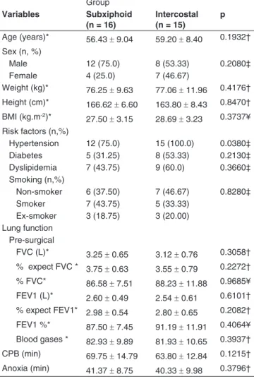
recommend-Table I - Comparison between Groups, Sociodemographic Variables, Risk Factors, Pre-surgical Pulmonary Function, Blood Gases
Group Variables Subxiphoid
(n = 16)
Intercostal (n = 15)
p
Age (years)* 56.43 ± 9.04 59.20 ± 8.40 0.1932†
Sex (n, %)
Male 12 (75.0) 8 (53.33) 0.2080‡
Female 4 (25.0) 7 (46.67)
Weight (kg)* 76.25 ± 9.63 77.06 ± 11.96 0.4176†
Height (cm)* 166.62 ± 6.60 163.80 ± 8.43 0.8470† BMI (kg.m-2)*
27.50 ± 3.15 28.69 ± 3.23 0.3737¥ Risk factors (n,%)
Hypertension 12 (75.0) 15 (100.0) 0.0380‡ Diabetes 5 (31.25) 8 (53.33) 0.2130‡ Dyslipidemia 7 (43.75) 9 (60.0) 0.3660‡ Smoking (n,%)
Non-smoker 6 (37.50) 7 (46.67) 0.8280‡ Smoker 7 (43.75) 5 (33.33)
Ex-smoker 3 (18.75) 3 (20.00) Lung function
Pre-surgical
FVC (L)* 3.25 ± 0.65 3.12 ± 0.76 0.3058†
% expect FVC * 3.75 ± 0.63 3.55 ± 0.79 0.2272† % FVC* 86.58 ± 7.51 88.23 ± 11.88 0.9685¥
FEV1 (L)* 2.60 ± 0.49 2.54 ± 0.61 0.6101† % expect FEV1* 2.98 ± 0.54 2.80 ± 0.65 0.2082†
FEV1 %* 87.50 ± 7.45 91.19 ± 11.91 0.4064¥ Blood gases * 82.93 ± 9.89 81.93 ± 10.65 0.3937†
CPB (min) 69.75 ± 14.79 63.80 ± 12.84 0.1215†
Anoxia (min) 41.37 ± 8.75 40.33 ± 9.98 0.3796†
ed by the WHO 15. BMI > 30 kg.m-2 was considered obesity;
patients who have smoked at least one cigarette per day were considered current smokers; patients who had stopped smok-ing for at least one year were considered ex-smoker; and pa-tients who had never used tobacco-derived substances were
considered non-smokers 16.
The sample size calculation was based on the variable FVC and a difference of at least 400 mL between groups or in relation to preoperative period was considered clinically rel-evant. Beta risk of 20%, alpha risk of 5% (p < 0.05), and a test power of 80% were considered to detect this difference and the sample calculation.
Data normality was assessed using the Shapiro-Wilk test. For intergroup comparison, Student the t test was used for parametric continuous variables and Wilcoxon-Mann-Whitney test for nonparametric continuous variables. Dichotomous
variables were analyzed using chi-square test. For intergroup comparison, the paired Student t test was used for normally distributed variables and Wilcoxon matched pairs test for the other. The significance level was 5%. Analyzes were per-formed using Stata 11.2 software for Windows.
RESULTS
We evaluated 31 patients in the study, 16 patients in SX group and 15 in IC group. The sociodemographic variables, risk fac-tors, measures of pulmonary function, blood gases, and dura-tion of cardiopulmonary bypass (CPB) showed no significant variation between groups, except for hypertension that was more prevalent in the IC group (Table I). Spirometry records were made postoperatively for three days and compared with
Table II - Inter and intragroup Comparison of the Studied Variables
Variables Pre-surgical
1st
Post-surgical
p 2nd
Post-surgical
p (pre x 2nd post)
p (1st x
2nd post)
3rd
Post-surgical
p (pre x 3rd post)
p (1st x
3rd post)
p (2nd x
3rd post)
Subxiphoid group
FVC (L)* 3.25 ± 0.65
0.99 ± 0.29 ¥
0.0001† 1.11 ± 0.29 ¥
0.0001‡ 0.0213‡ 1.24 ± 0.36 0.0001† 0.0009† 0.0043†
% FVC* 86.58 ± 7.51
26.92 ± 8.20 0.0001‡ 30.13 ± 8.29 ¥
0.0001‡ 0.0043† 33.47 ± 9.20 0.0001‡ 0.0007† 0.0068†
FEV1 (L)* 2.60 ± 0.49
0.86 ± 0.72 0.0001† 0.93 ± 0.26 ¥
0.0001‡ 0.0831‡ 1.03 ± 0.29 0.0001† 0.0116† 0.2101‡
FEV1 %* 87.50 ± 7.45
29.75 ± 9.63 0.0001† 32.03 ± 9.42
0.0001‡ 0.0768‡ 35.05 ± 9.50 0.0001† 0.0125† 0.2101‡
Blood
gases* 82.93 9.89 ± 94.04 17.32±
0.0126† 96.20 ± 15.04 ¥
0.0032† 0.3311† - - -
VAS before
- 6.37 ± 1.14 ¥
- 5.62 ±
1.02 ¥
- 0.0003† 4.62 ± 0.95 ¥ - 0.0001† 0.0001†
VAS after - 7.68 ± 1.19 ¥
- 6.68 ±
0.87 ¥
- 0.0008† 5.37 ± 1.08 ¥ - 0.0001† 0.0003†
Intercostal group
FVC (L)* 3.12 ± 0.76
0.79 ± 0.34 ¥
0.0001† 0.91 ± 0.35 ¥
0.0001‡ 0.3018‡ 1.08 ± 0.49 0.0001† 0.0089† 0.0196†
% LVC* 88.23 ± 11.88
22.18 ± 8.59 0.0001‡ 24.96 ± 6.61 ¥
0.0001‡ 0.1054† 29.82 ± 11.11 0.0001‡ 0.0050† 0.0181†
FEV1 (L)* 2.54 ± 0.61
0.72 ± 0.29 0.0001† 0.77 ± 0.28 ¥
0.0001‡ 0.4601‡ 0.90 ± 0.37 0.0001† 0.0140† 0.1796‡
FEV1 %* 91.19 ± 11.91
25.74 ± 10.03
0.0001† 27.32 ± 7.67
0.0001‡ 0.5000‡ 31.45 ± 11.49 0.0001† 0.0127† 0.3018‡
Blood
gases* 81.93 10.65± 97.86 27.50±
0.0202† 82.18 ±
4.20 ¥
0.4773† 0.0353† - - -
VAS before
- 8.33 ± 1.17 ¥
- 7.60 ±
1.05 ¥
- 0.0109† 6.20 ± 0.94 ¥ - 0.0001† 0.0001†
VAS after - 9.06 ± 0.88 ¥
- 8.86 ±
1.12 ¥
- 0.2551† 7.00 ± 1.13 ¥ - 0.0001† 0.0001†
preoperative values, whenever appropriate and a significant change in forced vital capacity and forced expiratory volume in the first second was found in both groups. In the analysis of postoperative variables (mean and standard deviation) in
order to compare the two groups, SX group showed lower loss
of FVC and FEV1 than group IC at all times (Table II).
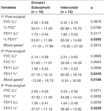
Post-operative lung function, when compared with
base-line values, were analyzed and showed that the loss in FEV1
and FVC was always lower in SX group than in IC group (Ta-ble III).
Postoperative pain evaluation was made before and af-ter expiratory effort in order to assess the influence of chest tube position. There was a significantly reduced intensity at all times in SX group compared to IC group (Figure 1).
PaO2 analysis in both groups, when compared with
preop-erative values, using the mean change in blood gas, showed that in the first postoperative day the mean was similar be-tween groups. However, in the second PO, the SX group had a PaO2 significantly greater than the IC group (Figure 2).
DISCUSSION
The insertion site of the non-toxic PVC chest tube had influence on lung function, which was shown by the decrease in FVC and FEV1 values recorded postoperatively. SX group showed better spirometric measurements and less pain compared to IC group. When chest tubes were removed, there was further improvement in measured values due to decreased pain, with
SX group values closer to preoperative values.
The change in pulmonary function after coronary artery by-pass grafting with CPB has a multifactorial dependency. In
addition to sternotomy, pleurotomy 17 and pain in the
postop-erative period 18 contribute to decline in lung function. Other
factors are also implicated in the reduction of postoperative ventilation, such as surgical manipulation, use of cardiopul-monary bypass (CPB), anesthesia, mechanical ventilation,
Table III - Analysis of Postoperative Values in Relation to Preoperative Periods of the Studied Groups
Groups† Variables Subxiphoid
(n = 16)
Intercostal (n = 15)
p
1st Post-surgical
FVC (L)* 2.26 ± 0.58 2.32 ± 0.70 0.3879
% FVC* 55.51 ± 11.26 62.88 ± 16.70 0.0789 FEV1 (L)* 1.73 ± 0.44 1.82 ± 0.53 0.3117
% FEV1* 53.61 ± 11.88 63.09 ± 14.69 0.0285
Blood gases* -11.10 ± 17.86 -15.92 ± 27.32 0.2815 2st Post-surgical
FVC (L)* 2.14 ± 0.58 2.21 ± 0.62 0.3686
FVC %* 51.83 ± 11.01 59.58 ± 16.39 0.0654 FEV1 (L)* 1.66 ± 0.42 1.76 ± 0.53 0.2699
FEV1 %* 51.10 ± 10.14 60.95 ± 16.16 0.0249
Blood gases* -13.26 ± 16.72 -0.24 ± 16.50 0.0188 3st Post-surgical
FVC (L)* 2.00 ± 0.55 2.04 ± 0.56 0.4371
FVC %* 47.93 ± 11.93 54.68 ± 14.53 0.0835 FEV1 (L) 1.56 ± 0.41 1.64 ± 0.48 0.3239
FEV1 %* 47.57 ± 11.13 56.99 ± 13.92 0.0228
*: Mean values ± standard deviation variation; †: comparisons by Student’s t test; FVC: forced vital capacity; FEV1: Forced Expiratory Volume in the first second.
Figure 1 Assessment of Pain before and after Expiratory Effort within Postoperative three Days. *p < 0.05; PO:Postoperative.
10.00 9.00 8.00 7.00 6.00 5.00 4.00 3.00 2.00 1.00 0.00
1st PO – Before 1st PO – After 6.37
5.62
4.65 8.33
7.60
6.20 7.68
6.68
5.37
9.06 8.86
7.00
2nd PO – Before 2nd PO – After 3nd PO – Before 3nd PO – After
and use of drains 19. Chest wall edema and change in
sur-factants are important factors in reducing lung volume and capacity 20,21.
The surgical technique used in coronary by-pass also af-fects the decrease in FVC, as demonstrated by some authors comparing the use of saphenous vein graft to the internal
mammary artery 22. It is known that the removal of internal
thoracic artery can reduce blood supply to the phrenic nerve, resulting in additional pulmonary dysfunction in the
postop-erative period 23. The incidence of pulmonary complications
after cardiac surgery is significant, and may worsen during the evolutionary picture of the patient, being the leading cause of morbidity and mortality 24.
A better understanding of the factors responsible for lung damage is needed to minimize the pulmonary dysfunction as-sociated with cardiac surgery and its repercussions.
Chest tube causes discomfort to the patient, facilitating the deterioration of respiratory mechanics 4,25. Cohen et al. 26
found that postoperative pain with the use on internal mam-mary artery is significantly higher than with saphenous vein, and may itself lead to a reduction in respiratory function. This is explained by the greater restriction on effective coughing,
deep breathing, and changes in position 26. Authors state that
the reduction in lung function when using the internal mam-mary artery for revascularization would be due to pleurotomy, greater thoracic manipulation, and pain 22,27. All these factors
contribute to a higher incidence of atelectasis, making patients more vulnerable to hypoxic pulmonary complications, mainly pneumonia 4.
Reduced lung function in the postoperative period is in-fluenced by different factors, and pain interference in the re-spiratory movements is evident. In our study, in addition to
evaluating the influence of the chest tube position, we made a counter-proof by measuring the lung volumes and capacities after removal of chest tubes.
Chest tubes placed in the subxiphoid region caused less pain than those in the intercostal space, results in agreement with literature 4,28. Some authors attributed chest pain to the
technique of tube insertion (major or minor tissue injury) and friction in the intrathoracic structures 2. Corroborating these
arguments, we noted lower values for respiratory function pa-rameters in both groups in the first postoperative day, with a gradual improvement until the third postoperative day, culmi-nating with chest tube removal. Although there was recovery
in both groups, the values of SX group were very close to the
preoperative period.
Guizilini et al. 10 conducted a similar study, but in patients
without CPB, and found results similar to ours, although they
did not study the immediate effect of chest tube removal 10.
Decreased lung volume and capacity persists in the fifth postoperative day, but due to other factors, such as CPB and surgical incision 29.
Clinically, the decrease in FVC leads to reduced peak ex-piratory flow, reducing the ability to cough, which hinders the movement of secretions predisposing to atelectasis and pneu-monia 30.
Different from Hagl et al. 4 who found no difference in PaO
2
between groups, we found a significant decrease in PaO2 in
the second PO in IC group. This may be explained by pro-cesses other than individual supplements of oxygen. In our
work this supplementation was set at 5 L.min-1 and not on
de-mand. Something that can also influence the respiratory pa-rameters is the presence of residual fluid in the chest, which could be assessed by ultrasound or by radiography. Studies
Figure 2 Distribution of Mean Percent Change of Gasometry according to Group and Postoperative Measurments. *p < 0.05; PO:Postoperative.
0.00 2.00 4.00 6.00 8.00 10.00 12.00 14.00 16.00 18.00 1o PO
2o PO M
o m e n t s
0.24
13.26
15.92
11.10
%
in this direction were made and showed that the efficiency of chest drainage depended on chest tube correct position-ing 31-33. In our study, the tube was placed under direct vision
before chest closure, ensuring the proper positioning. Analyz-ing the painful effects, Guizilini states that intercostal chest tube placement increases postoperative pain, with restriction of deep breathing, coughing, and changing patient’s position on the bed 10, findings that are consistent with our results.
Postoperative atelectasis is associated with decreased oxygenation and ventilation in dependent areas, increased pulmonary vascular resistance, as well as development of lung injury 34. Pain limits the rib cage voluntary expansion and,
consequently, increases the non-ventilated areas. The ability to cough is reduced, which may induce secretion retention fol-lowed by atelectasis progression with consequent hypoxemia. Ultimately, pain causes acid-base imbalances, which may contribute to increased morbidity and mortality 18,35.
According to Jakob et al. 36, the postoperative patient
evolves into a shallow inspiration due to constant irritation of the intercostal nerves and periosteum 36.
Noteworthy, there was improvement in FVC in the third postoperative day (without chest tube) compared with the second postoperative day (with chest tube) in both groups, which shows how much the permanence of chest tube influ-ences pulmonary function. This fact was also reported in other surveys of general thoracic surgery 36-38. Lima et al. 39 found
49.7% reduction in pain after tube removal.
Some factors may influence lung function by itself, such as age, chronic obstructive pulmonary disease, and duration of surgery. We consider all these factors as exclusion criteria, which made it difficult to find a higher sample in our study, as in the outpatient clinic of our hospital, the coronary patient is often a smoker, obese, and elderly. This fact, however, may be considered a bias.
CONCLUSION
REFERENCES
1. Imura H, Caputo M, Lim K et al. – Pulmonary injury after cardiopulmo-nary bypass: beneficial effects of low-frequency mechanical ventila-tion. J Thorac Cardiovasc Surg, 2009;137:1530-1537.
2. Pick A, Dearani J, Odell J – Effect of sternotomy direction on the in-cidence of inadvertent pleurotomy. J Cardiovasc Surg, 1998;39:673-676.
3. Taggart DP, el-Fiky M, Carter R, Bowman A et al. – Respiratory dys-function after uncomplicated cardiopulmonary bypass. Ann Thorac Surg, 1993;56:1123-1128.
4. Hagl C, Harringer W, Gohrbandt B et al. – Site of pleural drain insertion and early postoperative pulmonary function following coronary artery bypass grafting with internal mammary artery. Chest, 1999;115:757-761.
5. Peng MJ, Vargas FS, Cukier A et al. – Postoperative Pleural Changes after Coronary Revascularization – Comparison between Saphenous-Vein and Internal Mammary Artery Grafting. Chest, 1992;101:327-330.
6. Shapira N, Zabatino SM, Ahmed S et al. – Determinants of pulmonary function in patients undergoing coronary bypass operations. Ann Tho-rac Surg, 1990;50:268-273.
7. Guizilini S, Gomes WJ, Faresin SM et al. – Influence of pleurotomy on pulmonary function after off-pump coronary artery bypass grafting. Ann Thorac Surg, 2007;84:817-822.
8. Kollef MH, Peller T, Knodel A et al. – Delayed pleuropulmonary com-plications following coronary artery revascularization with the internal mammary artery. Chest, 1988;94:68-71.
9. Vargas FS, Uezumi KK, Janete FB et al. – Acute pleuropulmonary complications detected by computed tomography following myocardial revascularization. Rev Hosp Clin Fac Med São Paulo, 2002;57:135-142.
10. Guizilini S, Gomes WJ, Faresin SM et al. – Effects of the pleural drain site on the pulmonary function after coronary artery bypass grafting. Rev Bras Cir Cardiovasc, 2004;19:47-54.
11. Bjessmo S, Hylander S, Vedin J et al. – Comparison of three different chest drainages after coronary artery bypass surgery – A randomised trial in 150 patients. Eur J Cardiothorac Surg, 2007;31:372-375. 12. Frankel TL, Hill PC, Stamou SC et al. – Silastic drains vs conventional
chest tubes after coronary artery bypass. Chest, 2003;124:108-113. 13. Kollef MH, Dothager DW – Reversible cardiogenic shock due to chest
tube compression of the right ventricle. Chest, 1991;99:976-980. 14. Castro Pereira CA – Espirometria em diretrizes para testes de função
pulmonar 2002. J Bras Pneumol, 2002;28(Supl 3):S2-S82.
15. Eveleth PB, Andres R, Chumlea WC et al. – Uses and interpretation of antrhropometry in the elderly for the assessment of physical status. Report to the Nutrition Unit of the World Health Organization: the Ex-pert Subcommittee on the Use and Interpretation of Anthropometry in the Elderly. J Nutr Health Aging, 1998;2(1):5-17.
16. Oliveira MVC, Oliveira TR, Pereira CAC et al. – Tabagismo em pacien-tes internados em um hospital geral. J Bras Pneumol, 2008;34:936-941.
17. Berrizbeitia LD, Tessler S, Jacobowitz IJ et al. – Effect of Sternotomy and Coronary-Bypass Surgery on Postoperative Pulmonary Mecha-nics - Comparison of Internal Mammary and Saphenous-Vein Bypass Grafts. Chest, 1989;96:873-876.
19. Christenson JT, Aeberhard JM, Badel P et al. – Adult respiratory dis-tress syndrome after cardiac surgery. Cardiovasc Surg, 1996;4:15-21. 20. Taniguchi L, Pinheiro A – Particularidades do atendimento ao pacien-te em pós-operatório de cirurgia cardíaca, em: Regenga M - Fisiopacien-te- Fisiote-rapia em Cardiologia: Da UTI à Reabilitação. São Paulo, Roca, 2000, pp 121-154.
21. Regan K, Kleinfeld M, Erik P – Fisioterapia para pacientes com ci-rurgia abdominal ou torácica. Em: Irving S, Tecklin J – Fisioterapia cardiopulmonar. São Paulo, Manole, 1994, pp. 318-339.
22. Jenkins SC, Soutar SA, Forsyth A et al. – Lung function after coronary artery surgery using the internal mammary artery and the saphenous vein. Thorax, 1989;44:209-211.
23. O’Brien JW, Johnson SH, VanSteyn SJ et al. – Effects of internal mammary artery dissection on phrenic nerve perfusion and function. Ann Thorac Surg, 1991;52:182-188.
24. Brooks-Brunn JA – Postoperative atelectasis and pneumonia: risk fac-tors. Am J Respir Crit Care Med, 1995;4:340-9; quiz 350-351. 25. Galantier J – Particularidades da circulação extracorpórea. Em: Auler
Jr JOC & Oliveira AS – Pós-operatório de cirurgia torácica e cardio-vascular. Porto Alegre, Artmed, 2004, pp. 153-157.
26. Cohen AJ, Moore P, Jones C et al. – Effect of internal mammary har-vest on postoperative pain and pulmonary function. Ann Thorac Surg, 1993;56:1107-1109.
27. Burgess GE, Cooper JR, Marino RJ et al. – Pulmonary effect of pleu-rotomy during and after coronary artery bypass with internal mamma-ry artemamma-ry versus saphenous vein grafts. J Thorac Cardiovasc Surg, 1978;76:230-234.
28. Riebman JB, Olivenciayurvati AH, Laub GW – Improved Technique for Pleural Drain Insertion during Cardiovascular-Surgery. J Cardio-vasc Surg, 1994;35:503-505.
29. Giacomazzi CM, Lagni VB, Monteiro MB – Postoperative pain as a contributor to pulmonary function impairment in patients submitted to heart surgery. Rev Bras Cir Cardiovasc, 2006;21:386-392.
30. Oikkonen M, Karjalainen K, Kahara V et al. – Comparison of incentive spirometry and intermittent positive pressure breathing after coronary artery bypass graft. Chest, 1991;99:60-65.
31. Chon KS, vanSonnenberg E, D’Agostino HB, O’Laoide RM, Colt HG, Hart E – CT-guided catheter drainage of loculated thoracic air col-lections in mechanically ventilated patients with acute respiratory dis-tress syndrome. AJR Am J Roentgenol, 1999;173:1345-1350. 32. Lancey RA, Gaca C, Vander Salm TJ – The use of smaller, more
flexible chest drains following open heart surgery: an initial evaluation. Chest, 2001;119:19-24.
33. Nunez R, Munoz JA, Vazquez F et al. – Effects of several methods of thoracic drainage on respiratory function. Cir Pediatr, 1996;9:28-31. 34. Duggan M, Kavanagh BP – Pulmonary atelectasis: a pathogenic
pe-rioperative entity. Anesthesiology, 2005;102:838-854.
35. Wheatcroft M, Shrivastava V, Nyawo B et al. – Does pleurotomy during internal mammary artery harvest increase post-operative pulmonary complications? Interact Cardiovasc Thorac Surg, 2005;4:143-146.
36. Jakob H, Kamler M, Hagl S – Doubly angled pleural drain circumven-ting the transcostal route relieves pain after cardiac surgery. Thorac Cardiovasc Surg, 1997;45:263-264.
37. Gómez-Caro A, Roca MJ, Torres J et al. – Successful use of a single chest drain post-lobectomy instead of two classical drains: a randomi-zed study. Eur J Cardiothorac Surg, 2006;29:562-566.
38. Mueller XM, Tinguely F, Tevaearai HT et al. – Impact of duration of chest tube drainage on pain after cardiac surgery. Eur J Cardiothorac Surg, 2000;18:570-574.