online | memorias.ioc.fiocruz.br
Dengue is an acute viral disease transmitted by Aedes aegypti and Aedes albopictus mosquitoes and is considered the most important viral disease transmit-ted by arthropods worldwide (Guzmán & Kouri 2001, Guha-Sapir & Schimmer 2005, Simmons et al. 2012). The expansion of the geographic distribution and the overall incidence of dengue have increased steadily over the past six decades (Descloux et al. 2012). The disease is currently disseminated in over 100 countries from all continents, including Europe, which recently experienced an epidemic on Madeira Island (Portugal) (Sousa et al. 2012). Dengue outbreaks also impose high costs to health services and to the economic systems of these countries (Suaya et al. 2009, Baly et al. 2012). In the XXI century, Brazil has recorded most of the reported cases of dengue in the world, accounting for approximately 61% of all cases reported to the World Health Organization (WHO) (Teixeira 2009). Southeast Asia was the region most affected by dengue in the mid-1990s. Since then, Central and South American
coun-tries have been experiencing an increasing number of reported dengue cases (Teixeira 2009). The disease has been more prevalent in adults; however, in recent years, a progressive reduction in the ages of the patients affect-ed by dengue haemorrhagic fever (DHF) has been ob-served in Brazil (Teixeira 2009, Cavalcanti et al. 2011). A major weakness in the surveillance of severe dengue (SD) cases is the difficulty of categorising these cases according to the classification adopted by the WHO in 1997 [DHF/dengue shock syndrome (DSS) classifica-dengue shock syndrome (DSS) classifica- (DSS) classifica-tion] (WHO 1975, 1997). This classification is useful for the clinical management of patients, but SD cases do not frequently meet all of the criteria for classification according to this system (Bandyopadhyay et al. 2006, Narvaez et al. 2011). The emergence of dengue with an atypical involvement of specific organs, such as the cen-tral nervous system, liver, lungs and heart, highlighted this difficulty (Araújo et al. 2011, 2012a, b, Verna et al. 2011, Dussart et al. 2012). This disease presentation leads to an acute dengue infection with a severe multisystem manifestation (Borawake et al. 2011, Morrilo et al. 2011), which does not fit the criteria previously defined for se-verity surveillance.
Due to these limitations, Brazil chose to adopt the new category dengue with complications (DWC) in 2001. For the Brazilian Ministry of Health (MS), every suspected case of dengue fever (DF) progressing to a SD form that does not completely fit the DHF/DSS crite-ria should be considered DWC, especially in the pres-ence of one the following: neurological manifestation, cardiac dysfunction, liver failure, gastrointestinal bleed-doi: 10.1590/0074-0276140384
Financial support: PPSUS/REDE/MS/CNPq/FUNCAP/SESA 03/2012 (12535699-4), scientific initiation scholarship from UNICHRISTUS
+ Corresponding author: [email protected] Received 28 July 2013
Accepted 18 October 2013
Evaluation of the WHO classification of dengue disease severity
during an epidemic in 2011 in the state of Ceará, Brazil
Luciano Pamplona de Góes Cavalcanti1,2/+, Lia Alves Martins Mota2, Gustavo Porto Lustosa2,
Mayara Carvalho Fortes2, Davi Alves Martins Mota3, Antônio Afonso Bezerra Lima4,
Ivo Castelo Branco Coelho5,6, Maria Paula Gomes Mourão7
1Departamento de Saúde Comunitária 3Faculdade de Medicina 5Núcleo de Medicina Tropical Prof JE Alencar 6Departamento de Patologia, Universidade Federal do Ceará, Fortaleza, CE, Brasil 2Faculdade de Medicina, Centro Universitário Christus, Fortaleza, CE, Brasil 4Hospital São José de Doenças Infecciosas, Fortaleza, CE, Brasil
7Fundação de Medicina Tropical Dr Heitor Vieira Dourado, Manaus, AM, Brasil
In 2009, the World Health Organization (WHO) issued a new guideline that stratifies dengue-affected patients into severe (SD) and non-severe dengue (NSD) (with or without warning signs). To evaluate the new recommenda-tions, we completed a retrospective cross-sectional study of the dengue haemorrhagic fever (DHF) cases reported during an outbreak in 2011 in northeastern Brazil. We investigated 84 suspected DHF patients, including 45 (53.6%) males and 39 (46.4%) females. The ages of the patients ranged from five-83 years and the median age was 29. Ac-cording to the DHF/dengue shock syndrome classification, 53 (63.1%) patients were classified as having dengue fever and 31 (36.9%) as having DHF. According to the 2009 WHO classification, 32 (38.1%) patients were grouped as having NSD [4 (4.8%) without warning signs and 28 (33.3%) with warning signs] and 52 (61.9%) as having SD. A better performance of the revised classification in the detection of severe clinical manifestations allows for an improved detection of patients with SD and may reduce deaths. The revised classification will not only facilitate ef-fective screening and patient management, but will also enable the collection of standardised surveillance data for future epidemiological and clinical studies.
ing, pleural effusion, pericardial effusion and/or ascites, thrombocytopenia < 20,000/mm3, leukopenia < 1,000/
mm3 or fatal suspected cases of dengue (SVS/MS 2011a).
This new category is exclusively used in Brazil and makes evaluation very difficult when comparing cases with other dengue transmission areas worldwide.
Based on these difficulties and after a multicentre dengue study DENgue COntrol was conducted, a new classification of dengue cases was proposed in 2009 by the WHO. This proposal defines two main categories - SD and non-severe dengue (NSD) - and the NSD form can present with or without warning signs (WHO 2009, Alexander et al. 2011).
The aim of this study was to retrospectively evaluate a cohort of suspected DHF cases reported in a referral hospital in northeastern Brazil according to the 2009 WHO classification.
SUBJECTS, MATERIALS AND METHODS
This is a retrospective cross-sectional study of sus-pected DHF cases reported to a referral hospital for in-fectious diseases in the city of Fortaleza in the state of Ceará (CE), northeastern Brazil, during its largest den-gue epidemic of 2011.
Patients and definitions - All study patients were in-vestigated for being reported as suspected cases of DHF. However, some of the cases did not have laboratory con-firmation and did not undergo additional tests such as ul-trasound and chest X-ray, which would be needed to con-firm cavitary effusions. Notification data were acquired from the Notifiable Diseases Information System. This investigation included all of the cases reported as sus-pected DHF at the referral unit. Following the report of a suspected case, the epidemiological surveillance team performed the proper investigation for the confirmation or exclusion of each case (Cavalcanti et al. 2010, Araújo et al. 2011, SVS/MS 2011a). All reported cases were re-quired to be accompanied by a serum sample, but this was not always permitted by the patient.
DF was defined as an acute febrile illness (history of fever less than 7 days) accompanied by at least two of the following clinical findings: nausea, vomiting, head-ache, arthralgia, retro-orbital pain, rash, myalgia, haem-orrhagic manifestations and leukopenia. Because of the lack of specificity of these clinical signs and symptoms, laboratory evidence of dengue virus (DENV) infection was required for a confirmed diagnosis.
The definition of DHF consists of the presence of all of the five following criteria: fever (history of fever less than 7 days), haemorrhagic evidence (spontaneous bleeding or a positive tourniquet test), thrombocytope-nia (platelet count < 100,000 cells/mm3), plasma leakage
evidence (pleural effusion, ascites, hypoalbuminaemia or haemoconcentration greater than 20% related to base-line) and at least one positive test for dengue [the non-structural protein 1 (NS1), M antibody capture ELISA (MAC-ELISA), viral isolation or reverse transcription-polymerase chain reaction (RT-PCR)] (SVS/MS 2011b).
The definition of DWC was considered for patients that did not fulfil the DHF criteria, but presented with
at least one of the following: neurological manifesta-tion, cardiac dysfuncmanifesta-tion, liver failure, gastrointestinal bleeding, pleural effusion, pericardial effusion and/or ascites, thrombocytopenia < 20,000/mm3, leukopenia
< 1,000/mm3 or fatal suspected case of dengue (SVS/
MS 2011a).
The definition of NSD is somewhat similar to that of DF: a combination of two symptoms and signs in the acute febrile individual and coming from an area of known dengue endemicity. In addition, abdominal pain and tenderness, persistent vomiting, clinical fluid accumulation, mucosal bleeding, lethargy, restlessness and liver enlargement are considered warning signs for potentially SD.
The definition of SD was considered for cases in-cluding any of the following: plasma leakage leading to shock or respiratory distress, severe bleeding or organ failure (e.g., elevated liver enzyme levels, impaired con-sciousness or heart failure) (WHO 2009, Alexander et al. 2011, Srikiatkhachorn et al. 2011). A confirmed case of dengue was considered for those who had a positive laboratory result for at least one of the techniques used (MAC-ELISA, NS1, viral isolation or RT-PCR).
Data collection - Medical records, notification forms used by the MS and the results of laboratory tests were systematically retrieved for all reported cases during the study period. Data were collected from May 2011-March 2012 through a structured questionnaire includ-ing age, gender, signs and symptoms, warninclud-ing signs, evidence of shock (signs of poor perfusion and hypo-tension), effusions (ascites, pleural effusion, pleural ef-fusion and respiratory distress) and laboratory results. Leukopenia was considered as < 1,000/mm3,
hypoalbu-minaemia as serum albumin < 3 g/dL, liver injury as as-partate aminotransferase or alanine aminotransferase > 1,000/L and severe thrombocytopenia as platelet count < 20,000/mm3.
To address the sensitivity, specificity, positive pre-dictive values (PPV) and negative prepre-dictive values (NPV) of the DHF/DSS classification and WHO clas-sification of 2009 for the detection of SD, each classifi-cation was compared to the other. Patients classified as DHF or DSS were considered SD and those classified as DF were considered NSD. For the WHO classification of 2009, patients classified as having SD were considered severe and those with dengue, with or without warning signs, were considered NSD (Narvaez et al. 2011).
All data were stored in Microsoft Office Access v. 2003 and analysed using Epi Info v. 3.5.1 and terminated in accordance with both the classification used currently in Brazil [adapted from WHO (1997)] and the new clas-sification (WHO 2009).
RESULTS
In 2011, a large dengue epidemic occurred in CE. Disease transmission was recorded in 161 municipali-ties (87.5%), with an incidence of 670.98/100,000 in-habitants. Three different DENV serotypes were simul-taneously isolated: DENV-1 (98.7%), DENV-3 (0.4%) and DENV-4 (0.9%). Despite the recent introduction of DENV-4 in CE in 2011, that epidemic was mainly caused by DENV-1, which had only rarely been recorded in the region since 2002.
Among the 102 cases reported as DHF in this pe-riod, 84 (82.4%) were seen at the São José Hospital. Of the 84 patients investigated for suspected DHF, 45 (53.6%) were male and the median age was 29 years old (5-83) (Table I).
According to the adapted DHF/DSS classification (1997), one (1.2%) patient was classified as having DF, 31 (36.9%) as having DHF and 52 (61.9%) as having DWC (Tables II, III). The WHO classification of 2009 categorised the investigated patients as follows: 32 NSD [4 (4.8%) without warning signs and 28 (33.3%) with warning signs] and 52 (61.9%) SD (Table II).
The most frequently reported signs and symptoms were headache 74/77 (96.1%), nausea 44/47 (93.6%), myalgia 70/75 (93.3%), rash 60/68 (88.2%), retro-orbital pain 37/42 (88.1%), arthralgia 31/37 (83.8%), dizziness 47/57 (82.5%), abdominal pain 61/76 (80.3%), vomiting 57/73 (78.1%) and dyspnoea 20/64 (31.3%). Bleeding evidence was found in 70/80 patients (87.5%). Mucosal bleeding was detected in 28/69 (40.6%) and petechiae in 51/63 (81%) and the tourniquet test was performed in 44% of patients, with 21.6% positivity (Table I).
TABLE I
Demographic characteristics, signs and symptoms during a dengue epidemic in northeastern Brazil, 2011
Age (years) n/total (%)
≤ 15 16/84 (19)
> 15 68/84 (81)
Sex
Female 39 (46.4)
Male 45 (53.6)
Signs and symptoms
Headache 74/77 (96.1)
Nausea 44/47 (93.6)
Myalgia 70/75 (93.3)
Rash 60/68 (88.2)
Exanthema 60/68 (88.2)
Retro-orbital pain 37/42 (88.1)
Arthralgia 31/37 (83.8)
Dizziness 47/57 (82.5)
Abdominal pain 61/76 (80.3)
Vomit 57/73 (78.1)
Cough 30/50 (60)
Dyspnoea 20/64 (31.3)
Haemorrhagic manifestations 70/80 (87.5)
Petecchiae 51/63 (81)
Mucosal bleeding 28/69 (40.6)
Gastrointestinal bleeding 21/66 (31.8)
Haematemesis 14/63 (22.2)
Positive tourniquet test 8/37 (21.6)
Melena 9/59 (15.3)
Epistaxis 8/58 (13.8)
Haematuria 4/34 (11.8)
Extravasation of plasma
Pleural effusion 12/21 (57.1)
Cavitary effusion 16/31 (51.6)
Ascites 10/20 (50)
Plasma leakage 31/75 (41.3)
Laboratory values Median (min-max)
Platelet count 18,000 (12,000-85,000)
Leukocyte count 3,380 (900-9,800)
Haematocrit 39.2 (15.5-57.5)
TABLE II
Comparison between dengue haemorrhagic fever (DHF)/dengue shock syndrome (DSS) classification
for reporting severe cases during a dengue epidemic in northeastern Brazil, 2011
NSD n (%)
NSD with warning signs
n (%)
SD n (%)
Total n (%)
DF 3 (75) 18 (64.3) 32 (61.5) 53 (63.1)
DHF 1 (25) 10 (35.7) 20 (38.5) 31 (36.9)
DSS - - -
-Total 4 (100) (4.8 of total)
28 (100) (33.3 of total)
52 (100) (61.9 of total)
84 (100)
-DF: dengue fever; NSD: non-severe dengue; SD: severe dengue.
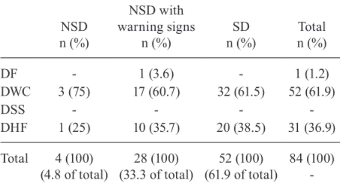
TABLE III
Comparison between dengue haemorrhagic fever (DHF)/dengue shock syndrome (DSS) classification
adapted by Brazilian Ministry of Health (2011) and the DHF/DSS classification for reporting severe dengue (SD) cases during a dengue epidemic in northeastern Brazil, 2011
NSD n (%)
NSD with warning signs
n (%)
SD n (%)
Total n (%)
DF - 1 (3.6) - 1 (1.2)
DWC 3 (75) 17 (60.7) 32 (61.5) 52 (61.9)
DSS - - -
-DHF 1 (25) 10 (35.7) 20 (38.5) 31 (36.9)
Total 4 (100) (4.8 of total)
28 (100) (33.3 of total)
52 (100) (61.9 of total)
84 (100)
Among patients evaluated with at least two haema-tocrit counts, the lowest result showed a median value of 35.5% (15.4-45.6%) and the highest of 43.5% (30.3-57.5%). Leukocyte count varied between 900-9,800/ mm3 and platelet count showed a median of 18,000/mm3
(1,200-85,000) (Table I).
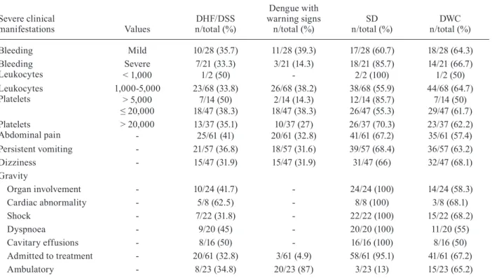
Plasma leakage was identified in 31/75 (41.3%) pa-tients, with cavity effusion in 16/31 (51.6%). A severe impairment of organs was reported in 24/83 (28.9%) cas-es, with an emphasis on cardiorespiratory changes pres-ent in 8/26 (30.8%) patipres-ents (Table IV). A positive IgM (MAC-ELISA) was detected in 75/84 (89.3%) cases.
Considering the DHF/DSS classification, which is still the current classification adopted in Brazil, only 31 (36.9%) out of 84 cases reported as suspected DHF ful-filled all DHF criteria. The other 52 cases were classi-fied as DWC, an exclusive category adopted in Brazil for SD cases not enrolled as DHF/DSS. All of the patients had fever, thrombocytopenia and laboratory confirma-tion of dengue infecconfirma-tion, but 22/44 (54.5%) showed no plasma leakage and 19/52 (36.5%) showed no evidence of bleeding. The main severity criterion for 27/52 (51.9%) patients was platelet count < 20,000/mm3.
Regarding the WHO classification of 2009, 52/84 cases were considered SD; 31 (100%) had been previ-ously classified as DHF and another 21 (40.4%) had been classified as DWC.
The WHO classification of 2009 showed 60.4% sen-sitivity [confidence interval (CI) 95% 46.0-73.5] and
35.5% specificity (CI 95% 19.2-54.6) to capturing SD cases. It also showed a PPV of 61.5% (CI 95% 47.0-74.7) and a low NPV of 34.4% (CI 95% 18.6-53.2) (Table V).
DISCUSSION
In this study, we compared two classifications of dengue - the DHF/DSS classification (and adapted by the MS) and the WHO classification of 2009 - through a retrospective analysis of cases diagnosed during the largest epidemic of dengue registered in CE. In the 1980s, a broad consensus emerged from dengue experts worldwide that the traditional DF/DHF/DSS classifica-tion was essentially retrospective and overly complex, limiting its usefulness for patient management and glob-al surveillance (Akbar et glob-al. 2012). Previous studies have addressed the difficulties in classifying dengue cases ac-cording to the DHF/DSS classification (Bandyopadhyay et al. 2006, Basuki et al. 2010, Barniol et al. 2011), but this is the first case series study evaluating the Brazil-ian scenario for potentially SD cases. Moreover, Sriki-atkhachorn et al. (2011) discuss the limitations of the WHO classification of 2009 because of its inclusion of a greater number of unspecific signs and symptoms and the difficulties associated with confirming the damage to organs in the absence of plasma leakage.
Considering the SD cases, the DHF/DSS classifica-tion and the WHO classificaclassifica-tion of 2009 would have identified 31 (36.9%) and 52 (61.9%) cases, respectively. The WHO classification of 2009 showed a higher
speci-TABLE IV
Severe clinical manifestations presents during a dengue epidemic in northeastern Brazil, 2011
Severe clinical
manifestations Values
DHF/DSS n/total (%)
Dengue with warning signs n/total (%)
SD n/total (%)
DWC n/total (%)
Bleeding Mild 10/28 (35.7) 11/28 (39.3) 17/28 (60.7) 18/28 (64.3)
Bleeding Leukocytes
Severe 7/21 (33.3) 3/21 (14.3) 18/21 (85.7) 14/21 (66.7)
< 1,000 1/2 (50) - 2/2 (100) 1/2 (50)
Leukocytes Platelets
1,000-5,000 23/68 (33.8) 26/68 (38.2) 38/68 (55.9) 44/68 (64.7)
> 5,000 7/14 (50) 2/14 (14.3) 12/14 (85.7) 7/14 (50)
≤ 20,000 18/47 (38.3) 18/47 (38.3) 26/47 (55.3) 29/47 (61.7)
Platelets Abdominal pain
> 20,000 13/37 (35.1) 10/37 (27) 26/37 (70.3) 23/37 (62.2)
- 25/61 (41) 20/61 (32.8) 41/61 (67.2) 35/61 (57.4)
Persistent vomiting - 21/57 (36.8) 18/57 (31.6) 39/57 (68.4) 36/57 (63.2)
Dizziness - 15/47 (31.9) 15/47 (31.9) 31/47 (66) 32/47 (68.1)
Gravity
Organ involvement - 10/24 (41.7) - 24/24 (100) 14/24 (58.3)
Cardiac abnormality - 5/8 (62.5) - 8/8 (100) 3/8 (68.1)
Shock - 7/22 (31.8) - 22/22 (100) 15/22 (68.2)
Dyspnoea - 9/20 (45) - 20/20 (100) 11/20 (55)
Cavitary effusions - 8/16 (50) - 16/16 (100) 8/16 (50)
Admitted to treatment - 20/61 (32.8) 3/61 (4.9) 58/61 (95.1) 41/61 (67.2)
Ambulatory - 8/23 (34.8) 20/23 (87) 3/23 (13) 15/23 (65.2)
ficity in capturing the most SD cases, as found in Taiwan (Tsai et al. 2013). This specificity may help to improve the risk classification and the health care provided to pa-tients upon first admission to health facilities. However, this classification system could lead to an overload in the health services network if the network is not well-prepared, as suggested by Srikiatkhachorn et al. (2011). From this perspective, the WHO classification of 2009 could also serve as a stimulus for a reorganisation of health services, making them ready to intervene strate-gically and promptly for patients with warning signs and those already considered severe, as suggested by Hor-stick et al. (2012).
Another relevant point of comparison is that the less rigid demand for laboratory criteria in the WHO classifi-cation of 2009 brings with it the possibility of concluding case reports in a timely manner by clinical and surveil-lance teams, bringing together experts and preventing discrepancies in clinical and epidemiological reports. An example of such a discrepancy was the creation, by the MS, of a new clinical category - DWC - to meet the request of clinicians who did not accept the fact that pa-tients were dying of SD in Brazil, without meeting any of the DHF criteria and had to be finally reported as having DF. This unique inclusion of DWC is already widespread in the service routine and epidemiological surveillance in Brazil, which is currently responsible for 60% of DF cases worldwide (Teixeira 2009). This situation impairs the ability to compare dengue cases with any other coun-try that uses the DHF/DSS classification. In 2011, CE reported the largest dengue outbreak in its history, with 457 DWC cases and a fatality rate exceeding 10%, higher than the estimated rate for DHF cases (7.5%). This fatal-ity rate suggests that more SD were escaping DHF/DSS classification and these deaths would most likely not be accounted for in the official statistics, as they did not fit as DHF or DSS. Considering this aspect, less than 40%
of these cases would have been classified as severe. The revised case classification, with its simplified structure, will facilitate effective screening, risk classification and patient management and improve comparative analyses of surveillance data from different countries (Basuki et al. 2010). The most severe manifestations of dengue, such as severe haemorrhage, severe leukopenia, throm-bocytopenia, severe shock, effusions and severe organ involvement, were more frequently found in cases of SD compared to cases of DHF, indicating a greater sensitiv-ity in the identification of cases that are more severe.
In the DHF/DSS classification (1997), among the 84 suspected cases, only 36.9% were confirmed as DHF. Moreover, the tourniquet test, which is considered a good predictor of risk (Kalayanarooj et al. 1999), was only used in 44% of patients, with very low positivity (21.6%). The severe impairment of organs was reported in 28.9% patients. Research carried out in Nicaragua (Balmaseda et al. 2005) noted difficulties in detecting patients with these severe manifestations, especially in adults; this challenge was confirmed by our findings, most likely due to an underreporting of medical evalua-tion in hospital files/charts.
Defining the plasma leakage is another great diffi-culty in dengue patients. In most cases, there is a need for repeated blood tests to identify the haemoconcentra-tion. This same difficulty was observed in another den-gue epidemic in Brazil in 2008, where 53.8% of patients with SD did not meet the haemoconcentration criteria and were not classified as DHF (Gupta et al. 2010). The identification of effusions through X-ray or ultrasonog-raphy assessment is rarely available in current practice and could be costly in public health settings (Cavalcanti et al. 2010). Gupta et al. (2010) also highlighted the diffi-culty in diagnosing signs of plasma extravasation using research methods that can be costly.
The revised WHO classification of 2009 for SD ap-pears to have higher sensitivity (60.4%), although it is no longer more specific (35.5%) than the DHF/DSS clas-sification. These data are similar to recent reports from Nicaragua (Narvaez et al. 2011, Gutiérrez et al. 2013) and are the first evidence from Brazil. However, the diversity of criteria used to define severity among the studies may explain the differences in sensitivity and specificity. The absence of a gold standard with which to compare these classifications requires caution when evaluating this information.
In our experience thus far, the 2009 WHO classifica-tion has been more sensitive in detecting and more help-ful for reporting SD cases in this epidemic. This clas-sification made it feasible to classify a greater number of SD cases, which could contribute to a better and timelier management of patients and thus avoid or decrease fatali-ties. Improvements in early diagnosis and risk prediction for severe disease are undoubtedly needed and research efforts in this area are on-going. However, the use of secondary data, even with research records and medical records, poses limitations to the quality and external va-lidity of our data. Prospective studies are needed to bet-ter define the warning signs and to evaluate the benefits of the new classification in different settings.
TABLE V
Concordance between dengue haemorrhagic fever (DHF)/ dengue shock syndrome (DSS) classification and WHO (2009) classification for reporting severe cases (SD)
during a dengue epidemic in northeastern Brazil, 2011
1997 classification
2009 WHO classification
Total Dengue
with/without
warning signs SD
Dengue fever 21 32 53
DHF/DSS 11 20 31
Total 32 52 84
ACKNOWLEDGEMENTS
To the healthcare professionals from São José Hospital, for their cooperation during data collection, and to Rivaldo Venâncio and Eric Martinez, for the criticisms and the final revision of the paper.
REFERENCES
Akbar NA, Allende I, Balmaseda A, Coelho ICB, Cunha RV, Datta B 2012. Regarding “dengue - how best to classify it”. Clin Infect Dis54: 1820-1821.
Alexander N, Balmaseda A, Coelho ICB, Dimaano E, Hien TT, Hung NY 2011. Multicentre prospective study on dengue classification in four South-east Asian and three Latin American countries.
Trop Med Int Health16: 936-948.
Araújo F, Nogueira R, Araújo MS, Perdigão A, Cavalcanti L, Bri- lhante R 2012a. Dengue in patients with central nervous system manifestations, Brazil. Emerg Infect Dis18: 677-679.
Araújo FMC, Araújo MS, Nogueira RMR, Brilhante RSN, Oliveira DN, Rocha MFG 2012b. Central nervous system involvement in dengue: a study in fatal cases from a dengue endemic area. Neu-rology78: 736-742.
Araújo FMC, Brilhante RSN, Cavalcanti LPG, Rocha MFG, Cordeiro RA, Perdigão ACB 2011. Detection of the dengue non-structural 1 antigen in cerebral spinal fluid samples using a commercially available enzyme-linked immunosorbent assay. J Virol Methods 177: 28-131.
Balmaseda A, Hammond SN, Pérez MA, Cuadra R, Soladno S, Rocha J, Idiaquez W, Harris E 2005. Short report: assessment of the World Health Organization scheme for classification of dengue severity in Nicaragua. Am J Trop Med Hyg73: 1059-1062.
Baly A, Toledo ME, Rodriguez R, Benitez JR, Rodriguez M, Boelaert M, Valerbergue V, Stuyft PV 2012. Costs of dengue prevention and incremental cost of dengue outbreak control in Guantanamo, Cuba. Trop Med Int Health17: 123-132.
Bandyopadhyay S, Lum LCS, Kroeguer A 2006. Classifying dengue: a review of the difficulties in using the WHO case classification for dengue haemorrhagic fever. Trop Med Int Health11: 1238-1255.
Barniol J, Gaczkowski R, Barbato EV 2011. Usefulness and applica-bility of the revised dengue case classification by disease: multi-centre study in 18 countries. BMC Infect Dis11: 106-118.
Basuki PS, Budiyanto, Puspitasari D 2010. Application of revised dengue classification criteria as a severity marker of dengue viral infection in Indonesia. Southeast Asian J Trop Med Public Health 41: 1088-1094.
Borawake K, Prayag P, Wagh A, Dole S 2011. Dengue encephalitis.
Indian J Crit Care Med15: 190-193.
Cavalcanti LP, Vilar D, Souza-Santos R, Teixeira MG 2011. Change in age pattern of persons with dengue, northeastern Brazil. Emerg Infect Dis17: 132-134.
Cavalcanti LPG, Coelho ICB, Vilar DCF, Holanda SGS, Escóssia KNF, Souza-Santos R 2010. Clinical and epidemiological char-acterization of dengue hemorrhagic fever cases in northeastern Brazil. Rev Soc Bras Med Trop43: 355-358.
Descloux E, Mangeas M, Menkes CE, Lengaigne M, Leroy A, Tehei T 2012. Climate-based models for understanding and forecasting dengue epidemics. PLoS Negl Trop Dis6: e1470.
Dussart P, Baril L, Petit L, Beniguel L, Quang LC, Ly S 2012. Clinical and virological study of dengue cases and the members of their households: The Multinational DENFRAME Project. PLoS Negl Trop Dis6: e1482.
Guha-Sapir D, Schimmer B 2005. Dengue fever: new paradigms for a changing epidemiology. Review. Emerg Themes Epidemiol2: 1-10.
Gupta P, Khare V, Tripathi S, Ng VL, Kumaar R, Khan MY 2010. Assessment of World Health Organization definition of dengue hemorrhagic fever in north India. J Infect Dev Ctries3: 150-155.
Gutiérrez G, Gresh L, Pérez MÁ, Elizondo D, Avilés W, Kuan G, Bal-maseda Á, Harris E 2013. Evaluation of the diagnostic utility of the traditional and revised WHO dengue case definitions. PLoS Negl Trop Dis 7: e2385.
Guzmán MG, Kouri G 2001. Dengue: an update. Lancet Infect Dis 2: 33-42.
Horstick O, Farrar J, Lum L, Martinez E, Martin JL, Ehrenberg J, Velayudhan R, Kroegerl A 2012. Reviewing the development, evidence base and application of the revised dengue case clas-sification. Pathoge Glob Health106: 94-101.
Kalayanarooj S, Nimmannitya S, Suntayakorn S 1999. Can doctors make an accurate diagnosis of dengue infections at an early stage? Dengue Bull23: 1-7.
Morrilo GA, Rivas CM, Reverol MM, Abreu MF, Larreal M, Nava GT 2011. Clinical and pathological correlation in fatal dengue cases found in Maracaibo, Venezuela. Rev Cubana Med Trop63: 44-51.
SVS/MS - Secretaria de Vigilância em Saúde/Ministério da Saúde Brasil 2011a. Dengue no Brasil: tendências e mudanças na epi-demiologia com ênfase nas epidemias de 2008 e 2010. In JB Siqueira Jr, LC Vinhal, RFC Said, JL Hoffmann, J Martins, SB Barbiratto, GE Coelho, Saúde Brasil 2010: uma análise da situa-ção de saúde e de evidências selecionadas de impacto de ações de vigilância em saúde, SVS/MS, Brasília, 15 pp.
SVS/MS - Secretaria de Vigilância em Saúde/Ministério da Saúde Brasil 2011b. Dengue: diagnóstico e manejo clínico: criança, SVS/MS, Brasília, 52 pp.
Narvaez F, Gutierrez G, Pérez MA, Elizondo D, Nuñez A, Balmaseda A, Harris E 2011. Evaluation of the traditional and revised WHO classifications of dengue disease severity. PLoS Negl Trop Dis 5: e1397.
Simmons CP, Farrar JJ, Chau NVV, Wills B 2012. Dengue. N Engl J Med366: 1423-1432.
Sousa CA, Clairouin M, Seixas G, Viveiros B, Novo MT, Silva AC, Escoval MT, Economopoulou A 2012. Ongoing outbreak of den-gue type 1 in the autonomous region of Madeira, Portugal: pre-liminary report. Euro Surveill17: 1-4.
Srikiatkhachorn A, Rothman AL, Gibbons RV, Sittisombut N, Mal-asit P, Ennis FA, Nimmannitya S, Kalayanorooj S 2011. Dengue - how best to classify it. Clin Infect Dis 53: 563-567.
Suaya JA, Shepard DS, Siqueira JB et al 2009. Costs of dengue cases in eight countries in the Americas and Asia: a prospective study.
Am J Trop Med Hyg80: 846-855.
Teixeira MG 2009. Dengue: twenty-five years since reemergence in Brazil. Cad Saude Publica 25: 7-18.
Tsai CY, Lee IK, Lee CH, Yang KD, Liu JW 2013. Comparisons of dengue illness classified based on the 1997 and 2009 World Health Organization dengue classification schemes. J Microbiol Immunol Infect46: 271-281.
Verna R, Sharma P, Garg RK, Atam V, Singh MK, Mehrotra HS 2011. Neurological complications of dengue fever: experience from a tertiary center of north India. Neurol India14: 272-278.
WHO - World Health Organization 1975. Technical guides for diag-nosis, treatment, surveillance, prevention and control of dengue haemorrhagic fever, WHO, Geneva, 42 pp.
WHO - World Health Organization 1997. Dengue hemorrhagic fever: diagnosis, treatment, prevention and control, 2nd ed., WHO, Ge-neva, 84 pp.