Daniela Brait Silva Ladeira(a) Adriana Dibo Cruz(b)
Deborah Queiroz Freitas(a) Solange Maria Almeida(a)
(a) Department of Oral Diagnosis, Piracicaba Dental School, Universidade Estadual de Campinas - Unicamp, Piracicaba, SP, Brazil.
(b) Department of Specific Formation, Dental School, Universidade Federal Fluminense - UFF, Nova Friburgo, RJ, Brazil.
Corresponding Author: Deborah Queiroz Freitas E-mail: [email protected]
Prevalence of C-shaped root canal in a
Brazilian subpopulation: a cone-beam
computed tomography analysis
Abstract: The aim of this study was to use cone-beam computed tomog-raphy (CBCT) images to evaluate the prevalence and conigurations of C-shaped canals in permanent mandibular second molars among members of a Brazilian subpopulation. CBCT exams of 214 patients (406 teeth) were evaluated for: presence of C-shaped root canals, number of canals and direction of the root grooves (buccal or lingual). Of the 214 CBCT images examined, 192 showed intact bilateral molars, which were used to analyze the distribution of unilateral and bilateral occurrences of C-shaped canals. The prevalence of these canals was correlated with gender and age, and the number of canals was correlated with the direction of the root grooves using the chi-squared test (α = 0.05). The prevalence of C-shaped canals was 15.3%. This prevalence did not differ with gender or age. Most of the C-shaped molars had three (43.5%) or two (37.1%) canals; 69.4% of the C-shaped canals were grooved in the buccal direc-tion; 68.3% of the C-shaped cases were unilateral. In conclusion, there was a signiicant prevalence of C-shaped canals in the mandibular sec-ond molars of the population studied; the C-shaped canal system varied in coniguration, with a higher prevalence of three and two canals and unilateral occurrence; CBCT is a useful tool for endodontic diagnosis and treatment.
Descriptors: Anatomy; Cone-Beam Computed Tomography; Molar.
Introduction
Preoperative radiographs may aid in visualizing and observing the anatomy of variant canal systems, since recognition of such variations is an essential prerequisite for successful endodontic diagnosis and treat-ment. One of these variations is the C-shaped canal system, so named because of the axial plane morphology of the root canal, irst document-ed in endodontic literature by Cooke and Cox1 in 1979. This variant is
seen mostly in mandibular second molars, although it can also appear in maxillary and mandibular premolars and molars.2,3 The main anatomic
feature of C-shaped canals is the presence of a in or web connecting individual mesial and distal canals, which causes the canal axial section to have a C-shape, and which hinders thorough cleaning, shaping, and obturation. Therefore, recognition of a C-shaped canal coniguration be-fore treatment can facilitate more effective management of the root canal system.3
Declaration of Interests: The authors certify that they have no commercial or associative interest that represents a conflict of interest in connection with the manuscript.
Submitted: Jun 03, 2013
Accepted for publication: Sep 01, 2013 Last revision: Sep 17, 2013
Although traditional radiographs are very use-ful in the preoperative assessment of canal anatomy, they are unlikely to show the extent of complexities of the root canal system, because of the image su-perimposition provided by the two-dimensional im-ages of conventional radiography.
Recently, cone-beam computed tomography (CBCT) was reported to be suficiently precise to perform morphological analyses.4,5 Its advantages
allow the clinician a more thorough understanding of the true morphology of root canal systems.6
The prevalence of a C-shaped canal in mandibu-lar second momandibu-lars has been estimated to range from 2.7% to 44.5%, depending on the population and ethnic group:
• 2.7%–7.6% for American,1,7
• 8% for Turkish,8,9
• 10.6% for Saudi Arabian,10
• 19.1% for Lebanese,11
• 31.5% for Chinese12 and
• 32.7%–44.5% for Korean populations.13,14
A study recently conducted in the Brazilian population evaluated the root canal coniguration of mandibular molars, and showed that the preva-lence of C-shaped canals in second molars was 3.5%; however, the authors did not report the char-acteristics of its coniguration, bearing in mind that the study was not limited to evaluating only the C-shaped canals.15 Therefore, the aim of this study was
to use CBCT images to evaluate the prevalence and conigurations of C-shaped canals in the permanent mandibular second molars of members of a Brazil-ian subpopulation.
Methodology
Patients and teeth
In this study, an evaluation was made of the man-dibular second molars of patients who were referred to the Oral Radiologic Clinic at Piracicaba Dental School, Universidade Estadual de Campinas - Uni-camp, Piracicaba, SP, Brazil, between February and November 2010, and who met the inclusion criteria described below, regardless of gender or age, thus characterizing a convenience sample. The exams were required for different treatment purposes not
exclusive to this study. For the purpose of obtaining clear images of the mandibular second molars, teeth with physiological and/or pathological defects were excluded. Teeth were selected according to the fol-lowing criteria:
• mandibular second molars with fully developed roots and with no periapical lesions;
• no root canal treatment;
• no root canals with open apices, resorption or calciication; and
• CBCT images of good quality, with ield of view in which the mandibular second molars could be seen adequately.
A total of 214 patients met the inclusion crite-ria; 22 had a unilateral intact molar, and 192 had bilateral molars; a total of 406 teeth were analyzed. Bilateral molar data were used to analyze the dis-tribution of unilateral and bilateral occurrences of C-shaped canals. The study group consisted of 130 females and 84 males, between 18 and 74 years of age (mean age of 29.9 years).
The study was approved by the Research Ethics Committee of the Universidade Estadual de Campi-nas - Unicamp (Protocol no. 53/2011) and a written consent of each patient was obtained.
Radiographic techniques
The CBCT images were performed using a Classic i-CAT scanner (Imaging Sciences Interna-tional, Inc., Hatield, USA), operating at 120 kVp and 8 mA, voxel 0.25 mm, 512×512 matrix, with no added iltration. All CBCT exposures were per-formed by an appropriately licensed radiologist.
Images analysis
canals was correlated with the direction of the root grooves. These correlations were determined and as-sessed by the chi-squared test—used to determine whether there is a signiicant difference between the expected frequencies and the observed frequencies in studied variables—using the Stata Statistics/Data Analysis version 11.0 software (StataCorp., Col-lege Station, USA) with a signiicance level set at 5%
(α = 0.05).
Results
Of the 406 mandibular second molars examined in CBCT images, 62 (15.3%) had a C-shaped root canal system. Considering the total 214 patients, 49 (22.9%) showed a C-shaped canal. Of the 192 patients with bilateral permanent mandibular sec-ond molars, analyzed to determine the distribution of unilateral and bilateral C-shaped canals, 41 pre-sented C-shaped canals, in that 28 patients (68.3%
of these cases) had a unilateral and 13 (31.7%) had a Determining of the C-shaped canal system in a
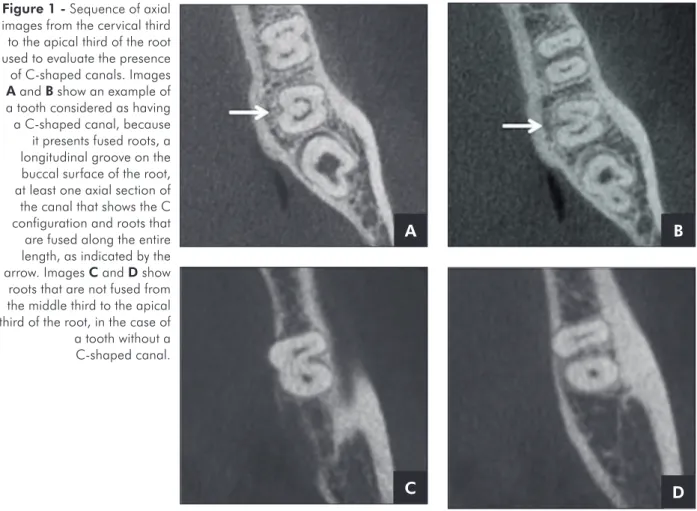
mandibular second molar required that the tooth exhibit all of the following features adapted from those described by Fan et al.:16
I. fused roots,
II. a longitudinal groove on the lingual or buccal surface of the root,
III. at least one axial section of the canal showing a C coniguration, and
IV. the roots fused along their entire length (Figure 1).
Accordingly, the images could be evaluated in axial, coronal and sagittal sections; however, to de-ine the canals as C-shaped, the evaluators had to examine all axial sections of the roots. In assessing each eligible tooth, all axial sections of the roots were also used to evaluate the number of canals and the direction of the root grooves (buccal or lingual).
The frequency of the C-shaped canals was cor-related with gender and age, and the number of the
Figure 1 - Sequence of axial images from the cervical third to the apical third of the root used to evaluate the presence of C-shaped canals. Images A and B show an example of a tooth considered as having a C-shaped canal, because it presents fused roots, a longitudinal groove on the buccal surface of the root, at least one axial section of the canal that shows the C configuration and roots that are fused along the entire length, as indicated by the arrow. Images C and D show roots that are not fused from the middle third to the apical third of the root, in the case of a tooth without a C-shaped canal.
A B
bilateral C-shaped canal.
Table 1 shows the distribution of C-shaped ca-nals in mandibular second molars by gender. No statistical difference was observed in this data. The distribution of C-shaped canals in mandibular sec-ond molars by age is shown in Table 2. No statisti-cal difference was observed in this data.
Table 3 shows the association between number of canals and the direction of the root grooves of teeth with a shaped root canal. Most of the C-shaped molars had three (43.5%) or two (37.1%) ca-nals, mainly with root grooves in a buccal direction (69.4%; p < 0.05), as illustrated in Figure 2.
Discussion
Many methods are used to investigate the root canal anatomy in vitro and in vivo.2 However, with
the exception of radiographic exams, these methods either destroy the specimen by grinding or splitting, or preclude further use of the specimen, from pro-cedures such as tooth clearing and dye assessment. An ideal technique is one that is accurate, simple, nondestructive, and most importantly, feasible in in vivo scenarios.4 Based on all of these factors,
radiog-raphy has been opted for as the most practical and frequently used method to predict the root canal anatomy in both laboratory and clinical studies.3
However, it is dificult to detect the canal sys-tems of mandibular molars with conventional
radi-ography because the images of the roots overlap, es-pecially in C-shaped roots, where the radiographic images may appear in diverse manners, depending on the exact nature and orientation of the root. The root may appear as a single, fused root or as two distinct roots with a communication. When it ap-pears as two distinct roots with a communication, the C-shaped canal may not be easily recognized on conventional periapical ilms.17 CBCT makes
this canal system more predictable, and is a good option clinically speaking, because it can detect C-shaped canal conigurations.5,18 Today, some studies
have been using micro-CT to investigate root canal morphology.3,19 This evaluation provides accurate
data, but micro-CT cannot be used clinically yet. Therefore, this study used CBCT to evaluate the canal systems of 406 mandibular second molars in Brazilian individuals to identify the prevalence of
C-Table 2 - Frequency of C-shaped canals in mandibular second molars by age [n (%)].
Direction of root grooves
Number of canals
Total
1 2 3 4
Buccal 2 (3.2) 18 (29.0) 22 (35.5) 1 (1.6) 43 (69.4)
Lingual 9 (14.5) 5 (8.1) 5 (8.1) 0 (0.0) 19 (30.6)
Total 11 (17.7) 23 (37.1) 27 (43.5) 1 (1.6) 62 (100.0)
Likelihood ratio test (p < 0.05). Table 3 - Correlation between
number of canals and direction of root grooves [n (%)].
Table 1 - Frequency of C-shaped canals in mandibular sec-ond molars by gender [n (%)].
C-shaped Gender Total
Female Male
Absent 205 (50.5) 139 (34.2) 344 (84.7)
Present 43 (10.6) 19 (4.7) 62 (15.3)
Total 248 (61.1) 158 (38.9) 406 (100.0)
Fisher’s exact test (p > 0.05).
C-shaped Age (years) Total
≤ 20 21–30 31–40 41–50 ≥ 51
Absent 50 (12.3) 190 (46.8) 64 (15.8) 16 (3.9) 24 (5.9) 344 (84.7)
Present 6 (1.5) 29 (7.1) 15 (3.7) 8 (2.0) 4 (1.0) 62 (15.3)
Total 56 (13.8) 219 (53.9) 79 (19.5) 24 (5.9) 28 (6.9) 406 (100.0)
shaped canals.
In the literature, the C-shaped canal was most common in a Korean subpopulation, with a 31%– 45% prevalence.13,14,18 The Chinese population also
presented high prevalence of C-shaped canals, as re-ported by Yang et al.,12 Zheng et al.20 and Zhang et al.5 (32%, 39% and 29%, respectively). C-shaped
root and canal conigurations were seldom found in Caucasian populations: Cooke and Cox,1 Weine7
and Cimilli et al.8 reported a prevalence of 2.7%,
7.6% and 8.1% of C-shaped canals, respectively. These data corroborate that canal shape was sig-niicantly related to race, with higher prevalence of C-shaped canals in Asians. This occurrence may explain the high number of studies in Asian popu-lations and the lack of studies in others. To our knowledge, only one study evaluated the canal con-igurations in a Brazilian subpopulation; the authors found a 3.5% prevalence of C-shaped canals in sec-ond mandibular molars.15 The present investigation
revealed a higher prevalence of C-shaped canals (15.3%). However, it is dificult to make a direct comparison between both studies, since the authors did not indicate the features adopted to classify the root canal as C-shaped; moreover, they did not men-tion where the populamen-tion studied was from. Even though these studies did not aim at gathering genet-ic-ethnic data, the fact that Brazil is a large country with extensive miscegenation could possibly explain the difference in results.
C-shaped canals present a complex and
irregu-lar space containing potentially infected soft-tissue remnants or debris that may escape normal clean-ing and illclean-ing procedures.21 Therefore, when
C-shaped canals are identiied, they may be debrided and obturated precisely to achieve successful root canal treatment. The access cavity and obturation for teeth with a C-shaped root canal system varies considerably and depends on the pulp morphology of the speciic tooth. However, in general, alterna-tive canal cleaning techniques, such as those using ultrasonics, would be more effective; an increased volume of irrigant and deeper penetration with small instruments using sonics or ultrasonics al-lows greater cleansibility in fan-shaped areas of the C-shaped canal. The obturation of C-shaped canals may require technique modiications: the mesial and distal canal spaces can be prepared and obturated as standard canals; however, sealing the buccal/lingual isthmus is dificult if lateral condensation is the only method used; therefore, application of thermoplasti-cized gutta-percha is more appropriate.21
In this study, no correlation was found between gender or age and the prevalence of C-shaped ca-nals. These data are similar to the indings of the study by Zheng et al.20 Sert and Bayirli22 claimed
that gender is an important factor to be considered in the preoperative evaluation of canal morphology for root canal treatment; however, the authors eval-uated second premolars.
The C-shaped roots showed wide variations in their canal coniguration, consistent with other
Figure 2 - Examples of C-shaped groove directions. A: a case with a groove in the lingual direction; B: a case in the buccal direction.
A
buccal buccal
reports.4,5,8,14,20 We found that 43.5%, 37.1% and
17.7% of the C-shaped molars had three, two and single canals, respectively. Only one second molar in the present study had four canals (1.6%). Man-ning23 also reported that C-shaped roots most
fre-quently had three canals. However, our results are opposite from those of the study by Zhang et al.,5
which revealed that 57%, 29% and 14% of the C-shaped roots had a single canal, two canals and three canals, respectively. Both studies used the same tool for diagnosis: CBCT images. Therefore, the differ-ence could not be attributed to the study method. We believe that the sample source may have caused this difference.
In the present study, most of the teeth with C-shaped canals were grooved in the buccal direction, which contrasts with the results by Helvacioglu-Yigit and Sinanoglu,9 Jin et al.14 and Zheng et al.20
Howev-er, these authors evaluated Turkish, Korean and Chi-nese populations, respectively. This could represent another anatomical variation related to ethnicity. The results also showed that there is a higher prob-ability of the lingual direction in single C-shaped ca-nal, whereas the buccal direction is more probable in C-shaped teeth with more than one canal. This data may be useful to guide instrumentation and illing, when a 3D image is not available.
Zheng et al.20 found that C-shaped canals were
bilaterally distributed in 81% of the sample, whereas we found that bilateral C-shaped canals were less frequent (31.7%). The fact that this anatomic varia-tion is unilateral adds yet another diagnostic chal-lenge, because when the method of exam provides less information to perform the diagnosis, it is
com-mon for the evaluator to use his previous knowledge and compare homologous anatomical structures to make his diagnosis. In any case, these data demon-strate that it is important for the professional to be aware of the likelihood that a C-shape canal may be present in the opposite molar, if a patient has a C-shaped canal in a mandibular second molar.
The importance of this study should be under-scored, insofar as it is the irst to evaluate the con-igurations of C-shaped canals in a sample of the Brazilian population; however, more studies are necessary, considering that a limitation of this study was that it was conducted on a local population. As indicated from the aforementioned studies, there is a strong relation between this type of anatomic variation and the characteristics of the population from which the sample was selected.2,8-15 We also
en-courage more studies to be conducted to determine the prevalence of anatomical variations in different ethnic groups, especially those that have not been evaluated previously.
Conclusion
There was a signiicant prevalence (15.3%) of C-shaped canals in the mandibular second molars in the population studied, with no predilection for gender or age. C-shaped canal systems varied con-siderably in regard to anatomical coniguration, with a higher prevalence of three and two canals. CBCT can be a useful clinical tool for performing en dodontic diagnosis and treatment when conven-tional radiographic views produce limited informa-tion and further radiographic details are required for endodontic diagnosis and treatment planning.
References
1. Cooke HG 3rd, Cox FL. C-shaped canal configurations in mandibular molars. J Am Dent Assoc. 1979 Nov;99(5):836-9. 2. Lu TY, Yang SF, Pai SF. Complicated root canal
mor-phology of mandibular first premolar in a Chinese population using the cross section method. J Endod. 2006 Oct;32(10):932-6.
3. Fan B, Gao Y, Fan W, Gutmann JL. Identification of a C-shaped canal system in mandibular second molars-part II: the effect of bone image superimposition and intraradicular
contrast medium on radiograph interpretation. J Endod. 2008 Feb;34(2):160-5.
4. Neelakantan P, Subbarao C, Ahuja R, Subbarao CV, Gut-mann JL. Cone-beam computed tomography study of root and canal morphology of maxillary first and second molars in an Indian population. J Endod. 2010 Oct;36(10):1622-7. 5. Zhang R, Wang H, Tian YY, Yu X, Hu T, Dummer PM.
6. Wang Y, Zheng QH, Zhou XD, Tang L, Wang Q, Zheng GN, et al. Evaluation of the root and canal morphology of mandibular first permanent molars in a western Chinese population by cone-beam computed tomography. J Endod. 2010;Nov;36(11):1786-9.
7. Weine FS. The C-shaped mandibular second molar: inci-dence and other considerations. Members of the Arizona Endodontic Association. J Endod. 1998 May;24(5):372-5. 8. Cimilli H, Cimilli T, Mumcu G, Kartal N, Wesselink P.
Spiral computed tomographic demonstration of C-shaped canals in mandibular second molars. Dentomaxillofac Radiol. 2005 May;34(3):164-7.
9. Helvacioglu-Yigit D, Sinanoglu A. Use of cone-beam com-puted tomography to evaluate C-shaped root canal systems in mandibular second molars in a Turkish subpopulation: a retrospective study. Int Endod J. 2013 Feb 27. DOI: 10.1111/iej.12094. Epub ahead of print.
10. Al-Fouzan KS. C-shaped root canals in mandibular sec-ond molars in a Saudi Arabian population. Int Endod J. 2002 Jun;35(6):499-504.
11. Haddad GY, Nehme WB, Ounsi HF. Diagnosis, classifi-cation, and frequency of C-shaped canals in mandibular second molars in the Lebanese population. J Endod. 1999 Apr;25(4):268-71.
12. Yang ZP, Yang SF, Lin YC, Shay JC, Chi CY. C-shaped root canals in mandibular second molars in a Chinese popula-tion. Endod Dent Traumatol. 1988 Aug;4(4):160-3. 13. Seo MS, Park DS. C-shaped root canals of mandibular
second molars in a Korean population: clinical observation and in vitro analysis. Int Endod J. 2004;Feb;37(2):139-44. 14. Jin GC, Lee SJ, Roh BD. Anatomical study of C-shaped
ca-nals in mandibular second molars by analysis of computed tomography. J Endod. 2006 Jan;32(1):10-3.
15. Silva EJ, Nejaim Y, Silva AV, Haiter-Neto F, Cohenca N. Evaluation of Root Canal Configuration of Mandibular Molars in a Brazilian Population by Using Cone-beam Computed Tomography: An In vivo Study. J Endod. 2013 Jul;39(7):849-52.
16. Fan B, Cheung GS, Fan M, Gutmann JL, Bian Z. C-shaped canal system in mandibular second molars: Part I–Anatomi-cal features. J Endod. 2004 Dec;30(12):899-903.
17. Huang RY, Cheng WC, Chen CJ, Lin CD, Lai TM, Shen EC, et al. Three-dimensional analysis of the root morphol-ogy of mandibular first molars with distolingual roots. Int Endod J. 2010 Jun;43(6):478-84.
18. Seo DG, Gu Y, Yi YA, Lee SJ, Jeong JS, Lee Y, et al. A bio-metric study of C-shaped root canal systems in mandibular second molars using cone-beam computed tomography. Int Endod J. 2012 Sep;45(9):807-14.
19. Liu N, Li X, Liu N, Ye L, An J, Nie X, et al. A micro-com-puted tomography study of the root canal morphology of the mandibular first premolar in a population from south-western China. Clin Oral Investig. 2013 Apr;17(3):999-1007.
20. Zheng Q, Zhang L, Zhou X, Wang Q, Wang Y, Tang L, et al. C-shaped root canal system in mandibular second molars in a Chinese population evaluated by cone-beam computed tomography. Int Endod J. 2011 Sep;44(9):857-62. 21. Jafarzadeh H, Wu YN. The C-shaped root canal
configura-tion: a review. J Endod. 2007 May;33(5):517-23. 22. Sert S, Bayirli GS. Evaluation of the root canal
con-figurations of the mandibular and maxillary permanent teeth by gender in the Turkish population. J Endod. 2004;Jun;30(6):391-8.
![Table 2 - Frequency of C-shaped canals in mandibular second molars by age [n (%)]. Direction of root grooves Number of canals Total 1 2 3 4 Buccal 2 (3.2) 18 (29.0) 22 (35.5) 1 (1.6) 43 (69.4) Lingual 9 (14.5) 5 (8.1) 5 (8.1) 0 (0.0) 19 (30.6) Tota](https://thumb-eu.123doks.com/thumbv2/123dok_br/15411639.586421/4.935.299.810.959.1077/table-frequency-mandibular-direction-grooves-number-buccal-lingual.webp)
