Gisele Maria Correr Daiana Iwanko Denise Piotto Leonardi Lucienne Miranda Ulbrich Melissa Rodrigues de Araújo Tatiana Miranda Deliberador
Mestrado Profissional em Odontologia Clínica, Universidade Positivo, Curitiba, PR, Brazil.
Corresponding author: Gisele Maria Correr
E-mail: [email protected]
Classification of bifid mandibular
canals using cone beam computed
tomography
Abstract: The objective of this study was to classify the morphology of biid mandibular canals and to evaluate their relationship with the roots of third molars, using cone beam computed tomography (CBCT) scans. The CBCT scans of 75 patients were analyzed and the bifurcations were classiied according to Langlais et al. (1985). The relationship of bifurca-tion and third molars was established according to the following classii-cation: class A - uninvolved, class B - close relationship, class C - intimate relationship and class D - absence of third molars. Data were submitted to descriptive statistics, and the results indicated that the patients’ mean age was 48.2 (± 13.2) years. Unilateral bifurcation (Type 1) was the most frequent type (72.6%), followed by unilateral Type 2 (19.3%). Class D was the most frequent (57.33%), followed by class C (21.33%), class B (13.33%) and class A (8%). It could be concluded that most cases pre-sented unilateral biid mandibular canals extending to the third molar or adjacent regions, and when present, the roots seemed to be a continua-tion of the biid mandibular canal.
Descriptors: Mandibular Nerve; Anatomy; Cone-Beam Computed Tomography.
Introduction
The mandibular canal is referred to as a bilateral single structure; however, reports in the literature clearly show the presence of a second or even a third accessory nerve branch.1-4 Greater knowledge of the
man-dibular canal anatomy may help in performing different procedures in the dental clinic. Because of the proximity of the inferior alveolar ner-ve to the mandibular canal, there are procedures that offer great risk of nerve injury, such as lower third molar extractions, placement of dental implants, orthognathic surgery, and ixation of mandibular fractures.5,6
Because many professionals are unaware of the anatomical variations of the mandibular canal, they often go unrecognized even though they may be recorded in panoramic radiographs.7
Various types of biid mandibular canals have been described and classiied in the literature according to anatomical location and conigu-ration, by using panoramic radiographs.8-11 However, few studies have
used computerized tomography for this purpose.12-15 According to Rouas
et al.,12 panoramic radiographs can only suggest the presence of biid
mandibular canals, but cannot conirm them. According to the authors, Declaration of Interests: The authors
certify that they have no commercial or associative interest that represents a conflict of interest in connection with the manuscript.
Submitted: May 20, 2013
Accepted for publication: Jul 31, 2013 Last revision: Aug 12, 2013
only a tridimensional image exam, such as the cone beam computed tomography (CBCT), can show the presence and morphology of the bifurcation path of the mandibular canal precisely.
Thus, the aim of this study was to classify the morphology of biid mandibular canals and evalu-ate their relationship with the roots of third molars, using CBCT scans of patients with this anatomical variation.
Methodology
This retrospective study was carried out after ap-proval by the Institutional Review Board (protocol # 114/11).
Seventy-ive CBCT scans previously diagnosed with bifurcation of the mandibular canal were ob-tained from the database of two private dental ra-diology clinics located in the cities of São Paulo, SP, and Joinville, SC, Brazil.
All exams previously diagnosed with bifurcation of the mandibular canal were taken from the data-base, and were selected by a radiologist with over 10 years of experience. The examinations were per-formed for various reasons (implant surgery, man-dibular fractures, etc.). All patients consented to us-ing CBCT images for research purposes. The scans used in this study corresponded to a sample of indi-viduals of both genders and different age groups. The study was conducted between May 2007 and May 2011. All exams were obtained using the same CBCT i-CAT (Imaging Sciences International, Heatield, USA), with technical parameters of 120 kVp, 5 mA, 0.25 mm voxel and 40 s acquisition. The images were analyzed by XoranCAT tomography software (ver-sion 3.1.62; Xoran Technologies, Ann Arbor, USA).
The CT images obtained were analyzed twice by the same calibrated examiner, with an interval of two weeks (intra-examiner Kappa test = 0.89). The analysis was performed in a quiet environment with adequate lighting and was evaluated in three spatial planes (axial, coronal and sagittal), and in transaxial or oblique slices formed from an outline of the axial cut and subsequent transversal cuts, al-ways following the mandibular canal path. Vertical inclination of the axial image made it possible to keep the sagittal cut more parallel to the long axis of
the mandibular canal, and represented an important resource for determining the path practically in its entirety. The sagittal cut allowed the best observa-tion of the mandibular canal bifurcaobserva-tion.
Image density and contrast were adjusted digital-ly for easy viewing. Images were classiied by gender and age, morphology of the bifurcation and rela-tionship of the biid mandibular canal to third mo-lar roots. Afterwards, the mandibumo-lar canal course was observed, and the bifurcation was classiied ac-cording to Langlais et al.9 (Table 1).
A classiication was assigned to each image for the purpose of evaluating the relationship of the bi-id mandibular canal and the roots of third molars, as follows:
a. no involvement of the biid mandibular canal with the third molar;
b. close relationship of the third molar root with the mandibular canal bifurcation (the bifurca-tion is close to the third molar root but does not touch it),
c. intimate relationship between the third molar
Table 1 - Mandibular canal bifurcation according to Lan-glais et al.9
Type of
bifurcation Description
Type 1
• Unilateral bifurcation extending to and border-ing (surroundborder-ing area) the region of the third molar (1U)
• Bilateral bifurcation extending to and bordering (surrounding area) (1B) the region of the third molar
Type 2
• Unilateral bifurcation extending along the main canal and then coming together in the mandibu-lar rami (2UR)
• Unilateral bifurcation extending along the main canal and then coming together in the mandibu-lar body (2UC)
• Bilateral bifurcation extending along the main canal and then coming together in the mandibu-lar rami (2BR)
• Bilateral bifurcation extending along the main canal and then coming together in the mandibu-lar body (2BC)
Type 3 Combination of the first two categories (Types 1 & 2)
Type 4
no involvement of third molar roots with bifurca-tion of the mandibular canal.
Discussion
Anatomical knowledge is imperative in dentistry. Variations of normal and/or pathological conditions should be properly diagnosed.16,17
Diagnostic errors in interpreting a biid mandib-ular canal in a panoramic radiograph may be attrib-uted to superimposition of structures, inadequate positioning of the patient, bone condensation pro-duced by the mylohyoid muscle in the loor of the mouth, distortion of the radiography and magniica-tion of the device.18
According to a panoramic radiograph,19 the
my-lohyoid groove anatomy, which frequently forms a bony canal and which sometimes starts from the man-dibular canal, could mimic bifurcation.20 However, an
analysis made with CBCT images would not be inter-preted in this way, since the bifurcation may be visual-ized in different spatial planes (axial, coronal or sag-ittal) and also in transaxial or oblique slices formed from an outline of the axial cut and subsequent trans-versal cuts, such as those performed in this study.
CBCT scans provide clearer images of the man-dibular canal when compared to digital panoramic radiographs, since CBCT images are free of overlap and other problems inherent to panoramic radio-graphs.21 Therefore, CBCT should be indicated for
surgical planning, to minimize the risk of misinter-pretation of the panoramic radiograph.
It is a consensus that the mandibular canal is an anatomical structure of extreme importance for clinical practice, and the recognition of its location is entirely responsible for determining the success of surgical procedures involving the mandible.8,9,22
Al-though many studies cited the canal as a single bilat-eral structure, there are sevbilat-eral reports in the litera-ture that clearly demonstrate the presence of a second or even a third accessory mandibular canal.4,9,13,14,23-26
The irst published studies reported that the in-cidence of biid mandibular canals in panoramic radiographs was 1%.8,9 These studies evaluated the
presence of biid mandibular canals using pano-ramic radiographs. However, more recent studies pointed to a broad variation of the incidence, ran-root and the mandibular canal bifurcation (the
bifurcation touches the third molar root) and
d. absence of third molars.
Data were tabulated and analyzed descriptively.
Results
The study sample consisted of 75 images (40 women, 53%, and 35 men, 47%). The patients’ ages ranged between 17 and 83 years; mean age was 48.2 (± 13.17) years. Table 2 shows the types of mandibu-lar canal bifurcations found in the study, as follows:
• Type 1 in 54 cases (72.6%);
• Type 2 in 14 cases (19.3);
• Type 3 in 7 cases (8%).
There were no biid mandibular canals of Type 4 (bifurcation originating from two foramina) or Type 2BR (bilateral bifurcation limited to the mandibular rami).
Figures 1 to 4 show representative images of the type of bifurcation found most frequently in the study sample.
Regarding the relationship of biid mandibular canals to the root of third molars, there were no third molars [class D - 43 (57.33%) cases] in most of the images. Of the images that did have third mo-lars, 16 (50%) cases revealed roots of the third mo-lars in an intimate relationship with bifurcation of the mandibular canal (class C, Figure 5), 10 (31%) cases showed roots in a close relationship with bi-furcation (class B, Figure 6), and 6 (19%) cases had
Table 2 - Classification of bifid mandibular canals found in the sample analyzed (%), according to Langlais et al.9
Type Code Number Percentage (%)
Type 1 1 28 37.3
1B 26 34.6
Type 2
2UR 1 1.3
2UC 11 14.10
2BR 0 0
2BC 2 2.6
Type 3 – 7 9.3
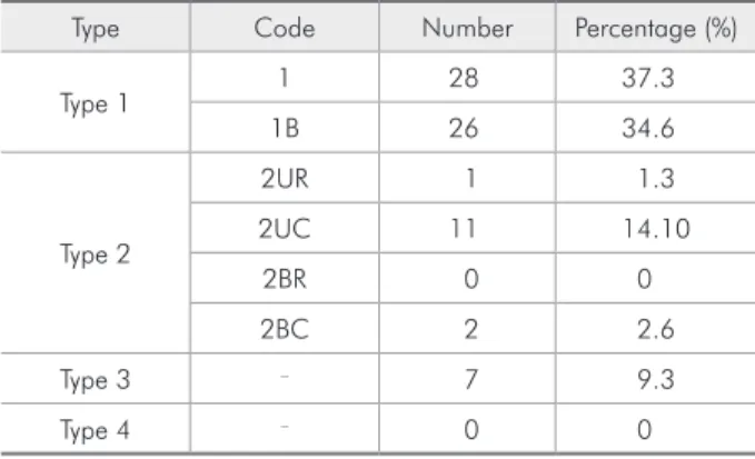
Figure 2 - Bilateral bifurcation extending to the region of the third molar and adjacencies (Type 1B). Right (A) and left (B) lateral and panoramic (C) views. The white arrows indicate the mandibular canal bifurcation.
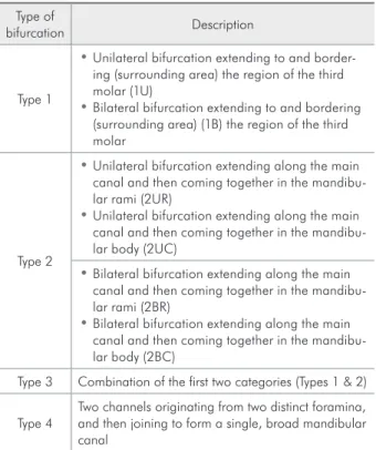
Figure 3 - Unilateral bifurcation (left side) extending along the main canal and united again in the mandibular body (Type 2UC). Left axial cut (A), lateral (B) and panoramic (C) views. The white arrows indicate the bifurcation and the black arrow indicates the mandibular canal. Figure 1 - Unilateral bifurcation (right
side) extending to the region of the third molar and adjacent structures (Type 1U). Axial cut (A), lateral (B) and panoramic (C) views. The white arrows indicate the bifurcation and the black arrow indicates the mandibular canal.
A
A
A
B
B
B C
C
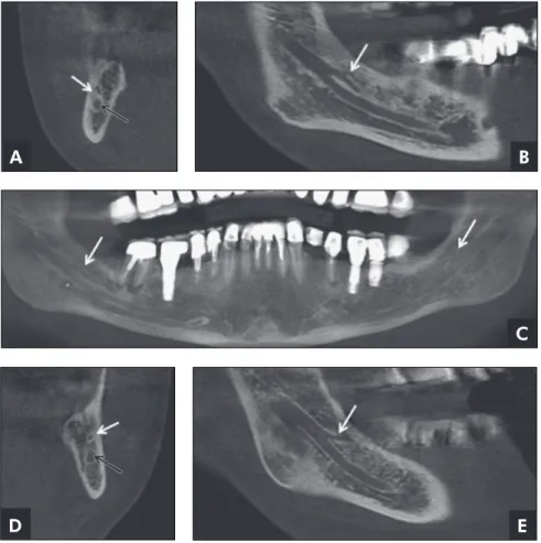
Figure 4 - Combination of the first two categories (Type 3). Right axial cut (A) and lateral view (B) of the bifurcation extending through the mandibular body (Type 2). Bilateral bifurcations in panoramic view (C). Left axial cut (D) and lateral view (E) of the bifurcation, limited to the rami (Type 1). The white arrows indicate the bifurcations of the mandibular canal and the black arrow indicates the mandibular canal.
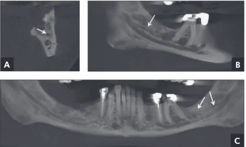
Figure 6 - Transaxial cuts of Type 1 mandibular canal bifurcation close to the roots of the left mandibular third molar. The white arrow indicates the bifurcation and the black arrow, the mandibular canal. The asterisk (*) indicates the third molar roots. Figure 5 - Different sagittal cuts of Type 1 mandibular canal bifurcation in intimate relationship with impacted left mandibular third molars. The red lines (light gray in print version) trace the mandibular canal and its bifurcation paths.
ging from 0.09% to 36%.10,11,24
This variation in incidence is due to differences in the sample size of each study, as well as to the type of examination performed. Studies carried out using
panoramic images show a lower incidence than stud-ies with CBCT images or dry mandibles.18,24,26
It is clear that with the advances in technology and the improvement of images as diagnostic tools,
A B
C
the number of anatomical variations of the mandib-ular canal is likely to increase considerably. Naitoh
et al.13 observed 65% of bifurcations of the
mandib-ular canal in 122 CBCT scans. On the other hand, Kuribayashi et al.14 observed 15% of bifurcated
ca-nals in 301 sides of the mandible, using computed tomography. Since this research work is a retrospec-tive analysis using CBCT previously diagnosed with bifurcations and using a relatively small sample, data could not be obtained on the incidence of bifurca-tions. These data in our population are still being collected for future comparisons of CBCT images.
Studies have found a higher prevalence of biid mandibular canals in females,9,13,14 as observed in
this sample. However, the gender difference was not statistically signiicant. A recent study15 found
a higher prevalence of bifurcation in males, in the Taiwanese population. This study was carried out using medical CT. However, the gender differences could be more related to the population observed rather than to the type of exam.
In the CBCT scans of this study, the biid man-dibular canals were classiied into four types ac-cording to the Langlais et al.9 classiication. Other
classiications have been proposed by Nortjé et al.8
and Naitoh et al.13 The Langlais classiication was
chosen because it is still the most cited in the litera-ture on bifurcation of the mandibular canal.10,11
The most frequent mandibular canal bifurca-tion was Type I (72.7%), which is in agreement with the indings of other studies,15,16 but our frequency
percentage was higher. Rossi et al.11 reported a
fre-quency of Type II mandibular canal bifurcation of 23.3%, which is close to our indings. Nevertheless, Type 2 mandibular canal bifurcation was the most frequent type referred to by Langlais et al.9 The
largest discrepancy observed in biid mandibular ca-nal types found in this study was Type III, account-ing for only 8% of the cases, and differing from the indings of other studies, which range from 0% to 3.92%. No Type IV mandibular canal bifurcation was found in this study. Withal Rossi et al.11
report-ed a high rate (34.9%) of this type of mandibular ca-nal bifurcation. Sample characteristics may explain these discrepancies between the incidences of bifur-cation types.
Biid mandibular canals may have important clinical implications.4,7,14,18,27,28 Third molar surgery
with mandibular canal bifurcation involvement may cause excessive bleeding and/or numbness, especial-ly in cases of Type I or III.9 Inadequate anesthesia
may occur, especially in cases of bifurcation of Type IV, which have two separate foramina.29
Bifurcations in the mandibular body may cause complications in procedures such as endodontics.2
They may inluence prosthesis support, orthodon-tics16,17 and insertion of dental implants.17 In surgical
procedures, such as mandibular osteotomy, surgery complexity increases with the addition of a second vascular-nerve bundle. Furthermore, in cases of oro-facial trauma, the reduction of mandibular fractures must be performed more carefully when there is mandibular canal bifurcation.7 If injured,
mandibu-lar canal and/or bifurcation may cause bleeding and compromise repair in the implant area.
In regard to third molar roots, most of the sam-ple (57.33%) did not show the presence of teeth 38 and/or 48, a fact probably due to the patients’ age and the possibility of these teeth having already been extracted.
The incidence of intimate contact of third molar roots with mandibular canal bifurcation was high (21.33%), followed by proximal contact of third mo-lar roots with the bifurcation (13.33%), and only 8%
of cases where the biid mandibular canal had no involvement with the roots of the third molars, indi-cating the importance of knowledge and identiica-tion of this variaidentiica-tion.30
Knowledge of both the anatomy and the anatom-ical variations of the mandibular canal are essential to the success of mandible-related dental proce-dures. It is important that these variations be identi-ied to prevent treatment complications. The CBCT provides a suitable tool for investigating, identifying and conirming such variations.
Conclusion
References
1. Carter RB, Keen EN. The intramandibular course of the in-ferior alveolar nerve. J Anat. 1971 Apr;108(3):433-40. 2. Moiseiwitsch JRD.. Position of the mental foramen in a North
American, white population. Oral Surg Oral Med Oral Pathol Oral Radiol Endod. 1998 Apr;85(4):457-60.
3. Wadu SG, Penhall B, Towsend GC. Morphological variabil-ity of the human inferior alveolar nerve. Clin Anat. 1997 Feb;10(2):82-7.
4. Nikzad S, Azari A, Sabouri S. Double mandibular foramina and canal: report a case with interactive CT-based planning software. Iran J Radiol. 2008 Feb;5(2):83-6.
5. Iizuka T, Lindqvist C. Sensory disturbances associated with rigid internal fixation of mandibular fractures. J Oral Maxil-lofac Surg. 1991 Dec;49(12):1264-8.
6. Teerijoki-Oksa T, Jääskeläinen SK, Forssel K, Forssel H, Vähätalo K, Tammisalo T, et al. Risk factors of nerve injury during mandibular sagital split osteotomy. Int J Oral Maxil-lofac Surg. 2002 Feb;31(1):33-9.
7. Claeys V, Wackens G. Bifid mandibular canal: literature review and case report. Dentomaxillofac Radiol. 2005 Jan;34(1):55-8. 8. Nortje CJ, Farmen AG, Grotepass FW. Variations in the
normal anatomy of the inferior dental (mandibular) canal: a retrospective study of panoramic radiographs from 3612 routine dental patients. Br J Oral Surg. 1977 Jul;15(1):55-63. 9. Langlais RP, Broadus R, Glass BJ. Bifid mandibular
canals in panoramic radiographs. J Am Dent Assoc. 1985 Jun;110(6):923-6.
10. Devito KL, Tamburús JR. [Anatomy of the mandibular canal: radiological classification of its variations]. Rev Assoc Paul Cir Dent. 2001 Jul-Aug;55(4):261-6. Portuguese.
11. Rossi PM, Brücker MR, Rockenbach MIB. [Bifid mandibular canals: panoramic radiographic analysis]. Rev Cienc Med. 2009 Mar-Apr;18(2):99-104. Portuguese.
12. Rouas P, Nancy J, Bar D. Identification of double mandibular canals: literature review and three case report with CT scans and cone beam CT. Dentomaxillofac Radiol. 2007 Jan;36(1):34-8. 13. Naitoh M, Hiraiva Y, Aimiya H, Ariji E. Observation of bifid
mandibular canal using cone beam computerized tomography. Int J Oral Maxillofac Implants. 2009 Jan-Feb;24(1):155-9. 14. Kuribayashi A, Watanabe H, Imaizumi A, Tantanapornkul
W, Katakami K, Kurabayashi T. Bifid mandibular canals: cone beam computed tomography evaluation. Dentomaxil-lofac Radiol. 2010 May;39(4):235-9.
15. Fu E, Peng M, Chiang CY, Tu HP, Lin YS, Shen EC. Bifid man-dibular canals and the factors associated with their presence: a medical computed tomography evaluation in a Taiwanese population. Clin Oral Implants Res. 2012 Nov 6. doi: 10.1111/ clr.12049. Epub ahead of print.
16. al Jasser NM, Nwoku AL. Radiographic study of the mental foramen in a selected Saudi population. Dentomaxillofac Ra-diol. 1998 Nov;27(6):341-3.
17. de Oliveira-Santos C, Souza PH, De Azambuja Berti-Couto SA, Stinkens L, Moyaert K, Rubira-Bullen IR, et al. Assessment of variations of the mandibular canal through cone beam com-puted tomography. Clin Oral Investig. 2012 Apr;16(2):387-93. 18. Sanchis JM, Peñarrocha M, Soler F. Bifid mandibular canal.
J Oral Maxillofac Surg. 2003 Apr;61(4):422-4.
19. Kim MS, Yoon SJ, Park HW, Kang JH, Yang SY, Moon YH, et al. A false presence of bifid mandibular canals in panoramic radiographs. Dentomaxillofac Radiol. 2011 Oct;40(7):434-8. 20. Arensburg B, Nathan H. Anatomical observation of the my-lohyoid groove, and the course of the mymy-lohyoid nerve and vessels. J Oral Surg. 1979 Feb;37(2):93-6.
21. Angelopoulos C, Thomas S, Hechler S, Parissis N, Hlavacek M. Comparison between digital panoramic radiography and cone-beam computed tomography for the identification of the mandibular canal as part of presurgical dental implant assessment. J Oral Maxillofac Surg. 2008 Oct;66(10):2130-5. 22. Xie Q, Wolf J, Soikkonen K, Ainamo A. Height of mandibular
basal bone in dentate and edentulous subjects. Acta Odontol Scand. 1996 Dec;54(6):379-83.
23. Auluck A, Pai KM, Shetty C. Pseudo bifid mandibular canal. Dentomaxillofac Radiol. 2005 Nov;34(6):387-8.
24. Bogdan S, Pataky L, Barabas J, Nemeth Z, Huszar T, Szabo G. Atypical courses of the mandibular canal: comparative examination of dry mandibles and x-ray. J Craniofac Surg. 2006 May;17(3):487-91.
25. Wadhawani P, Mathur RM, Kohli M, Sahu R. Mandibu-lar canal variant: a case report. J Oral Pathol Med. 2008 Feb;37(2):122-4.
26. Fukami K, Shiozaki K, Mishima A, Kuribayashi A, Hamada Y, Kobayashi K. Bifid mandibular canal: confirmation of limited cone beam computed tomography findings by gross anatomi-cal and histologianatomi-cal investigations. Dentomaxillofac Radiol. 2012 Sep;41(6):460-5.
27. Lew K, Townsend G. Failure to obtain adequate anaesthesia associated with a bifid mandibular canal: a case report. Aust Dent J. 2006 Mar;51(1):86-90.
28. Mizbah K, Gerlak N, Maal TJ, Bergé SJ, Meijer GJ. The clinical relevance of bifid and trifid mandibulars canals. Oral Maxillofac Surg. 2012 Mar;16(1):147-51.
29. DeSantis JL, Liebow C. Four common mandibular nerve anomalies that lead to local anesthesia failures. J Am Dent Assoc. 1996 Jul;127(7):1081-6.