The aim of this study was to evaluated the root surfaces modifications resulted by application of different chemicals agents, and their influence on the fibrin network and fibroblasts attachment. From 96 anterior mandibular human extracted incisor teeth, 192 dentin blocks of buccal and lingual surface were obtained and randomly divided into 6 groups: Cont- control group, which received no treatment; Root surface scaling and root planing (Srp); Citric acid-Srp; EDTA-Srp; Tetracycline capsule-Srp; Tetracycline gel-Srp. After dentin treatments the specimens were analyzed as follows: 1) demineralization level and residues of the product by scanning electron microscopy (SEM); 2) adhesion of blood components after 20 min of surface treatment by SEM; 3) fibroblast attachment after 24 h by SEM; 4) cell metabolism after 24 h by MTT assay. Data were analyzed using Fisher Exact, One-way ANOVA test followed by Dunn’s test, Tukey test and Dunnett test (α=0.05). Citric acid, EDTA and Tetracycline gel resulted in adequate demineralization with no completely smear layer and smear plug removal on root dentin surface. Tetracycline capsule produced great tetracycline residues with several demineralization areas. Tetracycline gel and EDTA groups presented more fibroblast fixation than other experimental groups. The highest mean blood clot adhesion score was observed in roots treated with tetracycline gel. EDTA and Tetracycline gel surface treatment removed the smear layer over dentin surface and promoted adhesion of fibrin network and fibroblast cells attachment.
Biological Effects of a Root Conditioning
Treatment on Periodontally Affected
Te e t h
– A n I n V i t r o
A n a l y s i s
Aline Cristina Silva1, Camilla Christian Gomes Moura2, Jessica Afonso Ferreira1, Denildo de Magalhães1, Paula Dechichi3, Priscilla Barbosa Ferreira Soares1
1Department of Periodontology
and Implantology, UFU -
Universidade Federal de Uberlandia, Uberlândia, MG, Brazil
2Department of Endodontics,
UFU - Universidade Federal de Uberlândia, Uberlândia, MG, Brazil
3Biomedical Sciences Institute,
UFU - Universidade Federal de Uberlândia, Uberlândia, MG, Brazil
Correspondence: Priscilla Barbosa Ferreira Soares, Av. Pará, nº 1720 - Bloco 4L Anexo A – Sala 4LA, 38400-902 Uberlândia, MG, Brasil. Tel: +55-34-3218-2255. e-mail: pbfsoares@yahoo.com.br
Key Words: fibroblasts, periodontal diseases, fibrin, scanning electron microscopy, smear layer.
Introduction
The traditional treatment of periodontally compromised roots have relied on mechanical instrumentation (1-4) that is not able to fully eliminate the infection due the formation of a smear layer of organic and mineralized debris (3-6). As a compensation for these limitations of mechanical therapy; chemical root conditioning has been introduced.
Chemical therapy may favor clot stabilization in the earlier stages of periodontal healing by increasing the adhesion of blood cells and fibrin to the root surface (7,8). Root conditioning is intended to detoxify the root surface by removing the smear layer and demineralize by exposing the collagen matrix that supports migration, proliferation, adhesion and matrix synthesis of the cells involved in periodontal healing (9). This is important for periodontal tissue repair and regeneration (2,9,10).
Chemical conditioning agents like citric acid, phosphoric acid, EDTA and tetracycline have been used in clinical practice (10,11). Nevertheless, the great variability of protocols employed by clinicians and researchers has prevented consistent comparisons among them. Citric acid and tetracycline hydrochloride have been extensively researched and used clinically because of higher tissue tolerance and easy storage (12). Also, etching at neutral pH
with agents like EDTA has been shown to have a comparable demineralizing potential (12).
Therefore, considering the controversies in the literature and the lack of new in vitro and in vivo controlled studies, the aim of this in vitro study was to evaluate the influence of different root conditioning agents on the adhesion of fibrin clot and fibroblast cells to root surfaces.
Material and Methods
Preparation of Human Dentin Blocks
Surface conditioning on periodontal teeth
phosphate-buffered saline (PBS) (pH 7.4) at 37 °C until the treatments were performed. Using a high-speed cylindrical bur under copious irrigation, two parallel grooves were made on the proximal root surface of each tooth: one at the CEJ and the other approximately 3 mm apical to the first groove. To create a smear layer, the area between the two grooves was scaled and planed with Gracey curettes 5/6 (Hu-Friedy, Chicago, IL, USA) through 50 apico-cervical traction movements by the same operator to remove the contaminated cement and expose dentin. The roots were cross cut in the first groove, separating them from the crown. All roots were immediately cut lengthwise in the buccolingual orientation and then in the mesiodistal orientation until the second groove was reached apically. Two dentin blocks, approximately 3x3x1 mm in size, were obtained from each tooth, one from the buccal and another from the lingual surface (192 specimens). The 192 specimens were divided into 6 groups, submitted to 4 experimental analyses: 1) presence of smear layer and residues of products (n=9); 2) cell metabolic activity (n=5); 3) fibroblast cell attachment (n=9); 4) blood component adhesion (n=9). The SEM micrographs were analyzed by blinded and calibrated examiner, which received codified images.
Chemical Treatment of Specimens
The specimens were randomly divided into 6 groups: Cont- control group, which received no treatment; Srp - root surface scaling and root planning with Gracey curettes (Hu-Friedy) to remove calculus deposits and cementum, creating a smear layer (it was the first step for all the others groups); SrpCi - after Srp, the dentin was etched with 30% citric acid pH 1.6 (Biopharma, Uberlândia, MG, Brazil) for 5 min; SrpEdta - after Srp the dentin was etched with 24% EDTA gel pH 7.0 (Biopharma) for 1 min; SrpTe - after Srp the dentin was etched for 3 min with a solution obtained by 500 mg tetracycline capsule (Tetraciclina, Medquimíca, São Paulo, SP, Brazil) dissolved in 2 mL of saline solution; SrpTeg - after Srp the dentin was etched with 50 mg/mL tetracycline gel pH 1.8 (Biopharma) for 1 min. Following, all specimens were rinsed for 1 min with 20 mL of saline solution and randomly used in 4 different methodologies.
Presence of Smear Layer and Residues of Products - Scanning Electron Microscopy (SEM)
Immediately after rinsing, the specimens (n=9) were dehydrated in an sequence ascending of 30%, 50% and 70% ethanol solutions for 30 minu, followed by washing with 95% and 100% ethanol for 60 min in a two times and immersed in 200 μL of 1,1,1,3,3,3-hexamethyldisilazane (HMDS; Sigma-Aldrich Inc., St. Louis, MO, USA) for 60 min at room temperature. The specimens were stored in a desiccator for 24 h, mounted on metallic stubs,
sputter-coated with gold and observed by field emission scanning electron microscopy (EVO MA 10, Carl Zeiss, London, UK) at a voltage of 20 kV. Images of the center of each specimen were obtained at 1000x magnification. Using a single-blind method, the SEM images were examined three times at 7-day intervals by an operator who was previously trained and calibrated. Each sample received the score that prevailed among the three readings. For specimens, a rating system for root biomodification (11) was employed:
Presence of smear layer:1- great amount of smear layer and smear plug; 2- absence of smear layer and smear plug; Presence of the product residues:1- great amount of product residues; 2- no product residues were left.
Cell Culture
Immortalized human mouth fibroblasts (Cell Bank of Rio de Janeiro, Rio de Janeiro, RJ, Brazil) were cultured in T-25 cell culture flasks containing Dulbecco’s Modified Eagle Medium (DMEM) (Sigma Chemical Co., St. Louis, MO, USA) supplemented with 10% fetal bovine serum (FBS; Gibco, Grand Island, NY, USA), with 100 IU/mL penicillin, 100 μg/mL streptomycin and 2 mM/L glutamine (Gibco) in a humidified incubator with 5% CO2 and 95% air at 37 oC (Isotemp; Fisher Scientific, Pittsburgh, PA, USA). Growth was permitted until the cells achieved 70% of confluence.
Cell Metabolic Activity (MTT Assay)
The specimens (n=5) were placed into 96-well plates (Coastar Corp., Cambridge, MA, USA). Because the square dentin slices did not fit into the rounded culture wells, 1x10⁵ number of cells per well was suspended in 50 μL of α-MEM medium, and the suspension was placed directly on the surface of the dentin block. After a 1-h incubation period, 50 μL of culture medium was added to the suspension, and it was further incubated for 24 h. The positive control corresponded to cells maintained at 10% DMEM, without additional treatment. After 24 h, the medium was removed and 100 μL of fresh medium and 20 µL of 3-[4,5-dimethylthiazol-2-yl]-2,5-diphenyl tetrazolium bromide (MTT) solution (Sigma-Aldrich) was added to each well in order to measure mitochondrial activity of the cells. After 4h, the MTT solution was removed, and 100 µL of dimethyl sufoxide (Sigma-Aldrich) was added to each well. Cell viability was determined by measuring the optical density at 540 nm on a microplate reader. The absorbance values (%) obtained from each specimen was measured after 24 h.
Fibroblast Cell Attachment - SEM Analysis
P
.B
.F
. Soares et al.
in the cell cytotoxicity section. The dentin slices with the fibroblasts cells adhered to dentin surface were immediately immersed in 2.5% glutaraldehyde solution for 60 min and post-fixed with 1% osmium tetroxide for 60 min. The cells that remained adhered to the dentin surface had their morphology examined with a SEM (EVO MA 10). Images of the center of each specimen were obtained at 3000x magnification. The attached fibroblasts were counted by using an Image J program. Two fields of 1 mm2 per specimen were counted and one mean value was calculated per specimen, which was used for analysis.
Blood Component Adhesion - SEM Analysis
Immediately after treatments, the specimens (n=9) were exposed to whole peripheral blood (7). After signed an informed consent form, fresh human whole peripheral blood from a healthy male donor was applied to the root surface. The blood was allowed to clot onto the root blocks for 20 min in a humidified chamber at 37 °C. Blocks were then rinsed three times for 5 min in PBS. Washes and rinses of the root blocks were performed in small Petri dishes with a gentle swirling motion using a rotating tabletop shaker at low speed. After rinsing, the blocks were immediately immersed in 2.5% glutaraldehyde solution for 60 min and post-fixed with 1% osmium tetroxide for 60 min each one. SEM images of representative areas of each specimen were obtained at 3000x magnification. Specimens received the score that prevailed among the three readings. A rating system for blood component adhesion (12) was used to analyze the morphologic characteristics obtained in the treatment: 0: absence of fibrin network and blood cells; 1: scarce fibrin network and/or blood cells; 2: moderate fibrin network and moderate quantity of blood cells; 3: dense fibrin network and trapped blood cells.
Statistical Analysis
Data of smear layer and product residue presence were analyzed using Fisher Exact test followed by Dunn’s test. The MTT assay and fibroblast cell attachment were analyzed using one-way ANOVA followed Tukey’s test and Dunnett test for comparison to control group. The fibrin clot adhesion data were analyzed using Fisher Exact test followed by Dunn’s test and Dunnett test for comparison to control group. Analysis of the statistical test power was verified with a minimum power of 0.75.All tests employed a 0.05 level of statistical significance and all statistical analyses were carried out with the statistical package Sigma Plot 13.1 (Systat Software Inc, San Jose, CA, USA).
Results
Presence of Smear Layer
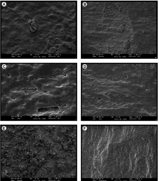
Figure 1 shows the surface of dentin treated with each
root conditioning agents. The smear layer presence on dentin (scores) for all groups are shown in Table 1. Fisher Exact test revealed a statistically significant difference among groups (p=0.001). Dunn’s test showed that Tetracycline gel, EDTA and Citric Acid groups had significantly lower smear layer level than scaling and root planing group (Table 1).
Presence of Product Residues
The presence of residue on dentin (scores) for all groups are shown in Table 1. Fisher Exact test revealed a statistically significant difference among groups (p=0.003). Dunn’s test showed that Tetracycline gel, EDTA and Citric Acid groups had significantly lower residue levels than Tetracycline capsule (Table 1).
Cell Metabolic Activity
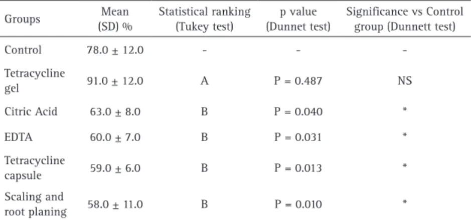
The MTT assay values (mean and standard deviation expressed in %) for all experimental groups and control group are shown in Table 2. ANOVA revealed a statistically significant difference among groups (p=0.001). Dunnett test showed that Tetracycline capsule, EDTA, Citric Acid and scaling and root planing groups resulted in significantly lower absorbance levels than control group. On the other hand, Tetracycline gel resulted in similar absorbance level with control group (Table 2). Tukey test showed that Tetracycline gel resulted in significantly more cell viability than other experimental groups (Table 2).
Fibroblast Cell Attachment
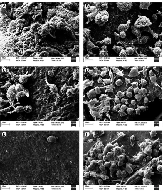
Figure 2 shows the attached fibroblasts cell of human dentin by SEM. The fibroblast cell values (mean and standard deviation) for all experimental groups and control group are shown in Table 3. ANOVA revealed a statistically significant difference among groups (p=0.001). Dunnett test showed that the number of fibroblast cells was significantly lower for Tetracycline capsule group than control group. Tetracycline gel and EDTA groups had significantly more fibroblast fixation than control group (Table 3). Tukey test showed that Tetracycline gel and EDTA groups had significantly more fibroblast fixation than other experimental groups. Tetracycline capsule group had significantly lower fibroblast fixation than scaling and root planing group (Table 3).
Fibrin Clot Adhesion
Surface conditioning on periodontal teeth
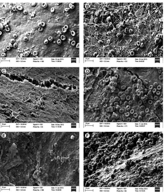
groups were significantly higher than control group (Table 4). Dunn’s test showed that Tetracycline gel group had significantly more fibrin clot adhesion than citric acid and Tetracycline capsule groups. Tetracycline capsule group had significantly lower fibrin clot adhesion than scaling and root planing and EDTA group (Table 4).
Discussion
This study was carried out to evaluate biological effects of different chemical agents used for root conditioning on periodontal therapy. For this purpose, substances largely studied by current literature, with broad access and low cost were chosen: tetracycline (1,5-7,11-19), EDTA (5,7,11,12,20) and citric acid (2,3,11,12,14). Despite the
large number of research involving such chemical agents (1-3,5-7,11-14,21), to date, no study has compared their effects on initial biological events related to the success of regenerative periodontal therapy. Considering that the removal of the smear layer and exposure of dentin matrix by acid agents is followed by the formation and adhesion of blood clot, which forms a network for migration and adhesion of fibroblasts and that the maintenance of the viability of those cells is essential for regenerative events, the following parameters were evaluated: smear layer removal, adhesion of blood components (22,23), fibroblast adhesion and metabolism (24).
In attempt to mimic the conditions found in vivo, prior to chemical treatment of the root surfaces, they were
P
.B
.F
. Soares et al.
scaled and root planned. However, as a result of mechanical therapy, is produced a thick smear layer of microcrystalline debris (14,20) which is intimately associated with the root surface, as confirmed by the present study in control group. Therefore, acid conditioning have been used to decontaminate and to demineralize the root surface, remove the smear layer, exposing some components of the extracellular matrix of dentin and cementum, such as type I collagen, and facilitating attachment between the root surface and the healing connective tissue (10,13,25). In agreement with previous studies (1,4,5), the method of application of the root conditioning used in the current research resulted in root surface demineralization, smear layer removal and exposing of dentinal tubules and collagen of intra- and peritubular dentinal matrix, which may helped the adhesion of the fibrin clot and fibroblasts cells on acid
treated groups, except in tetracycline in capsule.
The use of tetracycline have been associated with increased adhesion between glycoproteins and dentin, stimulation, proliferation and adhesion of fibroblasts (1,24), anticollagenase activity (16), inhibition of osteoclast function (15) and neutrophil function (17), as well as efficient substantively (1). In present study, regarding the parameter evaluated, favorable results were found in the group treated with tetracycline gel but not in the group treated with tetracycline capsule. In the further group, the high amount of residues showed on the root dentin surface are probably related to the components present into the capsules, which were not completely dissolved (11). The presence of residues of tetracycline particles during long time may result in continuous demineralization of the root dentin (11), creating gaps that could result
on lack of insertion of tissue (6). Furthermore, this procedure may cause irregular surface with many depressions after demineralization (11). Otherwise, tetracycline gel treatment promoted the most organized fibrin network and cell entrapment, probably related to the collagen fiber exposure, which was greater in this group. The analysis of cell attachment confirmed the superior ability of tetracycline gel created a favorable environment able to facilitate the initial events of healing as formation of fibrin clot and cell attachment (12). In agreement, other in vitro studies showed that tetracycline gel (1) and EDTA (5,25) demineralization might enhance fibroblasts cell adhesion to the root surface. Despite the methodological limitations to confirming the attachment of the fibrin clot and fibroblasts cells to the root surfaces, SEM analysis remains as a predictable way of assessing the attachment of fibrin clot and fibroblasts cells to the treated root surfaces.
Cell viability was indirectly evaluated by the metabolic analysis of attached cells using MTT assay. Considering that a material for use in dental tissues need to be atoxic, the present research evaluate the influence of demineralized agents
Table 1. Distribution of smear layer covering dentin and product residues scores - statistical significance regarding control group calculated by Dunn’s test.
Groups
Presence of smear layer Presence of product residues 1 2 Statistical Ranking
(Dunn’s test) 1 2
Statistical Ranking (Dunn’s test)
Control na na - na na
-Tetracycline capsule na na - 9 - B
Tetracycline Gel - 9 A - 9 A
EDTA - 9 A - 9 A
Citric Acid - 9 A - 9 A
Scaling and root planning 9 - B na na
-Different letter means statistically different in comparison to control group (p<0,05); na- not applied; presence of smear layer: 1-presence of great amount of smear layer and smear plug; 2-absence of smear layer and smear plug; presence of the product residues: 1-presence of great amount of product residues; 2- no product residues left use.
Table 2. Mean and standard deviations (SD) of cell viability, statistical categories by Tukey test and P values calculated by Dunnett test
Groups Mean (SD) % Statistical ranking (Tukey test) p value (Dunnet test)
Significance vs Control group (Dunnett test)
Control 78.0 ± 12.0 - -
-Tetracycline
gel 91.0 ± 12.0 A P = 0.487 NS
Citric Acid 63.0 ± 8.0 B P = 0.040 *
EDTA 60.0 ± 7.0 B P = 0.031 *
Tetracycline
capsule 59.0 ± 6.0 B P = 0.013 *
Scaling and
root planing 58.0 ± 11.0 B P = 0.010 *
Surface conditioning on periodontal teeth
Figure 2. Attached fibroblasts cell of human dentin seem by SEM. (A) Control group, (B) root surface scaling and root planning, (C) citric acid, (D) EDTA, (E) tetracycline capsule, and (F) tetracycline gel (Original magnification ×3000).
Table 3. Mean fibroblast cell values and standard deviations (SD) statistical categories by Tukey test and p values calculated by Dunnett test.
Groups Mean (SD) Statistical ranking (Tukey test)
p value (Dunnet test)
Significance vs Control group (Dunnett test)
Control 17.4 ±
10.5 - -
-Tetracycline gel 31.1 ± 8.2 A P = 0.002 *
EDTA 28.2 ± 6.3 A P = 0.025 *
Scaling and root planing 14.9 ± 7.7 B P = 0.972 NS
Citric Acid 9.7 ± 4.4 BC P = 0.201 NS
Tetracycline capsule 2.9 ± 2.0 C P = 0.001 *
P
.B
.F
. Soares et al.
Figure 3. Effect of surface treatment on fibrin clot formation on dentin. (A) Control group, (B) root surface scaling and root planning, (C) citric acid, (D) EDTA, (E) tetracycline capsule, and (F) tetracycline gel (Original magnification ×3000).
Table 4. Scores of fibrin clot adhesion on dentin and blood cells deposited over dentin; statistical significance in comparison with control group (Dunnett test) and among experimental groups (Dunn’s test)
Groups
Fibrin clot adhesion on dentin Blood cells deposited over dentin 0 1 2 3 Significance vs Control
group (Dunnett test) 0 1 2 3
Statistical Ranking (Dunn’s test)
Control - 7 2 -
-Tetracycline gel - - 3 6 * - - 3 6 A
Scaling and root planing - 1 4 4 * - 1 4 4 AB
EDTA - 7 2 - NS - 2 3 4 AB
Citric Acid - 7 2 - NS - 7 2 - BC
Tetracycline capsule 1 8 - - NS 1 8 - - C
Surface conditioning on periodontal teeth
on fibroblasts metabolic activity at 4h, which could be considered a short period. The present results confirmed the absence of cytotoxic of tetracycline gel (11). However, the low pH of citric acid solution and tetracycline gel or capsule suggested that such agents could interfere with cell viability (18) and the initial events of the healing process periodontal (19), which was confirmed by the current findings. Further studies need to evaluate the influence of demineralization agents in the long term, in other to evaluate the effects of initial cytotoxicity of those agents on other biological events as cell necrosis or apoptosis, and the capability of cell proliferation.
This study presents some limitations such as the use of immortalized cell culture, which have no interactions with other cell types, however the methodology employed allows variables isolation for mimicking the initial healing events after root conditioning. Other relative limitation of this study is the difference on the time application for each substance. The difference on the performance of the different products on the root dentin may impair on the results on this. However it is a relative limitation, because it is not possible to adjust the same time application for the different products (11).
Based on the methodology applied and the limitations of this study, the tetracycline gel was able to provide significant increase in the adhesion of fibrin network and fibroblasts cells to the root surfaces, when compared to the other group. Also, all the substances employed, except tetracycline capsule, were able to remove the smear layer when compared to the control group, but not all of them effectively cleaned the root surface, removing the smear layer and exposing collagen fibers.
Resumo
O objetivo deste estudo foi avaliar modificações nas superfícies radiculares sofridas pela aplicação de diferentes agentes químicos, e sua influência sobre a rede de fibrina e adesão de fibroblastos. A partir de 96 incisivos inferiores humanos, 192 blocos de dentina das superfícies vestibular e lingual foram obtidos e divididos aleatoriamente em 6 grupos: Cont-grupo controle, não recebeu nenhum tratamento; Raspagem e alisamento radicular (RAR); Ácido cítrico-SRP; EDTA-SRP; Tetraciclina em cápsula-SRP; Tetraciclina gel-SRP. Após o tratamento da dentina as espécimes foram analisadas: 1, nível de desmineralização e resíduos do produto por microscopia eletrônica de varredura (MEV); 2, adesão dos componentes sanguineos após 20 min na tratamento de superfície por SEM; 3, adesão de fibroblastos após 24h por SEM; 4, o metabolismo celular após 24 h por ensaio MTT. Os dados foram analisados por Fisher Exact, teste one-way ANOVA, seguido pelo teste de Dunn, teste de Tukey e teste de Dunnett (α = 0,05). O ácido cítrico, EDTA e gel tetraciclina resultaram na adequada desmineralização sem remoção completa de camada de smear layer e smear plug sobre a superfície da dentina radicular. Cápsula de tetraciclina produziu grandes resíduos de tetraciclina com várias áreas de desmineralização. Os grupos Gel de tetraciclina e EDTA apresentaram maior adesão de fibroblastos do que os demais grupos experimentais. O maior score de adesão de coágulo sanguineo foi observado nas superfícies tratadas com gel de tetraciclina. EDTA e Gel de tetraciclina removeram a camada de smear
layer sobre a superfície da dentina e promoveu adesão da rede de fibrina e de fibroblastos.
Acknowledgements
This study was financially supported by FAPEMIG and CAPES. This study was carried out in the CPBIO-FOUFU (Research Center at the School of Dentistry - Federal University of Uberlândia). The authors are indebted to Francielle Silva Batista (Chemical Engineering School - FEQ - UFU) for technical support in scanning electron microscopy.
References
1. Isik AG, Tarim B, Hafez AA, Yalçin FS, Onan U, Cox CF. A comparative scanning electron microscopic study on the characteristics of demineralized dentin root surface using different tetracycline HCl concentrations and application times. J Periodontol 2000;71:219-225. 2. Baker PJ, Rotch HA, Trombelli L, Wikesjö UM. An in vitro screening model to evaluate root conditioning protocols for periodontal regenerative procedures. J Periodontol 2000;71: 1139-1143.
3. Caffesse RG, De LaRosa M, Garza M, Munne-Travers A, Mondragon JC, Weltman R. Citric acid demineralization and subepithelial connective tissue grafts. J Periodontol 2000;71: 568-572.
4. Cavassim R, Leite FR, Zandim DL, Dantas AA, Rached RS, Sampaio JE. Influence of concentration, time and method of application of citric acid and sodium citrate in root conditioning. J Appl Oral Sci 2012;20:376-383.
5. Amaral NG, Rezende ML, Hirata F, Rodrigues MG, Sant’ana AC, Greghi SL et al. Comparison among four commonly used demineralizing agents for root conditioning: a scanning electron microscopy. J Appl Oral Sci 2011;19:469-475.
6. Ishi EP, Dantas AA, Batista LH, Onofre MA, Sampaio JE. Smear layer removal and collagen fiber exposure using tetracycline hydrochloride conditioning. J Contemp Dent Pract 2008;9:25-33.
7. Leite FR, Sampaio JE, Zandim DL, Dantas AA, Leite ER, Leite AA. Influence of root-surface conditioning with acid and chelating agents on clot stabilization. Quintessence Int 2010;41:341–349.
8. Amireddy R, Rangarao S, Lavu V, Madapusi BT. Efficacy of a root conditioning agent on fibrin network formation in periodontal regeneration: A SEM evaluation. J Indian Soc Periodontol 2011;15:228-234.
9. Fernyhough W, Page RC. Attachment, growth and synthesis by human gingival fibroblasts on demineralized or fibronectin-treated normal and diseased tooth roots. J Periodontol 1983;54:133-140.
10. Zandim DL, Leite FR, Silva VC, Lopes BM, Spolidorio LC, Sampaio JE. Wound healing of dehiscence defects following different root conditioning modalities: an experimental study in dogs. Clin Oral Investig 2013;17:1585–1593.
11. Soares PBF, Castro CG, Branco CA, Magalhães D, Neto AJF, Soares CJ. Mechanical and acid root treatment on periodontally affected human teeth - a scanning electronic microscopy. Braz J Oral Sci 2010;9:128-132.
12. Minocha T, Rahul A. Comparison of fibrin clot adhesion to dentine conditioned with citric acid, tetracycline, and ethylene diamine tetra acetic acid: An in vitro scanning electron microscopic study. J Indian Soc Periodontol 2012;16:333-341.
13. Trombelli L, Schincaglia GP, Zangari F, Griselli A, Scabbia A, Calura G. Effects of tetracycline HCl conditioning and fibrin-fibronectin system application in the treatment of buccal gingival recession with guided tissue regeneration. J Periodontol 1995;66:313-320.
14. Shetty B, Dinesh A, Seshan H. Comparitive effects of tetracyclines and citric acid on dentin root surface of periodontally involved human teeth: A scanning electron microscope study. J Indian Soc Periodontol 2008;12:8-15.
P
.B
.F
. Soares et al.
16. Golub LM, Ramamurthy N, McNamara TF, Gomes B, Wolff M, Casino A et al. Tetracyclines inhibit tissue collagenase activity. A new mechanism in the treatment of periodontal disease. J Periodontal Res 1984;19:651-655.
17. Gabler WL, Creamer HR. Suppression of human neutrophil functions by tetracyclines. J Periodontal Res 1991;26:52-58.
18. Alger FA, Solt CW, Vuddhakanok S, Miles K The histologic evaluation of new attachment in periodontally diseased human roots treated with tetracycline-hydrochloride and fibronectin. J Periodontol 1990;61:447-455.
19. Wang Y, Morlandt AB, Xu X, Carnes DL Jr, Chen Z, Steffensen B. Tetracycline at subcytotoxic levels inhibits matrix metalloproteinase-2 and -9 but does not remove the smear layer. J Periodontol 2005;76:1129-1139.
20. Martins Júnior W, De Rossi A, Samih Georges Abi Rached R, Rossi MA. A scanning electron microscopy study of diseased root surfaces conditioned with EDTA gel plus Cetavlon after scaling and root planing. J Electron Microsc 2011;60:167-175.
21. Kassab M, Cohen RE. The effect of root modification and
biomodification on periodontal therapy. Compend Contin Educ Dent 2003;24:31-34.
22. Theodoro LH, Sampaio JE, Haypek P, Bachmann L, Zezell DM, Garcia VG. Effect of Er:YAG and Diode lasers on the adhesion of blood components and on the morphology of irradiated root surfaces. J Periodontal Res 2006;41:381-390.
23. Leite FR, Moreira CS, Theodoro LH, Sampaio JE. Blood cell attachment to root surfaces treated with EDTA gel. Braz Oral Res 2005;19:88–92. 24. Baker DL, Stanley Pavlow SA, Wikesjö UM. Fibrin clot adhesion to dentin
conditioned with protein onstructs: an in vitro proof-of-principle study. J Clin Periodontol 2005;32:561-566.
25. Cekici A, Maden I, Yildiz S, San T, Isik G. Evaluation of blood cell attachment on Er: YAG laser applied root surface using scanning electron microscopy. Int J Med Sci 2013;10:560-566.


