CEREBROVASCULAR DISEASE IN NEWBORN INFANTS
REPORT OF THREE CASES WITH CLINICAL
FOLLOW-UP AND BRAIN SPECT IMAGING
MARIA VALERIANA L. DE MOURA-RIBEIRO*, SYLVIA MARIA CIASCA**,
MARIZA VALE-CAVALCANTI***, ELBA C. S. C. ETCHEBEHERE****, EDWALDO E. CAMARGO*****
ABSTRACT - The clinical and neurological findings of three neonates with the diagnosis of cerebrovascular disease are reported. The neuropsychological evaluation disclosed impairment of fine motor function, coordination, language, perception and behavioral disturbances. Brain SPECT imaging revealed perfusional deficits in the three cases.
KEY WORDS: neonates, stroke, cerebrovascular disease, brain SPECT imaging.
Doença cerebrovascular em recém-nascidos : relato de três casos com estudo clínico evolutivo e SPECT cerebral
RESUMO - Os resultados dos estudos clínico e neurológico de três recém-nascidos com diagnóstico de doença cerebrovascular são apresentados. A avaliação neuropsicológica demonstrou alterações na motricidade fina, coordenação, linguagem, percepção e comportamento. O SPECT cerebral mostrou alterações perfusionais nos três recém nascidos.
PALAVRAS-CHAVE: recém-nascido, icto neonatal, doença cerebrovascular, SPECT cerebral.
In the past decade, new methods of cerebral function evaluation using different imaging modalities have acquired expressive development and clinical relevance. In cerebrovascular diseases (CVD) the demonstration of blood flow and metabolic abnormalities in local and remote areas (diaschisis)1,2 is possible. In spite of this progress, brain SPECT imaging has not been used routinely
in newborns and infants, especially those with confirmed diagnosis of CVD in the first days of life. A better understanding of the regional cerebral metabolism may be achieved using brain SPECT imaging as a complementary diagnostic modality.
The clinical and laboratory data of three newborns with neonatal CVD have been reviewed with the purpose of: a) to determine the magnitude of anatomic and functional impairment caused by the acute event (stroke); b) to correlate prospectively the neurologic and cognitive findings with brain SPECT imaging data.
Disciplina de Neurologia Infantil do Departamento de Neurologia e Divisão de Medicina Nuclear do Departamento de Radiologia, Faculdade de Ciências Médicas (FCM) da Universidade Estadual de Campinas (UNICAMP): *Professor Associado, **Professor Assistente Doutor, ***Médica Estagiária, ****Médica Assistente da Medicina Nuclear, *****Professor Titular. Aceite: 4-agosto-1999.
The patients were submitted to brain SPECT imaging with [Tc-99m] ECD. Tomographic images of the brain were obtained 20 minutes after an intravenous administration of 740 MBq (20 mCi) of [Tc-99m] ECD, using a scintillation camera with a fan beam collimator. The images were reconstructed in the transaxial, coronal and sagittal planes to improve detection of perfusion abnormalities in the cerebral cortex, basal ganglia, thalami and cerebellum. Visual analysis of the studies was performed by two experienced Nuclear Medicine physicians.
The neurological evaluation in the neonatal period and in the first years of life was performed according to the protocol of the Division of Pediatric Neurology, Campinas State University School of Medical Sciences (Unicamp), similar to the recommendations of Lefèvre3 and Diament4. The
neurological evaluation and brain SPECT images were correlated with the childhood assessment of cortical functions using the Luria-Nebraska test 5 .
CASE REPORTS
Case 1.
ALM, a white male, was born to term by breech delivery, with 2,700 g, 46 cm in length, head perimeter of 35 cm and an Apgar index of 1 and 3, in the first and fifth minutes, respectively. Two hours after birth the newborn developed clonic seizures of the left upper limb of short duration and by three hours a right-sided tonic seizure occurred. There was also hyperexcitability in the first 24 hours of life, and he was admitted into the Neonatal Intensive Care Unit for 13 days, with progressive improvement. A transfontanelar ultrasound performed on the third day revealed a left subependymal hemorrhage. The EEG performed on the third week recorded a focal epileptiform activity with projections into the left fronto-temporal region. The patient had good clinical and neurological evolution until 2 months of age, when a single convulsive disorder of the “pedaling” type occurred and a left-sided hemiparesis with brachial prevalence was noted and confirmed in the evaluation at 7 and 11 months of age. At the age of 2 years and 5 months, and at 3 years of age, he presented dyslalia of change and suppression and irritability with normal social behavior.
At the age of 4.5 years the patient did not attend school, had convoluted speech but, according to his mother, everyone could understand him. He had neuropsychomotor development retardation and frequently, an irritable behavior. The neuropsychological evaluation, using Luria-Nebraska, guestaltic, visual motor (Bender) and drawing of human figure (Goodnough) tests, demonstrated: generalized difficulties with simple motor tasks and fine or global coordination; ill-defined laterality; difficulty with tactile ability; expressive and receptive languages with changes and suppression; severe visual perception deficit with inability in drawing simple patterns. These abnormalities demonstrated a significant loss in all simple motor activities (mediated by the secondary area of the frontal lobe) and characterized a specific motor dysfunction.
In fact, brain SPECT imaging performed at 4 years of age showed hypoperfusion in the left fronto-temporal region and the left cerebellar hemisphere (Fig 1).
Case 2.
GSC, a white male, was born with 2,990 g, 47 cm in length, head perimeter of 35 cm and an Apgar index of 8 and 9, in the first and fifth minutes, respectively. In the 7th month of pregnancy the mother, who was under
treatment for hypertension, began a spontaneous labor and, due to fetal distress, a cesarean section was performed and a thick meconial fluid was noted. The venous hematocrit at 4 hours of life was of 65%, indicating a high risk for thrombosis. With 22 hours of life, the baby presented a hyperexcitability syndrome, with tremors of extremities and widespread cyanosis. With 42 hours of life he presented a right-sided seizure, with generalization and, then, on the left side. A transfontanellar ultrasound at 50 hours of life revealed a hyperechogenic signal on the left hemisphere, involving the fronto-parietal-occipital regions and the left ventricle. A CSF tap on the 5th day demonstrated moderate
pleocytosis predominantly with neutrophils, acute bacterial meningitis was ruled out . In the 8th day of life, a CT
scan of the head showed blood surrounding the left centrum semiovale and left peri- and intra-ventricular regions. On the 18th day, another transfontanellar ultrasound showed hemorrhage and cavitation in the left occipital region.
The neurological evaluation revealed a right-sided hemiparesis. The EEG recorded in the 5th month of life showed
abnormal left cerebral hemisphere electrogenesis, which subsided in the next two recordings.
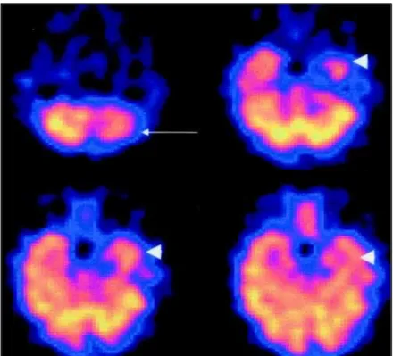
Fig 2. Case 2.Transaxial images show mild hypoperfusion in the left parietal lobe (arrow heads) and moderate hypoperfusion in the right parietal lobe (arrows).
A brain SPECT study was then performed and showed a focus of mild hypoperfusion in the left parietal lobe, close to the left ventricle and also an area of moderate hypoperfusion in the right parietal lobe (Fig 2).
Case 3.
MFGL, a white female, was born by spontaneous delivery with 3,600 g, 52 cm in length, a head perimeter of 33 cm and an Apgar index of 9 and 10, in the first and fifth minutes, respectively. With 36 hours of life she developed a right-sided tonic clonic seizure, for one minute. Three additional similar episodes were seen in the subsequent 24 hours, with mild jaundice, without recognizable etiology, despite several laboratory tests performed. Tansfontanellar ultrasound on the second day demonstrated an extensive hypoechogenic area on the left cerebral hemisphere. The CT scan in the 5th day identified a left fronto-parietal vascular hemorrhage and the
angioMRI was normal. The EEG recorded on the 7th day revealed continuous slow waves on the left cerebral
hemisphere, with epileptiform activity in the left fronto-temporal region. There was progressive clinical improvement and, after discharge from nursery, the patient was reevaluated at 2, 4 and 6 months of age, with a good neuropsychomotor development and a slight right hemiparesis. At the age one year and 10 months, no motor deficits were detected, and the EEG revealed a focal paroxysm in the left temporal region. The assessment of higher cortical functions demonstrated that the patient’s neuropsychomotor development was adequate for her age (picking up objects, piling up objects with appropriate fine movements; walking up and down stairs; imitating actions; pointing to parts of the body and to simple objects). She could use both hands with ill-defined handedness, but with some preference for the left side, probably due to her previous hemiparesis on the right. She could speak appropriately up to 8 words and to associate two. However, she had difficulty controlling her social behavior.
Taking her age and development into account, her performance was acceptable, without significant deficits in the cognitive, perceptive and motor functions. Only her behavior still needed long term evaluation.
The brain SPECT imaging performed at one year and 4 months, demonstrated a small area of mild hypoperfusion in the left fronto-parietal region and another one in the right temporal lobe (Fig 3).
DISCUSSION
The characterization of cerebral metabolism and perfusion abnormalities in neurologic and psychiatric diseases have been investigated in adults and children. The incidence of CVD in term newborns is low, sometimes variable, and dependent on the clinical recognition and imaging modalities used for diagnostic confirmation. Balcom and Redmond 6, in 1997,estimated CVD in term newborns as
being 1/ 10,000, while in the nursery of the Division of Pediatric Neurology, Campinas State University School of Medical Sciences, the incidence is 2.8 / 10,000 term newborns. These data confirm the importance of recognizing CVD in neonates 7. The neurological and imaging studies in the present
investigation demonstrated the presence of hemorrhage in two newborns (Cases 1 and 2) and ischemia evolving to hemorrhage in one (Case 3). In these three patients, the CT scan showed CVD on the left cerebral hemisphere (subependymal region, left fronto-parietal-occipital region and left fronto-parietal region), causing temporary motor changes on the right side (Cases 1 and 3), and a persistent lesion in case 2. However, the long term evaluation of the higher cortical functions in these three patients disclosed permanent motor and behavioral abnormalities. The investigation of cerebral electrogenesis showed left sided abnormalities in the acute phase (Cases 1 and 2), as well as in the follow-up (Cases 2 and 3), which correlated with the findings of the ultrasound and CT scan (Cases 2 and 3).
Brain SPECT imaging is an important method in the diagnosis of several neurologic and psychiatric diseases, but has not been used in newborns with proven diagnosis of ischemia or hemorrhage or ischemia evolving to hemorrhage. Also, brain SPECT imaging is essential for a better understanding of the pathophysiology of newborns with CVD and for a better correlation of the clinical, ultrasound and CT scan findings.
A progressive motor improvement and disappearance of the left hemiparesis was seen in Case 1. However, brain SPECT imaging performed at 4 years of age revealed a persistent hypoperfusion in the left fronto-temporal region and in the left cerebellar hemisphere (ipsilateral cerebellar diaschisis). Diaschisis is defined as reduction of the blood flow and neuronal metabolism in a remote area from the primary cerebral lesion, which leads to failure of the facilitation mechanisms, as a consequence of a destructive lesion in the interconnecting pathways1,7-9. In supratentorial lesions, this abnormality can be
frequently detected in the contralateral cerebellar hemisphere due to destruction of the cortico-ponto-cerebellar pathways. However, cortico-ponto-cerebellar diaschisis can also be found on brain SPECT imaging as a transient phenomenon where there is clearly decreased function of a brain area, but without destruction of interconnecting pathways. Ipsilateral cerebellar diaschisis, which occurred in Case 1, has been detected in children with cerebral lesions occurring before 3 years of age 9,10.
The neuropsychological evaluation contributed to demonstrate several deficits involving fine motricity, alterations in coordination, language, perceptual abilities and behavioral disturbance, in agreement with the findings of brain SPECT imaging. In fact, the results of the neuropsychological tests in Case 1 are indicative of a more significant deficit of the cognitive function, disclosing the difficulties in verbal understanding, as well as in normal visual ability on the non-affected side. These findings are in agreement with the results of Koelfen et al.11. The demonstration of the effect
of the acute lesion upon other interconnecting regions is well demonstrated in Case 2, hypometabolism is noted in the right parietal lobe and the acute lesion occurred in the left parietal lobe.
The data presented are in agreement with the findings of brain SPECT imaging and demonstrated that a normal cerebral development depends on the full ability of the cerebral vascular system to function properly 12.
REFERENCES
1. Sztrilha L, Sulhil A, Pras V, Nurk M. Regional cerebral blood perfusion in children with hemiplegia: a SPECT study. Neuropediatrics 1996;27:178-183.
2. Bowler JV, Costa DC, Jones BE, Steiner TJ, Wade JPH. High resolution SPECT, small deep infarcts and diaschisis.J R Soc Med 1992;85:142-146.
3. Lefèvre AB. Exame neurológico do recém-nascido. In Diament AJ (ed.). Neurologia infantil: semiologia, clínica, tratamento. 3Ed. São Paulo: Sarvier; 1996:33-62.
4. Diament AJ. Exame neurológico do lactente. In Lefèvre AB, Diament AJ (eds).Neurologia infantil: semiologia, clínica, tratamento. São Paulo: Sarvier; 1980:4-39.
5. Teeter PA, Semrud-Clikeman M. Child Neuropsychology: assessment and intervention for neurodevelopmental disorders. Boston: Allyn and Bacon, 1998:69.
6. Balcom TA, Redmond BG. Cerebral infarction as multifocal clonic seizures in a term neonate. J Am Board Fam Pract 1997;10:43-49.
7. Moura-Ribeiro MVL, Pessoto MA, Marba STM. Cerebrovascular disease in neonates: evaluation of four cases. Arq Neuropsiquiatr 1999;57:84-87.
8. Mountz JM, Zhang B, Liu HG, Inampudi CH. A reference method for correlation of anatomic and functional brain images: validation and clinical application. Semin Nucl Med 1994;24:256-271
9. Hamano SI, Nara T, Nakanishi Y, Horita H, Kumagi K, Mackawa K. Secondary changes in cerebellar perfusion (diaschisis) in hemiplegia during childhood: SPECT study of 55 children. Pediatr Neurol 1993;9:435-443.
10. Flores LG, Futami S, Hoshi H, et al.. Crossed cerebellar diaschisis: analysis of iodine 123-IMP SPECT imaging. J Nucl Med 1995;36:399-402.