PRIMARY PROGRESSIVE APHASIA
Analisys of 16 cases
Márcia Radanovic
1, Mirna Lie Hosogi Senaha
2, Letícia Lessa Mansur
3,
Ricardo Nitrini
4, Valéria Santoro Bahia
5, Maria Teresa Carthery
6,
Flávia Nóbrega Freire Aires
7, Sandra Christina Mathias
8, Paulo Caramelli
9ABSTRACT - Primary progressive aphasia (PPA) is an intriguing syndrome, showing some peculiar aspects that differentiate it from classical aphasic pictures caused by focal cerebral lesions or dementia. The slow and progressive deterioration of language occurring in these cases provides an interesting model to better understand the mechanisms involved in the linguistic process. We describe clinical and neuroimaging aspects found in 16 cases of PPA. Our patients underwent language and neuropsychological evaluation, magnetic resonance imaging (MRI) and single photon emission computerized tomography (SPECT). We observed a clear distinction in oral expression patterns; patients were classified as fluent and nonfluent. Anomia was the earliest and most evident symptom in both groups. Neuroimaging pointed to SPECT as a valuable instrument in guiding the differential diagnosis, as well as in making useful clinical and anatomical correlations. This report and a comparison to literature are an attempt to contribute to a better understanding of PPA.
KEY WORDS: primary progressive aphasia, focal cortical degeneration, cerebral perfusion, neuroimaging.
Afasia progressiva primária: análise de 16 casos
RESUMO - A afasia progressiva primária (APP) é uma síndrome que tem despertado grande interesse devido a aspectos particulares que a diferenciam das afasias clássicas (secundárias a lesões cerebrais focais) e dos quadros demenciais. A deterioração lenta e progressiva da linguagem presente nesses casos fornece um interessante modelo de observação dos mecanismos subjacentes ao processamento linguístico. Descrevemos as características clínicas e de neuroimagem de 16 casos de APP. Os pacientes foram submetidos a exame de linguagem, neuropsicológico, ressonância magnética (RM) e tomografia computadorizada por emissão de fóton único (SPECT). Clinicamente pudemos observar uma nítida distinção nos padrões de produção oral, sendo os pacientes agrupados em fluentes e não-fluentes. Anomia foi o sintoma mais precoce e evidente nos dois subgrupos. Os achados de neuroimagem permitem destacar a sensibilidade do SPECT como instrumento diagnóstico. O registro e a comparação destes casos aos da literatura são uma tentativa de contribuir para a compreensão da APP.
PALAVRAS-CHAVE: afasia progressiva primária, degeneração focal cortical, perfusão cerebral, neuroimagem.
Behavioral and Cognitive Neurology Unit, Department of Neurology (DN); Department of Physiotherapy, Speech Pathology and Occupa-tional Therapy (DP), University of São Paulo School of Medicine, São Paulo SP, Brazil: 1M.D., Consultant Neurologist (DN); 2MSc in
Neurosciences (DN); 3PhD, Assistant Professor (DP); 4MD, Associate Professor (DN); 5Post graduate student (DN); 6MSc in Neurosciences
(DN); 7Speech Pathologist (DP); 8M.D., Consultant Neurologist (DN); 9M.D., Assistant Professor (DN)
Received 22 January 2001, received in final form 26 April 2001. Accepted 30 April 2001.
Dra. Márcia Radanovic - R. Cristiano Viana, 163 / 92 - 05411-000 São Paulo SP Brasil. E-mail: [email protected]
Progressive language impairment in patients with relative preservation of other cognitive functions is called primary progressive aphasia (PPA); it is a clini-cal syndrome with several aspects still open to ques-tion, for example language profile characterization and clinical and neuroimaging diagnostic criteria.
The first description of isolated progressive loss of language was reported by Pick, in 18921,
descri-bing a patient with severe language disturbance associated with atrophy in the left temporo-polar
region and posterior two thirds of the frontal lobe. Later, the same author described other patients with temporal2, parietal and frontal lobe atrophy3. The
term Picks disease was later used for any similar progressive atrophic diseases, especially those with predominant behavioral disturbances. Other authors have also described predominant language alter-ations, under the name aphasic dementia or pure verbal deafness evolving to sensorial aphasia4-6.
dis-ease was liberally diagnosed to many clinical condi-tions, including those that could be considered PPA. In 1982, Mesulam7 described six patients with
iso-lated and long lasting language deterioration, nam-ing these clinical pictures as slowly progressive apha-sia without dementia, later called PPA. Some of these patients, in spite of serious limitations to oral and written communication, were able to maintain au-tonomy in daily life activities. More than a hundred similar cases have been reported in literature8. In
Brazil, Oliveira et al.9 were the first to report a case
of PPA.
To date, some 33 cases of PPA have undergone pathologic examination, showing predominantly (in approximately 60% of cases) signs of non-specific focal degeneration with neuronal loss, gliosis and spo-ngiform alterations in superficial cortical layers10-12.
Alterations compatible with Picks disease were seen in about 20% of the patients13-15, whereas the
re-maining 20% had Alzheimers disease16. There is a
report of Creutzfeldt-Jakob disease manifesting it-self as PPA in its fluent form17. It is nowadays
ac-cepted that PPA constitutes a syndrome including different etiologies and whose diagnosis is clinical and based on the presence of progressive language deficit, which during the first two years is either the only affected cognitive function or is the most im-portant function involved. Although usually isolated, PPA may coexist with disorders such as acalculia, ideomotor or constructional apraxia, indicative of left hemisphere damage18,8. These clinical manifestations
emerge in the absence of morphologic alterations (excluding atrophy) both in computerized tomogra-phy (CT) and magnetic resonance imaging (MRI). Atrophy usually appears in left frontal, temporal or perisylvian cortices19.
Clinical presentations similar to PPA can occasion-ally be found such as in pure progressive verbal deaf-ness, described in a patient with left superior tem-poral atrophy20 and pure progressive aphemia21.
The purpose of this paper is to report clinical, linguistic and neuroimaging characteristics found in 16 PPA cases diagnosed and accompanied by the Behavioral and Cognitive Neurology Unit of the Neu-rological Division, Hospital das Clínicas of the Uni-versity of São Paulo School of Medicine.
METHOD
Sixteen patients were studied, whose demographic data are presented in Table 1. Language evaluation was performed using Montreal-Toulouse (BETA version)22 and
Boston Diagnostic Aphasia Examination (BDAE)23
proto-cols, with patients classified as fluent and nonfluent. Seven
patients were evaluated longitudinally. The remaining cog-nitive functions were evaluated using the Mini-Mental State Examination (MMSE)24 and complementary
neurop-sychological tests. Cranial MRI was performed on all pa-tients. Ten patients were also submitted to single photon emission computerized tomography (SPECT).
RESULTS
Our series comprised nine females and seven ma-les, with ages varying between 39 and 83 years (av-erage 62.8 years) and an educational level between two and 20 years (average 10.7 years). Six patients spoke other languages besides Portuguese (see Table 1). Neuropsychological evaluation results are shown in Table 2. Performance in language tests, repeated after a one to two year interval in seven cases, is presented in Table 3. Neuroimaging exam results are described in Table 4.
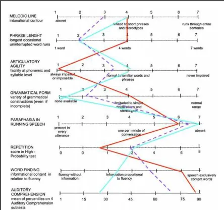
Figure 1 shows a comparison in language perfor-mances between fluent and nonfluent groups. Fig-ures 2 and 3 show the speech characteristics profile scales for each group and its evolution over time. These profiles are useful in identifying similarities and differences between fluent form and Brocas apha-sia (Fig 2) or between nonfluent form and Wernickes aphasia (Fig 3). Figures 4 and 5 illustrate the neuro-imaging (SPECT and MRI) findings in a fluent and a nonfluent patient, respectively. These figures include speech samples from the same patients.
Differential diagnosis and evolution of cases
The clinical course, although restricted in some cases, can cast further light on the differential diag-nosis between correlate syndromes such as seman-tic dementia, as observed in case 14.
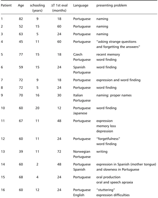
Table 1. Demographic data and presenting symptoms.
Patient Age schooling ∆T 1st eval Language presenting problem (years) (months)
1 82 9 18 Portuguese naming
2 52 15 60 Portuguese naming
3 63 5 24 Portuguese naming
4 45 11 60 Portuguese asking strange questions and forgetting the answers
5 77 15 18 Czech recent memory
Portuguese word finding
6 59 15 24 Spanish word finding
Portuguese
7 72 9 18 Portuguese expression and word finding
8 72 5 24 Portuguese word finding
9 70 16 30 Italian naming: proper names
Portuguese
10 60 20 12 Portuguese word finding
Japanese
11 67 11 48 Portuguese expression
memory loss depression
12 60 11 24 Portuguese forgetfulness
word finding
13 39 11 72 Norwegian writing
Portuguese
14 60 2 48 Portuguese expression in Spanish (mother tongue) Spanish and slowness in Portuguese
15 68 4 24 Portuguese oral production
oral and speech apraxia
16 60 12 24 Portuguese stuttering
English expression difficulties
,T 1st eval : time from onset of symptoms to first evaluation
Table 2. Neuropsychological evaluation and deficit evolution.
Patient MMSE / NP alterations (first evaluation) deficit evolution
1 NT / mild memory impairment oral comprehension and reading after one year 2 22 / verbal and non-verbal memory oral comprehension after one year
memory, apathy, irritability, obsessive behavior after five years
FTD after eight years
3 9/ digit span anomia
4 9/ memory, digit span and calculation pragmatic communication disturbance
5 20 / normal oral comprehension
depression
6 26 / verbal memory anomia
7 22 / NT memory
transient behavioral disturbances (nocturnal fears, strangeness of environment) after three years
8 25/ NT recent memory
9 23 / anterograde episodic memory and visuospatial abilities naming in general 10 22 / initiation, perseveration, verbal and visual memory reading and calculation
and conceptualization impaired lexical access, phrase reformulation 11 12 / Mattis34: 87/144 (attention, memory, constructional symptoms worsening
praxis and executive functions)
12 25/ Mattis: 116/144 (executive functions and verbal agraphia
episodic memory) slowness in talking
13 24 / constructional praxis, acalculia and non-verbal memory agraphia after four years
expression and recent memory after five years
14 NT / normal oral production in both languages
15 29 / normal dementia after five years
16 20/ Mattis: 97/144 (attention, constructional praxis and expression difficulties worsening executive functions)
NP: neuropsychological evaluation; NT: not tested; FTD: frontotemporal dementia.
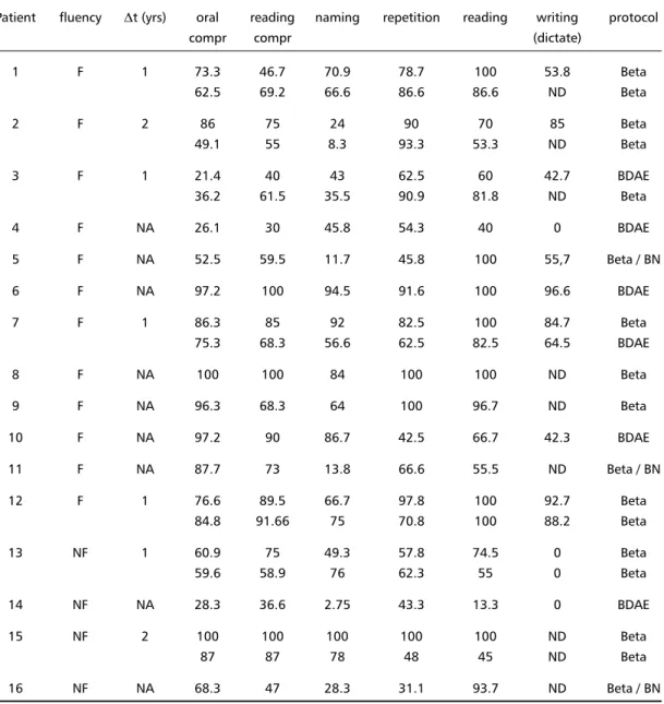
Table 3. Language examination. Performance in tests is expressed in terms of percentage of correct answers.
Patient fluency ,t (yrs) oral reading naming repetition reading writing protocol
compr compr (dictate)
1 F 1 73.3 46.7 70.9 78.7 100 53.8 Beta
62.5 69.2 66.6 86.6 86.6 ND Beta
2 F 2 86 75 24 90 70 85 Beta
49.1 55 8.3 93.3 53.3 ND Beta
3 F 1 21.4 40 43 62.5 60 42.7 BDAE
36.2 61.5 35.5 90.9 81.8 ND Beta
4 F NA 26.1 30 45.8 54.3 40 0 BDAE
5 F NA 52.5 59.5 11.7 45.8 100 55,7 Beta / BN
6 F NA 97.2 100 94.5 91.6 100 96.6 BDAE
7 F 1 86.3 85 92 82.5 100 84.7 Beta
75.3 68.3 56.6 62.5 82.5 64.5 BDAE
8 F NA 100 100 84 100 100 ND Beta
9 F NA 96.3 68.3 64 100 96.7 ND Beta
10 F NA 97.2 90 86.7 42.5 66.7 42.3 BDAE
11 F NA 87.7 73 13.8 66.6 55.5 ND Beta / BN
12 F 1 76.6 89.5 66.7 97.8 100 92.7 Beta
84.8 91.66 75 70.8 100 88.2 Beta
13 NF 1 60.9 75 49.3 57.8 74.5 0 Beta
59.6 58.9 76 62.3 55 0 Beta
14 NF NA 28.3 36.6 2.75 43.3 13.3 0 BDAE
15 NF 2 100 100 100 100 100 ND Beta
87 87 78 48 45 ND Beta
16 NF NA 68.3 47 28.3 31.1 93.7 ND Beta / BN
,t: interval between first and second evaluation ; compr: comprehension; NA: not applicable; ND: not done; BN: Boston naming test.
Neuroimaging
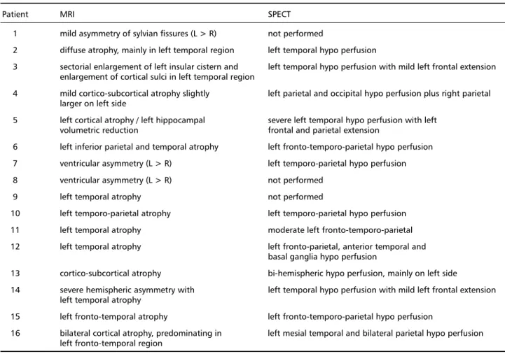
Considering the MRI findings, we noted that 15 patients in our series presented predominant left hemi-sphere atrophy. In three cases (2, 4 and 16), the atro-phy was bilateral, although predominant in the left side. The temporal region was the most affected, with atrophy present in 11 cases; one with temporo-pari-etal atrophy (Case 6), two with fronto-temporal at-rophy (Cases 15 and 16) and one with volumetric reduction of left hippocampus (Case 5). Perisylvian atrophy was reported in three cases (1, 7 e 8), thus implying frontal and/or temporal degeneration (these cases are considered separately because we can not describe the site of anomaly accurately). One case (13) presented diffuse cortical and subcortical atrophy.
Table 4. Neuroimaging results.
Patient MRI SPECT
1 mild asymmetry of sylvian fissures (L > R) not performed
2 diffuse atrophy, mainly in left temporal region left temporal hypo perfusion
3 sectorial enlargement of left insular cistern and left temporal hypo perfusion with mild left frontal extension enlargement of cortical sulci in left temporal region
4 mild cortico-subcortical atrophy slightly left parietal and occipital hypo perfusion plus right parietal larger on left side
5 left cortical atrophy / left hippocampal severe left temporal hypo perfusion with left volumetric reduction frontal and parietal extension
6 left inferior parietal and temporal atrophy left fronto-temporo-parietal hypo perfusion 7 ventricular asymmetry (L > R) left temporo-parietal hypo perfusion 8 ventricular asymmetry (L > R) not performed
9 left temporal atrophy not performed
10 left temporo-parietal atrophy left temporo-parietal hypo perfusion 11 left temporal atrophy moderate left fronto-temporo-parietal 12 left temporal atrophy left fronto-parietal, anterior temporal and
basal ganglia hypo perfusion
13 cortico-subcortical atrophy bi-hemispheric hypo perfusion, mainly on left side
14 severe hemispheric asymmetry with left temporal hypo perfusion with mild left frontal extension left temporal atrophy
15 left fronto-temporal atrophy left fronto-temporo-parietal hypo perfusion
16 bilateral cortical atrophy, predominating in left mesial temporal and bilateral parietal hypo perfusion left fronto-temporal region
L: left; R: right
DISCUSSION
Our results disclosed different and peculiar pat-terns of language impairment among the patients, to be expected, especially considering the differing progressions of the disease over time. The anatomi-cal distribution of abnormalities in neuroimaging is similar to that described in literature .
The first possible consideration, analyzing lan-guage data, is including these cases in the classical aphasic syndromes. Using generic taxonomy, we cho-se to divide the cacho-ses into fluent and nonfluent and to identify the isolated aphasic symptoms, given that one of the difficulties in regard to classification, stems from the fact that the patients were diagnosed when the intensity, combination and sum of alterations surpassed the classical aphasic syndrome definitions. The attempt to recoup evolution from clinical his-tory provided by family proved unsuccessful, given the overlapping interpretations of memory and lan-guage alterations, exemplified by frequent indica-tions of word forgetting for anomia, or the ge-neric descriptions of pronunciation alterations.
As new cases of PPA were being described, it be-came evident that the clinical features are complex
and heterogeneous. This has led several authors to attempt a classification of the patients into subgroups with similar clinical characteristics. One of the first attempts was to categorize into nonfluent (where expression alterations such as agrammatism, emis-sion reduction and phonologic alterations predomi-nate) and fluent, where the semantic impairment is more evident. One of the most intriguing discussions arises from similarities and differences that may ex-ist between fluent PPA and semantic dementia25,26.
In the latter condition, however, the impairment of cognition seems to be more prevalent, affecting the recognition of words and objects, traits usually as-sociated with bilateral temporal lobe dysfunction.
The subdivision into nonfluent and fluent groups proved insufficient to include all new data observed by researchers. Principally due to the overlapping of symptoms between patients in both groups, as well as features shared by PPA and other degenerative diseases with lobar atrophy, such as frontotemporal dementia (FTD) 27,25. The existence of pathologic
al-terations that are present both in FTD and PPA rein-forces the hypothesis of both manifestations repre-senting a non-Alzheimer degenerative process, with the occurrence of a different distribution of the
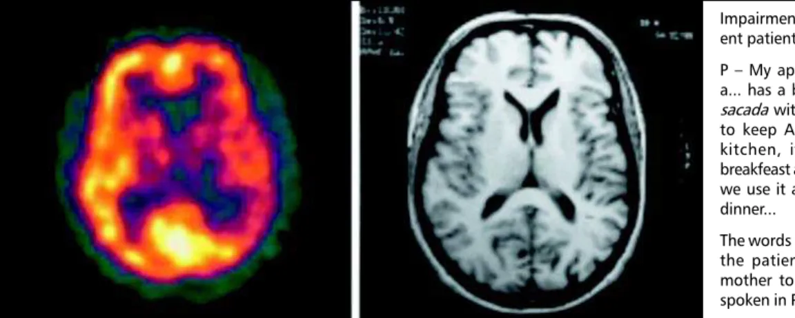
pa-Fig 4. Case 6 - SPECT and MRI showing mild hypoperfusion and atrophy in left parieto-temporal region.
Impairment of lexical access in a flu-ent patiflu-ent (case 6)
P My apartment has 3 rooms has a... has a big living room, has a big sacada with a...churrasqueira that is to keep Argentine habits. It has a kitchen, it has a small room for breakfeast and also when we are alone we use it as...as...to have lunch and dinner...
The words in italic refer to names that the patient couldnt access in his mother tongue (Spanish) and were spoken in Portuguese.
Agrammatism found in a nonfluent pa-tient (case 15)
Illustrations sequence: in this story, a woman leaves her car with two children inside. The children start driving and the car crashes in a lamppost. Patients description: car guide womun (woman) car exits womun
car break fall car fall break
the boy guide aumobile (automobile) womun.. (unintelligible).. count (crash) pasts (posts)
womun beak (break)
thological process, either predominantly in the fron-tal or temporal lobes. Due to this overlapping of clini-cal and pathologiclini-cal characteristics, Kertesz et al. 28,29
proposed the term Pick complex to include the clinical conditions currently described as FTD, PPA, Picks disease and corticobasal degeneration.
Weintraub and Mesulam30 proposed a
classifica-tion of degenerative dementia based on four clini-cal profiles, each in turn having more probability of association with a specific cerebral disease. This clas-sification takes into account the neuropsychological profile exhibited by the patient during the first two years of disease. PPA is one of these profiles (Pro-gressive Language Network Syndrome).
In our series, it was not possible to classify the patients into classical aphasic syndromes, although a resemblance to predominantly frontal dysfunction (agrammatism), or temporal (marked comprehen-sion disturbances) is admissible. That said, symptoms also described in dementia occurred alongside the language primary deficit.
Regarding fluency, which is a referential for sub-group identification, we did not notice any remark-ably positive characteristics, such as hyper fluency. Reduction in fluency and quantitative emission pre-dominated, and were shared by the patients with clinical symptoms compatible with both frontal and temporal dysfunction. Absence of language produc-tion occurred when extra-language cognitive symp-toms intensified. Anomia was the earliest and most notable symptom, being present in all patients. Dis-turbances of repetition were also frequent and could be found in the fluent and nonfluent groups.
We can notice in Figures 2 and 3 that the non-fluent group presents greater similarities to Brocas aphasia profile as the disease evolves, except for com-prehension, which is more impaired in our data. In the fluent group, independent of evolution time, the profile is more akin to that found in Wernickes apha-sia, except for the occurrence of fewer semantic para-phasias in language production.
One of the main goals in the research field of degenerative cerebral diseases is to find a method with enough specificity to allow the elaboration of a prognosis, bearing in mind the great number of years necessary to reach a clinical diagnosis with rea-sonable acuity. Toward this goal, several groups have striven to establish relations between perfusion and neuroimaging, and the probable clinical evolution for each profile. Talbot et al.31 described the
correla-tion between standard perfusion with SPECT in 363
patients studied prospectively for 3 years. They found positive correlation between the presence of poste-rior bilateral alterations and Alzheimers disease, and between anterior bilateral alterations and FTD. Other perfusion patterns, such as patchy distribution and generalized or unilateral (anterior or posterior) al-terations, showed less correlation and therefore were less useful for the characterization of a diagnosis.
Neuroimaging findings in PPA keep certain cor-relation with the clinical variant. In nonfluent cases, there is left frontal and perisylvian atrophy in cranial CT and MRI, with CBF abnormalities in the same ar-eas in SPECT. In fluent cases, the cortical atrophy and CBF abnormalities are located predominantly in the left temporal lobe, hippocampus and parahippo-campal gyrus32. In a review article of 112 cases,
West-bury and Bub33 found the following distribution: of
59 cases that performed PET or SPECT, 39 (66.1%) had CBF abnormalities exclusively in the left hemi-sphere and 18 (30.5%) had bilateral CBF abnormali-ties; perfusion was normal only in two cases (3.4%). In the left hemisphere, for 27.3% of cases the CBF abnormalities were located in the fronto-temporal region, 21.2% in the temporal region, 12.1% in the frontal region, and the remaining 39.4% were dis-tributed between frontal, temporal and parietal le-sions combined. In those cases for which MRI was performed (105), 49 (46.7%) had atrophy in the left side only, 38 (36.2%) had bilateral atrophy and in 17 (16.2%) the exams were normal.
The anatomical distribution found in our cases was similar to that documented in literature, namely: predominance in the left hemisphere and, within this, in the temporal and frontal regions or combi-nation of the two. We observed that the abnormali-ties were more apparent in SPECT than in MRI find-ings, generally showing areas of CBF abnormalities that were larger and more evident than those seen in MRI. We think this feature of SPECT might be help-ful, especially in the earlier stages of the disease when MRI shows only mild diffuse atrophy or asymmetry of Sylvian fissures, in providing a useful clue for cor-rect diagnosis.
CONCLUSION
the symptoms that are typical of this phase. Further-more, this description can aid to identification of those symptoms that overlap in language disorders and other cognitive losses. Regarding the neuroima-ging exams, we believe that SPECT is a valuable in-strument in guiding the differential diagnosis, as well as in making useful clinical and anatomical correla-tions.
REFERENCES
1. Pick A. Über die Beziehungen der senilen Hirnatrophie zur Aphasie. Prager Med Wochenschr 1892;17:165-167 apud Poeck K, Luzzatti C, 1988:160.
2. Pick A. Zur Symptomatologie der linksseitigen Schläfenlappenatrophie. Monatsschr Psychiatr Neurol 1904;16:378-388 apud Poeck K, Luzzatti C, 1988:160.
3. Pick A. Über einen weiteren Symptomenkomplex im Rahmen der De-mentia senilis, bedingt durch umschriebene stärkere Hirnatrophie (gemischte Apraxie). Monatsschr Psychiatr Neurol 1906;19:97-108 apud Poeck K, Luzzatti C, 1988:160.
4. Sérieux P. Sur un cas de surdité verbale pure. Rev Med 1893;13:733-750. 5. Franceschi F. Gliosi perivascolare in un caso di demenza afasica. Annali di Nevrologia 1908; 26:281-290 apud Poeck K, Luzzatti C, 1988:160. 6. Mingazzini G. On aphasia due to atrophy of the cerebral convolutions.
Brain 1914; 36:493-524 apud Poeck K, Luzzatti C, 1988:160.
7. Mesulam MM. Slowly progressive aphasia without generalized demen-tia. Ann Neurol 1982;11:592-598.
8. Mesulam MM. Aging, Alzheimer’s disease, and dementia. In Mesulam MM (ed) Principles of behavioral and cognitive neurology. 2.Ed. New York: Oxford University Press, 2000: 455-458.
9. Oliveira SAV, Castro MJO, Bittencourt PRM. Slowly progressive aphasia followed by Alzheimer’s dementia. Arq Neuropsiquiatr 1989;47:72-75.
10. Kirshner HS, Tanridag A, Thurman L, Whetsell WO Jr. Progressive aphasia without dementia: two cases with focal spongiform degenera-tion. Ann Neurol 1987;22:527-532.
11. Lippa CF, Cohen R, Smith TW, Drachman D. Primary progressive apha-sia with focal neuronal achromaapha-sia. Neurology 1991;41:882-886. 12. Deruaz JP, Assal G, Peter-Favre C. Un cas clinico-pathologique
d’aphasie progressive. Rev Neurol (Paris) 1993;149:186-191. 13. Holland Al, McBurney DH, Moosey J, Reinmuth OM. The dissolution
of language in Pick’s disease with neurofibrillary tangles: marries her study. Brain Lang 1985;24:36-58.
14. Graff-Radford NR, Damasio AR, Hyman BT, Hart MN, Tranel D, Damasio H et al. Progressive aphasia in patient with Pick’s disease: neuropsychological, radiologic, and anatomic study. Neurology 1990;40:620-626.
15. Kertesz A, Munoz D. Clinical and pathological characteristics of pri-mary progressive aphasia and frontal dementia. J Neurol Transm Suppl 1996;47:133-141.
16. Kempler D, Metter EJ, Riege WH, Jackson CA, Benson DF, Hanson WR. Slowly progressive aphasia: three cases with language, memory, CT and PET data. J Neurol Neurosurg Psychiatry 1990;53:987-993. 17. Mandell AM, Alexander MP, Carpenter S. Creutzfeldt-Jakob disease
presenting isolated aphasia. Neurology 1989;39:55-58.
18. Poeck K, Luzzatti C. Slowly progressive aphasia in three patients – the problem of accompanying neuropsychological deficit. Brain 1988;111: 151-168.
19. Turner RS, Kenyon LC, Trojanowsky JQ, Gonatas, N, Grossman, M. Clinical, neuroimaging, and pathologic features of progressive nonfluent aphasia. Ann Neurol 1996;39:166-173.
20. Otsuki M, Sum Y, Sato M, Homma A, Tsuji S. Slowly progressive pure word deafness. Eur Neurol 1998;39:135-140.
21. Cohen L, Benoit N, Van Eeckhout P, Ducarne B, Brunet P. Pure pro-gressive aphemia. J Neurol Neurosurg Psychiatry 1993;56:923-924. 22. Nespoulous JL, Lecours AR, Lafond D, Reads May A, Puel M, Joanette
Y et al. Protocole Montreal-Toulouse d’examen linguistique of l’Aphasie: module standard initial (version Beta). Montréal: Laboratoire Théophile-Alajouanine, 1986.
23. Goodglass H, Kaplan E. The assessment of aphasia and related disor-ders. 2.Ed. Malvern: Lea & Fabiger; 1983.
24. Folstein MF, Folstein IF, McHugh PR. Mini-mental state. J Psychiatr Res 1975;12:189-198.
25. Snowden JS, Griffiths HL, Neary D. Progressive language disorder as-sociated with frontal lobe degeneration. Neurocase 1996;2:429-440. 26. Hodges JR, Garrard P, Patterson K. Semantic dementia. In Kertesz
A, Munoz DG. (eds). Pick’d disease and Pick complex. Wiley-Liss, 1998:83-103.
27. Snowden JS, Neary D. Progressive language dysfunction and lobar at-rophy. Dementia 1993;4:226-231.
28. Kertesz A, Hudson L, Mackenzie IRA, Munor DG. The pathology and nosology of primary progressive aphasia. Neurology 1994;44:2065-2072. 29. Kertesz A, Davidson W, Munoz DG. Clinical and pathological overlap between frontotemporal dementia, primary progressive aphasia and corticobasal degeneration: Pick complex. Dement Geriatr Cogn Disord 1999;10:45-49.
30. Weintraub S, Mesulam MM. Four neuropsychological profiles in de-mentia. In Boller F, Grafman J (eds.). Handbook of Neuropsychology. Amsterdam: Elsevier, 1993;8:253-282.
31. Talbot PR, Lloyd JJ, Snowden JS, Neary D, Testa HJ. A clinical role for 99Tc-HMPAO SPECT in investigation of dementia? J Neurol Neurosurg Psychiatry 1998;64:306-313.
32. Abe K, Ukita H, Yamagihara T. Imaging in primary progressive apha-sia. Neuroradiology 1997;39:556-559.
33. Westbury C, Bub D. Primary progressive aphasia: review of 112 cases. Brain Lang 1997;60:381-406.