UNIVERSIDADE DE LISBOA
FACULDADE DE FARMÁCIA
NANOFORMULATIONS OF A POTENT AQUAPORIN-3
INHIBITOR WITH CYTOTOXIC EFFECT AGAINST CANCER
CELL LINES
Mariana Vieira de Almeida Nave
Dissertação de Mestrado
Mestrado em Ciências Biofarmacêuticas
UNIVERSIDADE DE LISBOA
FACULDADE DE FARMÁCIA
NANOFORMULATIONS OF A POTENT AQUAPORIN-3
INHIBITOR WITH CYTOTOXIC EFFECT AGAINST CANCER
CELL LINES
Mariana Vieira de Almeida Nave
Dissertação de Mestrado orientada por:
Prof. Doutora Graça Soveral, Faculdade de Farmácia, Universidade de Lisboa
Doutora Maria Manuela Gaspar, Faculdade de Farmácia, Universidade de Lisboa
Doutor Rui E. Castro, Faculdade de Farmácia, Universidade de Lisboa
Mestrado em Ciências Biofarmacêuticas
iii
Communications in scientific meetings
Nave MA, Gaspar MM, Castro RE, Rodrigues CM, Soveral G. (2013) “Nanoformulations of a
potent Aquaporin-3 inhibitor with cytotoxic effect against cancer cell lines”, poster presented at
the 5th iMed.UL Post-Graduate Students Meeting, Faculdade de Fármacia Universidade de
v
A ti minha pequenina… mãe com açúcar. Das pessoas que mais me amou e sem dúvida a que mais vibrou com cada conquista minha, por mais pequena que fosse. Sei que estás comigo. Por tudo… esta “vitória” também é tua.vii
Agradecimentos
À Professora Teresa Moura. Foi a primeira pessoa que, ainda antes de terminar a licenciatura, depositou confiança em mim. Foi o meu primeiro passinho ao encontro da realização profissional e claro, pessoal. Foi também a pessoa que me apresentou as AQPs e...a Professora Graça. Muito grata por tudo Teresa.
Logo de seguida quero agradecer à Professora Graça pela oportunidade de me receber no grupo e claro, avançar com este projecto que desde logo abracei com entusiasmo. Agradeço por tudo o que me ensinou, por toda a disponibilidade, confiança, opinião e orientação.
À Professora Cecília Rodrigues agradeço pela oportunidade de colaboração, tendo-me permitido realizar parte do trabalho no seu grupo.
Ao Professor Rui, pela orientação e apoio ao longo desta minha etapa. Para além da partilha de conhecimento, um obrigado pela boa disposição e pensamento positivo constantes.
À Doutora Manuela Gaspar… “Chefinha”, o meu eterno obrigado por toda a disponibilidade, partilha, orientação e dedicação! Agradeço também a compreensão demonstrada num momento menos feliz da minha vida.
Quero deixar um agradecimento também às várias pessoas com quem partilhei oxigénio, algumas sábias palavras (muitas!) e saudáveis gargalhadas nestes últimos meses. Mesmo a brincar há sempre alguma coisa a aprender e quando estamos dispostos a aprender todas as pessoas com quem nos cruzamos têm algo a ensinar.
Cláudia agradeço-te o conhecimento que partilhaste e a paciência que demonstraste quando comecei o trabalho no laboratório. À Paulinha e Ana Madeira agradeço principalmente pela paciência para os meus constantes e típicos “porquês”. Ambas me ensinaram muito! Necas... não me esqueço de ti! Tens aqui uma amiga-jukebox!
Susana, o que teria sido de mim nos primeiros tempos que passei no laboratório se não fosse a tua constante disponibilidade?! Obrigado pela partilha, boa disposição e pelos momentos de descontracção e pura parvoíce! Maria, Joana, Rui, Xana, Ana… Obrigado a cada um de vocês. Proporcionaram bons momentos!
Aos meus amigos de mestrado, principalmente Anocas, Ceci e Vasco, quero deixar um agradecimento muito especial. Ter-vos conhecido foi um dos grandes pontos de viragem neste meu caminho. A vossa amizade importa muito!
Hugo...faço questão de deixar um beijinho enorme. Foste um dos pilares da minha sanidade mental durante este último ano. Um obrigado bem lamechas para ti pela tua dedicação e
viii
amizade sincera. Das várias pessoas com quem me cruzei até hoje, és sem dúvida uma das que maior coração tem (e paciência!).
Bruno, a ti agradeço todo o amor e carinho… toda a amizade e dedicação mas acima de tudo por toda a paz com a qual constantemente me envolves. Obrigado do fundo do coração!
Quero agradecer também a duas pessoas que foram essenciais para ser o que hoje sou: ao meu “amigo do coração” André… não preciso de te dizer muito. Ensinaste-me a ver a vida de uma maneira muito mais cheia e iluminada. Hoje sinto-me mais preenchida e ao mesmo tempo mais leve. Sim, a nossa amizade perdurará. A ti Miguel, agradeço a paciência, dedicação e amizade que dispensaste ao longo de vários anos. Tiveste um papel central na minha formação tanto a nível pessoal como profissional. É impossível esquecer o que por mim fizeste. A vocês os dois o meu eterno e sentido obrigado.
A todos os meus restantes amigos, não menos importantes para o meu crescimento e evolução constantes... Obrigado pelo respeito, carinho, amor, sinceridade e compreensão.
Aos meus tios e primos que sempre me apoiaram. Destaco o meu tio Lipi que, apesar de não me ver terminar esta etapa, me ensinou que a “sorte vale sempre muito pouco”.
Agradeço também do fundo do coração à minha tia Maria Helena sem a qual esta tinta nestas folhas, não faria qualquer sentido. Eternamente grata minha tia!
Aos meus pais… obrigado por toda a confiança que em mim depositam desde pequena. Obrigado pelo apoio, amor, carinho e amizade incondicionais! Obrigado por todos os “Não!”, raspanetes e castigos. Neste mesmo parágrafo incluo-te a ti meu irmão. És o meu maior e mais bonito reflexo… a melhor parte de mim. Porque são e serão sempre o meu porto de abrigo, a minha família mas acima de tudo os meus melhores amigos. Amo-vos!
ix
Table of Contents
Agradecimentos ... vii
Table of Contents ... ix
List of Figures ... xi
List of Tables ... xiii
Resumo ... xv
Abstract ... xvii
Abbreviations and Symbols ... xix
1. Introduction ... 1
1.1. Aquaporins ... 3
1.1.1. Structure of aquaporins ... 4
1.1.2. Pore Structure of AQPs ... 5
1.2. AQP selectivity ... 7
1.2.1. Orthodox aquaporins ... 7
1.2.2. Aquaglyceroporins ... 7
1.3. Aquaporinopathies – AQP-related human diseases ... 8
1.3.1. AQPs in cancer ... 8
1.3.1.1. AQP-facilitated cellular migration ... 9
1.3.1.2. AQP3 in skin tumorigenesis ... 10
1.4. AQP inhibitors: pharmaceutical opportunities ... 12
1.4.1. Metal-based agents as AQP3 inhibitors ... 12
1.4.2. Metallodrugs as antitumoral agents ... 13
1.5. Drug Delivery Systems (DDS) ... 15
1.5.1. Liposomes... 15
1.6. Aims and goals ... 19
2. Materials and Methods ... 21
2.1. Materials ... 23
2.2. Methods ... 24
2.2.1. Preparation of Cuphen liposomal formulations ... 24
2.2.2. Characterization of Cuphen liposomal formulations ... 24
2.2.2.1. Cuphen quantification ... 25
2.2.2.2. Phospholipid quantification ... 25
x
2.2.2.4. Zeta potential determination... 26
2.2.2.5. Stability of Cuphen liposomes ... 27
2.2.3. Cell Culture ... 27
2.2.4. Evaluation of cellular viability ... 28
2.2.4.1. Hoechst Staining ... 28
2.2.4.2. Trypan Blue assay ... 28
2.2.5. MTS assay ... 28
2.2.5.1. Metallodrugs cytotoxicity screening ... 29
2.2.5.2. Cytotoxic effect of Cuphen formulations against cancer cells ... 29
2.2.6. Statistical analysis ... 30
3. Results and Discussion ... 31
3.1. Metallodrugs cytotoxicity screening ... 33
3.1.1. Trypan Blue assay and Hoechst staining ... 33
3.1.2. MTS assay ... 34
3.2. Optimization of methodologies for Cuphen quantification ... 37
3.3. Cuphen liposomes ... 38
3.3.1. Characterization of Cuphen liposomes ... 38
3.4. In vitro cytotoxicity of Cuphen formulations ... 43
3.4.1. Cytotoxicity of Cuphen against A431 cells ... 44
3.4.2. Cytotoxicity of Cuphen against C26 cells ... 48
3.5. Storage stability of Cuphen liposomes ... 52
4. Conclusions and Future Work ... 55
xi
List of Figures
Figure 1.1. Structural organization of an AQP1 monomer. In the membrane, the six α helices form a right-handed twisted arrangement. However and to simplify, the helices 3, 1 and 2 are drawn separated from helices 5, 4 and 6. Adapted from (Zeuthen, 2001). ... 4
Figure 1.2. Representation of hAQP1 monomers (side view). Generated on Chimera. PDB ID: 1H6I. A| All the helices and loops are shown. B| Representation of loops B and E with the Proline residues of the NPA motif shown in dark blue. ... 5
Figure 1.3. Detailed view of a bAQP1 (bos taurus) pore region (red mesh). Half helices dipoles and the hydrophilic and hydrophobic residues lining the pore are depicted in red and yellow, respectively. (Soveral G, 2011) ... 6
Figure 1.4. Proposed mechanism of AQP-facilitated cell migration. Water entry into protruding lamellipodia in migrating cells. Modified from (Verkman, 2012). ... 9
Figure 1.5. Reduced skin hydration in AQP3 deficiency. A| Schematic representaion of skin layers; B| Proposed mechanism of AQP3 function in the skin. Modified from (Verkman, 2005). ... 10
Figure 1.6. Schematic representation of the proposed mechanisms of AQP3-dependent skin hydration, wound healing, and tumorigenesis. Modified from (Hara-Chikuma, 2008c; Verkman, 2009). ... 11
Figure 1.7. Structures of several metallodrugs described as AQP3 inhibitors by Martins and colleagues. Modified from (Martins, 2013). ... 13
Figure 1.8. Liposomes – versatile structures. A| classic liposomes with hydrophilic drug (a) at the inner aqueous compartment and hydrophobic drug (b) within the lipid bilayer. B| Immunoliposomes possess specific antibodies (or antibody fragments) at surface to enhance a specific targeting. C| Long-circulating liposomes present modified surface by the presence of polymers such as PEG (c) that allows the increase of circulation time in bloodstream and protects the carrier from opsonizing proteins (d). D| Long-circulating immunoliposome. Adapted from (Torchilin, 2005). ... 16
Figure 1.9. Extravasation and accumulation of liposomes in tumor tissue due to EPR effect. (Saetern, 2004) ... 17
Figure 3.1. Cellular death induced by the different AQP3 inhibitors at 5, 15 and 50 µM (shown in ascending order), after incubation in A431 cells for 24h. Results are expressed as mean ± SD. *p<0.05; **p<0.01; ***p<0.001. ... 33
Figure 3.2. Concentration-dependent inhibition of A431 cellular viability by different metallodrugs after A| 48h and B| 72h of incubation. Results are expressed as mean percentage (%) of control ± SD. ... 34
Figure 3.3. UV spectra obtained for Cuphen solution at two different concentrations and wavelength ranges. Spectrum traced with a Cuphen solution of A| 20uM; B| 10uM. Absorbance maximum recorded at 272nm. ... 37
Figure 3.4. Graphical representation of data sets of calibration curves for Cuphen. Data are represented as mean ± S.D. of several independent experiments (n=12). R2 - linear correlation coefficient: 0.9983; Slope: 0.0331 ± 0.0017; Y-intercept (x=0): 0.0001 ± 0.0020. ... 37
xii
Figure 3.5. Influence of the bilayer rigidity on Cuphen incorporation parameters: I.E. (%) (white columns) and [Cuphen/Lip]f (grey columns). Comparison between A| PC, DMPC and DPPC (F1, F5 and F9); B| PC:Chol:PEG, DMPC:Chol:PEG and DPPC:Chol:PEG (F2, F6 and F10). Results are expressed as mean ± S.D. ... 43
Figure 3.6. Concentration-dependent inhibition of A431 cells proliferation after different times of incubation with Cuphen in the free form. (●) – 48h; (○) – 72h. Results are expressed as mean percentage (%) of control ± SD. ... 44
Figure 3.7. Graphical representation of A431 cells proliferation inhibition induced by Cuphen (5 μM) in free form 48 and 72h after incubation. Results are expressed as mean percentage (%) of control ± SD. ***p<0.001. ... 45
Figure 3.8. Concentration-dependent inhibition of 431 cells proliferation after 72h of incubation. A| Unloaded liposomes; B| Cuphen formulations. (○) - Free Cuphen; (▲) – PC Cuphen liposomes; (x) – PC:Chol:PEG Cuphen liposomes. Results are expressed as mean percentage (%) of control ± SD. ... 46
Figure 3.9. Graphical representation of A431 cells proliferation inhibition induced by Cuphen in free and liposomal forms at 15 μM. Different incubation times were tested A| 48 and B| 72h. Results are expressed as mean percentage (%) of control ± SD. *p<0.05; **p<0.01; ***p<0.001; n.s.: not statistically significant. ... 47
Figure 3.10. Concentration-dependent inhibition of C26 cells proliferation after different times of incubation with Cuphen in the free form. (□) – 24h; (●) – 48h; (○) – 72h; (♦) – 96h. Results are expressed as mean percentage (%) of control ± SD. ... 48
Figure 3.11. Concentration-dependent inhibition of C26 cells proliferation after 72h of incubation. A| Unloaded liposomes; B| Cuphen formulations. (○) - Free Cuphen; (▲) – PC Cuphen liposomes; ( x ) – PC:Chol:PEG Cuphen liposomes. Results are expressed as mean percentage (%) of control ± SD. ... 49
Figure 3.12. Graphical representation of C26 cells proliferation inhibition induced by Cuphen in free and liposomal forms at 10 μM. Different incubation times were tested: A| 48h; B| 72h and C| 96h. Results are expressed as mean percentage (%) of control ± SD. ***p<0.001; n.s.: not statistically significant. ... 51
Figure 3.13. Cuphen liposomes stability after storage at 4ºC for 10 days. Data from two independent experiments. Results are expressed as mean ± SD. ... 52
xiii
List of Tables
Table 1.1. Permeability and occurrence of human aquaporins. Modified from (Castle, 2005). ... 3
Table 1.2. AQP expression in different human tumors. Modified from (Verkman, 2008). ... 8
Table 3.1. Half-inhibitory concentrations for the cellular proliferation in A431 cells. ... 35
Table 3.2. Cuphen physical properties. ... 36
Table 3.3. Physicochemical characterization of Cuphen liposomes: PC-based vesicles. ... 39
Table 3.4. Physicochemical properties of unloaded PC-based vesicles. ... 40
Table 3.5. Physicochemical characterization of Cuphen liposomes: DMPC-based vesicles. ... 40
Table 3.6. Physicochemical properties of unloaded DMPC-based vesicles... 41
Table 3.7. Physicochemical characterization of Cuphen liposomes: DPPC-based vesicles. ... 42
Table 3.8. Inhibition of the cellular proliferation of A431 cells by Cuphen in free form. Half-inhibitory concentrations. ... 45
Table 3.9. Inhibition of the cellular proliferation of A431 cells by Cuphen liposomes. Half-inhibitory concentrations. ... 46
Table 3.10. Inhibition of the cellular proliferation of C26 cells by Cuphen in free form. Half-inhibitory concentrations. ... 48
Table 3.11. Inhibition of the cellular proliferation of C26 cells by Cuphen liposomes. Half-inhibitory concentrations. ... 50
xv
Resumo
As aquaporinas (AQPs) são proteínas transmembranares responsáveis pelo transporte de água e de outros solutos, como o glicerol, através das membranas plasmáticas.
Estes transportadores entraram recentemente na lista de possíveis alvos terapêuticos na área da oncobiologia uma vez que a sua sobre-expressão está associada a diferentes tipos de cancro. Em particular a aquaporina-3 (AQP3), uma aquagliceroporina abundantemente expressa ao nível da epiderme, é agora tida como “chave” na tumorigénese e quimioresistência em casos de cancro de pele. Deste modo, as AQPs estão a ganhar relevância enquanto alvos biológicos na terapia do cancro e os seus modeladores a reunir interesse por parte da indústria farmacêutica.
Recentemente, o nosso grupo descreveu diferentes compostos baseados em iões metálicos como inibidores potentes e selectivos da AQP3 humana. Em particular, o composto derivado de cobre(II) da fenantrolina – Cuphen – demonstrou ter efeito inibitório selectivo sobre a permeabilidade ao glicerol quando testado em eritrócitos humanos, mostrando-se assim promissor para administração in vivo.
Por conseguinte, este trabalho teve como principal objectivo o desenvolvimento de um sistema de veiculação adequado, baseado em estruturas lipídicas artificiais, nomeadamente lipossomas, de modo a poder permitir uma estabilização de inibidores da AQP3, alterando o seu perfil de biodistribuição in vivo, proporcionando um direccionamento preferencial para as áreas de interesse terapêutico aquando da sua administração.
Assim, e recorrendo à linha celular tumoral A431, derivada de carcinoma epidermoide humano, e que apresenta sobre-expressão endógena de AQP3, foi avaliado o potencial citotóxico de diferentes compostos metálicos inibidores da AQP3. Após identificação do composto mais citotóxico, o Cuphen (([Cu(phen)Cl2]Cl (phen = 1,10-fenantrolina)), já descrito como inibidor
selectivo da AQP3 em eritrócitos humanos, foi seleccionado para incorporação em lipossomas
de escala nanométrica (inferior a 0.2 µm). Usando diferentes composições lipídicas foram obtidas eficiências de incorporação de cerca de 50%.
O efeito citotóxico do Cuphen, nas formas livre e lipossomal, foi avaliado na linha celular A431 e numa linha de cancro de cólon de murganho (C26). Para a forma livre, obtiveram-se valores de IC50 de 3.0 ± 0.4 µM e 1.8 ± 0.1 µM para as linhas A431 e C26, respectivamente, após 72h
de incubação. A incorporação do Cuphen em lipossomas permitiu a preservação do seu efeito citotóxico (IC50 ≤ 10 µM após 72h de incubação).
É ainda de referir que os lipossomas vazios não apresentaram qualquer efeito a nível da viabilidade das células testadas.
xvi
Com base nestes resultados, o estudo de formulações lipossomais para encapsular este inibidor deve ser aprofundado e o estabelecimento de um modelo animal de melanoma humano deve ser considerado, por forma a avaliar o efeito terapêutico deste composto metálico.
xvii
Abstract
Aquaporins (AQPs) are a family of small transmembrane proteins that facilitate the transport of water and other solutes, such as glycerol, across the cell plasma membrane. AQPs are now part of the expanding list of effectors in cancer biology after establishment of positive correlations between the histological tumor grade and their aberrant expression in different tumor types. In particular, the AQP3 aquaglyceroporin, which is abundantly expressed in skin keratinocytes, is now seen as a key player in skin tumorigenesis and chemoresistance. Therefore, AQPs are gaining relevance as drug targets for cancer therapy and AQPs' modulators are gathering interest from the pharmaceutical industry.
Our group recently reported different metallodrugs as potent and selective human AQP3 inhibitors for further exploitation on in vivo studies. Pursuing this idea, this work had as major aim the development of appropriate drug carrier systems based on artificial closed structures formed by lipid bilayers – liposomes – that may stabilize AQP3 inhibitors and improve in vivo delivery.
With this aim, a cytotoxic screening using different AQP3 inhibitors against a human epidermoid carcinoma cell line (A431), presenting endogenous overexpression of AQP3, was performed. Cuphen ([Cu(phen)Cl2]Cl (phen = 1,10-phenantroline)), previously shown to
selectively inhibit AQP3 glycerol transport in human red blood cells, was selected as the most promising inhibitor and incorporated in liposomes in a nanometric scale (below 0.2 µm). Using different lipid compositions, incorporation efficiencies of approximately 50% were achieved. The in vitro cytotoxic effect of Cuphen, in both free and liposomal forms, was assessed in the A431 and the C26 murine colon cancer cell lines. In the free form, the IC50 obtained was 3.0 ±
0.4 µM and 1.8 ± 0.1 µM for the A431 and for C26, respectively, after 72h of incubation. The incorporation of Cuphen in liposomes was able to preserve the cytotoxic effect of this AQP3 inhibitor (IC50 ≤ 10 µM after 72h of incubation). Moreover, unloaded liposomes did not exert
any effect on the viability of these cancer cells.
In view of these in vitro results, more liposomal formulations should be tested and the establishment of a murine melanoma model to evaluate the therapeutic effect of Cuphen formulations should be considered.
xix
Abbreviations and Symbols
(Cuphen/Lip)i Initial Cuphen to lipid ratio
(Cuphen/Lip)f Final Cuphen to lipid ratio
t 1/2 Half-life time
AQP Aquaporin
Au Gold
AU Arbitrary Units
bipy 2,2’-bipyridine; bipyridyl
Chol Cholesterol
Cisplatin cis-diammine-dichloro platinum(II)
Cu Copper
dien Diethylendiamine
DMEM Dulbecco’s modified Eagle’s medium
DMPC Dimyristoyl phosphatidylcholine
DMSO Dimethyl sulfoxide
DPPC Dipalmitoyl phosphatidylcholine
DRV Dehydration-rehydration vesicles
I.E. Incorporation Efficiency
EtOH Absolute ethanol
GI Gastrointestinal
HEPES 4-(2-hydroxyethyl)-1-piperazineethanesulfonic acid
IC50 Half-inhibitory concentration
LCL Long-circulating liposomes
Lip Lipid
MPS Mononuclear phagocytic system
MTS
xx
MW Molecular weight
NaCl Sodium chloride
n.d. Not determined
NK Not known
n.s. Not significant
nm Nanometer
RPMI Roswell Park Memorial Institute
PBS Phosphate buffered saline
PC Phosphatidylcholine
PEG Polyethylene glycol
Phen 1,10-phenantroline; phenantroline
P.I. Polydispersity index
PO 4 Phosphate
R2 Linear correlation coefficient
RBCs Red blood cells
SA Octadecylamine; n-Stearylamine
S.D. Standard deviation
S.E. Standard error
Tc Phase transition temperature
terpy 2,2':6',2''- terpyridine; terpyridine
Zeta Pot. Zeta Potential
1
1. Introduction
3
1.1. Aquaporins
Aquaporins (AQPs) are a family of small (≈30 kDa) ubiquitous transmembrane proteins that facilitate the bidirectional transport of water and small uncharged molecules such as glycerol or urea and, in particular cases, ammonia, carbon dioxide and hydrogen peroxide, across the lipid bilayer. (Agre, 1993; Wu, 2007)
The first water channel (AQP1) was reported in 1992 by Preston and co-workers, after purification and subsequent characterization of a protein isolated from red blood cells (RBCs) in
Xenopus oocytes (Preston, 1992) and its reconstitution into liposomes. (Zeidel, 1992) Since
then, several AQPs were identified from yeast, plants and animals. (Kruse, 2006)
Table 1.1. Permeability and occurrence of human aquaporins. Modified from (Castle, 2005). AQP Permeants Major tissue distribution
AQP0 ↓ Water Lens (eye)
AQP1 ↑ Water
CO2; NO (Carbrey, 2009)
Erythrocytes, lung, kidney, brain, eye and vascular endothelium
AQP2 ↑ Water Kidney
AQP3
↑ Water ↑ glycerol; urea
H2O2 (Miller, 2010)
Skin, kidney, lung, eye and GI tract
AQP4 ↑ Water Kidney, brain, lung, GI tract and muscle
AQP5 ↑ Water Salivary, lacrimal and sweat glands, lung and
eye
AQP6 ↓ Water Anions (NO
3> Cl-) (Carbrey, 2009) Kidney
AQP7
↑ Water
↑ glycerol; urea (Wspalz, 2009) Arsenite (Carbrey, 2009)
Adipose tissue, kidney and testis
AQP8
↑ Water
urea; ammonia (Wspalz, 2009)
H2O2 (Wu, 2007)
Kidney, liver, pancreas, GI tract and testis
AQP9
↓ Water
↑ glycerol; urea (Wspalz, 2009) Arsenite (Carbrey, 2009)
Liver, leukocytes, brain and testis
AQP10 ↓ Water ↑ glycerol; urea (Wspalz, 2009) GI tract
AQP11 NK Brain, liver, kidney
AQP12 NK Pancreas (Ishibashi, 2009)
NK – not known.
1. Introduction
4
In humans, 13 different AQP isoforms are known (AQP0-AQP12) and they are classified into two different subgroups according to their ability to strictly transport water or also other small molecules such as glycerol: the orthodox aquaporins (AQP0, AQP1, AQP2, AQP4, AQP5, AQP6, AQP8) and the aquaglyceroporins (AQP3, AQP7, AQP9, AQP10). Additionally, AQP11 and AQP12 were found in intracellular membranes but their function is yet not defined. (Magni, 2006; Ishibashi, 2009)
Mammalian AQPs are organ, tissue and localization specific, being differentially expressed in epithelial tissues involved in fluid transport (e.g. epithelia from kidney and intestine) but also in other non-fluid transporting tissues such as skin, fat and brain. Table 1.1 shows the tissue distribution of AQPs in mammals.
1.1.1. Structure of aquaporins
AQPs are homotetrameric proteins (featuring four independent pores) with approximately 270 amino acids per monomer, each monomer behaving as a water channel. All AQPs share a similar structure (Figure 1.1) where, in general, each monomer comprises six membrane-spanning helical domains (H1-H6), organized in two distinct halves (H2, H1, H3 and H5, H4, H6) that interact to form a pore. These six highly hydrophobic transmembrane spanning helices are connected by five loops: three extracellular - loops A, C and E, and two intracellular – loops B and D. Both amino- and carboxyl- terminal ends are cytoplasmatic. Loops B and E present a consensus motif highly conserved, asparagine–proline–alanine (NPA; single letter code for amino acid) considered the channel signature and crucial for selectivity, namely the water/solute specificity.
Figure 1.1. Structural organization of an AQP1 monomer. In the membrane, the six α helices form a right-handed twisted arrangement. However and to simplify, the helices 3, 1 and 2 are drawn separated from helices 5, 4 and 6. Adapted from (Zeuthen, 2001).
1. Introduction
5
Loops B and E, which are shaped into small α helices as shown in Figure 1.2, fold back into the membrane and interact with the entire protein structure through hydrogen-bonds and several ion pairs. They also interact with each other by Van der Waal interactions between the proline residues of the NPA motifs. Thus, they are essential for the physiological role of AQPs in different ways: NPA motifs have both functional and positional relevance.Figure 1.2. Representation of hAQP1 monomers (side view). Generated on Chimera. PDB ID: 1H6I. A| All the helices and loops are shown. B| Representation of loops B and E with the Proline residues of the NPA motif shown in dark blue.
Viewed from top of the extracellular surface, the six α helices form a right-handed, twisted arrangement. AQPs structure is achieved and maintained from the large crossing angles between helices, local fits between helices - ridges and grooves, and from interactions of highly conserved residues of Glycine at the crossing sites. (Zeuthen, 2001; Kruse, 2006)
1.1.2. Pore Structure of AQPs
Essential differences are found between aquaporins and aquaglyceroporins. In general, the pores of AQPs are roughly 25 Å long and exhibit two sites which interact strongly with water molecules: a constriction site and the NPA motif.
The first site of “selectivity” is located close to the relatively wide extracellular top of the pore: the diameter of this opening in orthodox aquaporins is approximately 2.8 Å, i.e. identical to that of a water molecule, while in aquaglyceroporins this opening is about 3.4 Å, matching the diameter of carbon hydroxyl groups of polyols such as glycerol. This constriction site is known as aromatic residue/arginine (ar/R) and is generally more hydrophobic in aquaglyceroporins.
1. Introduction
6
The second interaction site is the NPA motif, the key for water/solute specificity. This region is larger than the ar/R, and it is located in the center of the pore (Figure 1.3). The small α helices at the end of loops B and E are capped with two polar uncharged amino-acids, two asparagines. These residues act like hydrogen donors to the oxygen atoms of the permeants. In addition, the water molecules that enter the pore are reoriented by the dipoles of the half helices (loops B and E), avoiding the formation of hydrogen bonds between adjacent water molecules. This dipole is also responsible for the exclusion of protons from the central region of the channel, through the free energy barrier that is generated.
Figure 1.3. Detailed view of a bAQP1 (bos taurus) pore region (red mesh). Half helices dipoles and the hydrophilic and hydrophobic residues lining the pore are depicted in red and yellow, respectively. (Soveral G, 2011)
The NPA motif also confers size selectivity. In orthodox AQPs, the central region of the pore presents a leucine residue opposite to a phenylalanine (bulky side chain), while in aquaglyceroporins two leucine residues oppose to two asparagines. Thus, the pores of aquaglyceroporins are suitable for solutes larger than water.
The remaining residues, from the NPA to the intracellular top of the pore, exhibit hydrophobic side chains, with the carbonyls of the backbone exposed to the pore surface. The oxygen atoms, lined-up along one side of the pore, serve as hydrogen bond acceptors leading to an efficient transport of small hydrogen bond donors, such as water, urea or glycerol. (Wspalz, 2009) Regarding the AQP gating, many factors are now known and well described, such as pH, osmotic stress, and even membrane tension or (de)activation through phosphorylation. (Soveral G, 2011)
1. Introduction
7
1.2. AQP selectivity
1.2.1. Orthodox aquaporins
AQP1 is mainly found in erythrocytes and renal proximal tubules. (King, 2004) AQP1 water channels allow water to move freely and bidirectionally across the cell membrane, but exclude all ions including hydroxide, hydronium ions and protons, the later being essential to preserve the electrochemical potential across the membrane. It was recently described that AQP1 is also permeable to gases, such as carbon dioxide (CO2) and nitric oxide (NO). However, the
mechanism and the physiological relevance of this permeability (namely to CO2) is still under
debate and, thus far, this was the only AQP reported to be permeable to this gas. (Carbrey, 2009)
AQP0, AQP2, AQP4 and AQP5 are reported as strict water channels. (Takata, 2004)
AQP6 has been considered a special case, since it showed a low affinity for water (being only slightly permeable) but in contrast, is permeable to anions, namely nitrate and chloride. (Carbrey, 2009)
AQP8 is another particular case. Although with high affinity for water this channel also permeates hydrogen peroxide (Wu, 2007; Bertolotti, 2013) and was reported as a transporter for uncommon solutes such as ammonia derivates. (Liu, 2006)
1.2.2. Aquaglyceroporins
Aquaglyceroporins transport glycerol or other uncharged solutes in addition to water.
Compared with other mammalian orthodox aquaporins, namely with AQP1, AQP3 is moderately permeable to water, but highly permeable to glycerol and, possibly, to urea. Although unexpected, it was recently proposed to also act as a hydrogen peroxide channel, alongside with AQP8. (Miller, 2010; Bertolotti, 2013)
When expressed in Xenopus oocytes, the isoforms AQP7 and AQP10 were shown to transport water, glycerol and urea (Wspalz, 2009), while AQP9 was reported to transport all of these solutes plus a wide range of other solutes such as arsenite and antimonite. AQP7 is also permeable to these trivalent versions of the toxic metalloids, arsenic and antimony. (Carbrey, 2009)
1. Introduction
8
1.3. Aquaporinopathies – AQP-related human diseases
AQPs are extensively expressed in human body and play several roles in different physiological processes, such as the urinary concentrating mechanism, glandular secretion or skin hydration. AQP0, for example, being exclusively expressed in the eye lens, plays a central role in maintaining lens transparency. This water channel constitutes 60% of lens membrane proteins and, when absent, leads to the development of cataracts in humans and mice. (Ishibashi, 2009) Although there is still much to learn on the pathological processes arising from AQPs mutations or dysfunction, several unexpected pathologies have already been associated with/or attributed to the impaired function of water channels. Among them are brain edema, epilepsy, obesity, diabetes and cancer, where AQPs play unpredictable roles. (Verkman, 2009)
1.3.1. AQPs in cancer
Until 2008, twelve different tumor cell types in humans and mice were reported to express AQPs and for the majority, histological tumor grade showed a positive correlation with AQPs expression levels. (Verkman, 2008)
As shown in Table 1.2, several AQPs are found in different tumor types. Among them is AQP1, which is strongly expressed in tumor microvessels (Endo, 1999; Vacca, 2001), with an impressive presence in several cancers.
Table 1.2. AQP expression in different human tumors. Modified from (Verkman, 2008). Tumor type AQP Expression Glioma AQP1, 3, 4, 5, 9 ↑ Laryngeal cancer AQP1 ↑ Lung adenocarcinoma AQP1, 3 ↑ Renal cell AQP1
AQP3 ↓ ↑ Colorectal AQP1,3,5 AQP8 ↑ ↓
1. Introduction
9
1.3.1.1. AQP-facilitated cellular migration
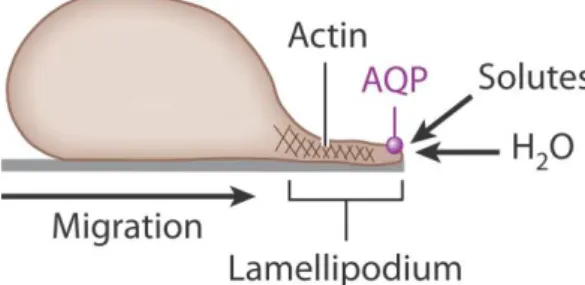
Studies with AQP1-null mice for investigation of a possible role of AQP1 in tumor angiogenesis, revealed a lower density of microvessels and consequently, an impaired angiogenic process. The overall result was a slowed tumor growth and improved survival. Other studies with AQP1-null mice reported a remarkable impairment in cellular migration, suggesting a strong correlation between AQP1 overexpression and migration. (Verkman, 2005) The same authors proposed a three step mechanism for AQP-facilitated cell migration, regardless of the cell type and AQP isoform (Verkman, 2008), as shown in Figure 1.4.
In the first step of cell migration, actin is cleaved leading to transient formation of membrane protrusions (lamellipodia and membrane ruffles). Ion uptake occurs at the tip of a lamellipodium (anterior end of the cell) creating local osmotic gradients. Consequently, a rapid water influx occurs increasing the local hydrostatic pressure, leading to the expansion of the cell membrane. If present, AQP polarizes to the leading edge of the cell membrane, facilitating water influx. Finally, actin repolymerizes leading to the stabilization of the protrusion. (Verkman, 2011a)
Figure 1.4. Proposed mechanism of AQP-facilitated cell migration. Water entry into protruding lamellipodia in migrating cells. Modified from (Verkman, 2012).
When expressed in tumor cells, AQP increases their ability to extravasate across blood vessels and to invade local tissues. (Hu, 2006) AQP-facilitated cell migration thus appears to be important not only in angiogenesis but also in tumor cell metastasis and spread. These findings may explain the high expression levels of AQPs in different tumor types and the correlation between AQP expression levels and tumor grade. (Verkman, 2012)
1. Introduction
10
1.3.1.2. AQP3 in skin tumorigenesis
AQP3 is an aquaglyceroporin widely expressed in many human tissues. Recently, analysis of AQP3-knockout mice has provided interesting information on the glycerol transport through this channel. In addition, different skin pathologies have been associated to AQP3 misexpression, namely skin cancer. (Hara-Chikuma, 2008a,b)
Mammalian skin is composed by three different layers, as shown in Figure 1.5A. The deepest layer of skin is rich in adipocytes. Above lies the dermis, rich in capillaries and composed by collagen fibers and elastin (among other components), that acts as a support layer for the third and most superficial skin layer, the epidermis. This region contains several different cell layers, being the external layer the stratum corneum (SC), that consists in terminally differentiated keratinocytes. These cells provide the outermost barrier against loss of body fluids. Thus, adequate hydration of the SC is essential for the maintenance of the skin health, allowing its flexibility and decreased vulnerability to external aggressions. (Rojek, 2008)
In mammalian skin, AQP3 is strongly expressed at the basal membrane of keratinocytes (Figure 1.5B). Studies with hairless mice lacking AQP3 exhibit reduced SC hydration (Verkman, 2005), reduced skin elasticity, delayed wound healing and delayed biosynthesis of SC (after removal by tape-stripping) (Hara, 2002). Studies using AQP3-null mice have also shown that water transport through AQP3 is not a rate-limiting factor in the trans-epidermal water loss. (Verkman, 2005)
Figure 1.5. Reduced skin hydration in AQP3 deficiency. A| Schematic representaion of skin layers; B| Proposed mechanism of AQP3 function in the skin. Modified from (Verkman, 2005).
The AQP3 deficiency in the skin reflects reduced epidermal glycerol permeability and reduced glycerol content in SC and epidermis, while normal glycerol content in serum and dermis. Thus, a reduced glycerol transport from blood into the epidermis, through the basal keratinocytes, is pointed as the plausible responsible for the skin phenotype in AQP3-null mice. (Verkman, 2005)
1. Introduction
11
More recently, studies in mice with disrupted AQP3 gene, revealed reduced epidermal pools of glycerol, glucose and ATP. (Hara-Chikuma, 2008b) Hara-Chikuma and co-workers also found a positive correlation between glycerol and ATP content in AQP3+/+ mice keratinocytes, suggesting the involvement of AQP3-mediated glycerol transport in ATP synthesis. This fact, together with a positive correlation between ATP content and cell proliferation, brought new clues about the importance of AQP3 in states of epidermal hyperproliferation, such as psoriasis, atopic dermatitis, wound healing, ichthyosis and even tumorigenisis, where it is upregulated. Summarizing, AQP3 plays central roles in skin hydration, wound healing and tumorigenesis (Hara-Chikuma, 2008b,c) although the implicit mechanism of tumorigenesis resistance observed for AQP3-null phenotype is still not clear. Figure 1.6 illustrates the proposed pathways for AQP3-dependent skin hydration, wound healing and AQP3-dependent cell proliferation during skin tumorigenisis. According to this mechanism, the overexpression of AQP3 leads to an increased glycerol uptake. As a rich energetic substrate, a high glycerol pool leads to increased ATP synthesis and, consequently, cell proliferation. Notwithstanding, and regarding AQP3 in particular, the triggering pathway for tumorigenesis is still obscure.Figure 1.6. Schematic representation of the proposed mechanisms of AQP3-dependent skin hydration, wound healing, and tumorigenesis. Modified from (Hara-Chikuma, 2008c; Verkman, 2009).
It must be noted that in 2012, Gao and co-workers described the contribution of AQP3 to the chemoresistance of melanoma to arsenite. (Gao, 2012) In addition, other studies identified an unexpected permeant for AQP3, the hydrogen peroxide (Miller, 2010; Bertolotti, 2013). This fact may unravel new unexpected roles for AQP3 in tumor biology through oxidative stress.
1. Introduction
12
1.4. AQP inhibitors: pharmaceutical opportunities
Several pathologies have been associated and/or attributed to the impaired functioning of AQPs. Consequently, the potential utility of aquaporin modulators for the treatment of several pathologies such as kidney diseases, obesity, glaucoma, brain edema, epilepsy and cancer should be considered. (Verkman, 2009)
As an example, AQP1-null xenograft models of subcutaneous melanoma tumors showed decreased tumor growth and reduced angiogenesis when compared to wild-type controls. These observations were associated with AQP1 gene disruption. (Lopez-Campos, 2011; Machida, 2011) Moreover, a chemical down-regulation of AQP1 expression was reported to block angiogenesis and tumor growth. (Bin, 2011)
Due to the central roles of AQP3 in epidermal cell migration and proliferation (Hara-Chikuma, 2008b,c) this aquaglyceroporin can also be seen as a potential target for cancer therapy. Recently, AQP3 inhibition by copper(II) ions reduced cell growth rates and increased the therapeutic efficacy of Cisplatin. (Huber, 2012)
Therefore, pharmacotherapy via AQP modulation should be considered as a valuable strategy for treating several and different human diseases. Despite AQP-based therapy for human diseases still being considered a distant reality, recent progresses were achieved with AQP4 in neuromyelitis optica. (Verkman, 2011b; Huber, 2012)
1.4.1. Metal-based agents as AQP3 inhibitors
Recently, gold complexes were described as new potent inhibitors of AQP3, being four times more effective, for the same concentration, than the common mercurial compounds used for AQP inhibition studies in vitro. (Martins, 2012)
In 2012, Martins and co-workers reported [Au(phen)Cl2]Cl (phen= phenantroline, Auphen) and
[Au(dien)Cl]Cl2 (dien= diethylendiamine, Audien) as potent and highly selective inhibitors of
the glycerol transport in human RBCs (90% inhibition) with half-inhibitory concentration (IC50)
values in the low-micromolar range. In addition, these Au(III) compounds showed to be non-toxic in RBC during the time span of channel inhibition. Their water solubility, plus the previous considerations, makes them suitable drugs for in vivo studies. (Martins, 2012) More recently, the same group tested other Au(III) compounds as AQP3 inhibitors. In order to evaluate and compare the influence of metal substitution on the inhibitory potency, the Cu(II) compound Cuphen was also tested.
1. Introduction
13
As result, the following compounds were also identified as potent and selective AQP3 inhibitors: [Cu(phen)Cl2]Cl (phen= phenantroline, Cuphen), [Au(terpy)Cl]Cl2 (terpy =terpyridine, Auterpy), [Au(bipy(R,R’))Cl2]PF6 (bipy = bipyridyl) where R=R’= H, Me, NH2
(Aubipy, AubipyMe, AubipyNH2 respectively). (Martins, 2013)
Figure 1.7 displays the structures of several metallodrugs described as AQP3 inhibitors.
Figure 1.7. Structures of several metallodrugs described as AQP3 inhibitors by Martins and colleagues. Modified from (Martins, 2013).
At this point, it is interesting to refer that all of these gold complexes were already described as antiproliferative agents in vitro, being considered as promising candidates to anticancer drugs. (Messori, 2000; Casini, 2009; Serratrice, 2012)
1.4.2. Metallodrugs as antitumoral agents
Metal compounds have been widely used in medicine for several decades. Among them we can find bismuth (anti-ulcer), silver (anti-microbial) and iron (anti-malarial) compounds and, in terms of anti-tumor activity, the earliest reports date from the sixteenth century. (Desoize, 2004) In the 1960s, B. Rosenberg discovered a platinum-based compound – cisplatin - with antitumor activity (Rosenberg, 1965, 1969), which became approved by the FDA in 1978 and still continues to be the first line of treatment for some types of cancer. (Guidi, 2012) Thus, metallodrugs are still a promising research area, drawing increased attention within the medicinal chemistry communities due to their antiproliferative and antitumor properties, obtained in both in vitro and in vivo studies. (Nobili, 2010; Berners-Price, 2011)
Since 1890, when Robert Koch discovered that gold cyanide was able to inhibit the growth of
M. tuberculosis, gold compounds started to be developed as therapeutic agents. (Benedek, 2004)
In the mid 1980s, auranofin, an orally active Au(I) which was approved for rheumatoid arthritis treatment, was described as an inhibitor of tumor cells growth in vitro. (Mirabelli, 1985)
1. Introduction
14
This was the beginning of a great advance in medicinal inorganic chemistry and metal-based compounds started to gain relevance in this field. This fact is due to the variety of properties that metal-based compounds can present like the coordination number, redox state or different geometries in addition to the intrinsic properties of the metal ion itself, dictating alterations in the compound reactivity.
By this time, Cisplatin was already marketed and Au(III) compounds, with square planar geometry, were seen as possible mimetics, since they present the same electronic configuration. The AQP3 inhibitors described above, including the Au(III), are examples of metallodrugs with cytotoxic activity. It is important to highlight that several Au(III) compounds are already described as being more potent than Cisplatin, overcoming the recurrent resistance problems associated to this chemotherapeutic. (Messori, 2000)
Despite the cytotoxic potential presented by metallodrugs, and in particular Au(III) compounds, this type of compounds have some drawbacks, namely instability under physiological conditions. As reactive molecules, they present high hydrolysis rates and reduction potential giving rise to the need of an adequate stabilization of these drugs at oxidation state +3. (Messori, 2004)
Several efforts have been made in order to synthesize gold complexes with increased stability under physiological conditions for pursuing Au(III) potential in cancer therapy. One approach consists on the stabilization of the metal center by nitrogen donors, to which Au(III) shows preference. Examples are Auphen and Auterpy (see Figure 1.7). (Nobili, 2010) Regarding Au(III) reactivity, it is also important to consider the variety of biological targets that this class of compounds may have in vivo. Human RBCs, in which glycerol movement across membranes is mostly mediated by AQP3, constitute a good example. (Martins, 2013)
In order to minimize the drawbacks mentioned above (i.e. drug instability and reactivity) appropriate carriers must be designed. In addition to a possible protection of the drug from inactivating events in vivo, an ideal carrier, among other characteristics, should be biocompatible and improve the therapeutic index of the associated compound.
1. Introduction
15
1.5. Drug Delivery Systems (DDS)
For the last decades, several efforts have been made pursuing the improvement on pharmacokinetic, biodistribution and pharmacodynamic profiles of drug candidates, which in the free form, present pharmaceutical properties far from the ideal.
As briefly referred above, a drug carrier should provide the release of the drug within the therapeutic window at the site of action, must be biodegradable and/or easily excreted after exerting its therapeutic effect, should present low immunogenicity and prevent premature degradation of the drug. Ideally, it should also be stable upon storage and present low production costs. (Gaspar, 2008)
Regarding cancer therapy, and particularly solid tumors, the DDS should also provide the ability to penetrate into the tumor interstial space. Based on these previous considerations, the reason of nanotechnology growth in the recent years is evident. Within this area, and gathering all the properties of an ideal DDS, liposomes are considered one of the most promising and successful drug carriers for cancer therapy. (Allen, 2004)
1.5.1. Liposomes
Liposomes were described for the first time by Dr Alec D. Bangham roughly 50 years ago. (Bangham, 1965) These systems were firstly developed as models of biological membranes due to their architecture that mimics the molecular structure of natural cell membranes. As an example, the identification of the first AQP was performed after protein reconstitution into liposomes in 1992. (Zeidel, 1992)
In the 70’s, these lipid systems started to be used for drug delivery (Gregoriadis, 1972) and nowadays, several liposomal formulations are already available in the clinic or in advanced development stages, for treatment of different pathologies including cancer. (Slingerland, 2012) These vesicles may present mean sizes ranging from few nanometers (50 nm) to more than 1 µm, and are colloidal particles generally constituted by naturally occurring phospholipids and cholesterol, conferring biodegradability properties. These spherical vesicles are organized in bilayers, separated by aqueous compartments, mimicking cellular membranes, and possessing the ability to incorporate different substances/compounds independently of their properties such as molecular weight, solubility or charge. (Cruz, 2009)
Due to the liposomes typical structure, they are able to encapsulate/incorporate molecules with distinct properties, which is an advantage per se. Hydrophilic molecules can be entrapped in the aqueous compartments while compounds with hydrophobic properties can be partial or totally accommodated within the lipid bilayers, as shown in Figure 1.8A. (Bei, 2010)
1. Introduction
16
Figure 1.8. Liposomes – versatile structures. A| classic liposomes with hydrophilic drug (a) at the inner aqueous compartment and hydrophobic drug (b) within the lipid bilayer. B| Immunoliposomes possess specific antibodies (or antibody fragments) at surface to enhance a specific targeting. C| Long-circulating liposomes present modified surface by the presence of polymers such as PEG (c) that allows the increase of circulation time in bloodstream and protects the carrier from opsonizing proteins (d). D| Long-circulating immunoliposome. Adapted from (Torchilin, 2005).
Focusing on their therapeutic action, liposomes as DDS present several advantages over non-encapsulated drugs.
In addition to the fact that liposomes are able to entrap a therapeutic molecule in their structure, several other advantages should also be mentioned. Liposomes are able to enhance drug solubility and to protect the incorporated drug from premature degradation or metabolization, while keeping its therapeutic activity, affording to a partial reduction of possible side-effects in healthy tissues and/or organs. Moreover, liposomes present a high versatility. They can be constructed according to the physicochemical properties of each particular drug or to the desired target tissue or organ. This may be performed by i) varying the lipidic constituents, ii) changing bilayer rigidity, iii) adding surfactants to lipid composition to alter superficial charge and, consequently, their stability and/or interaction with affected cells or organs, iv) attaching different polymers as shown in Figure 1.8C (such as PEG) to enhance the half-time of the vesicle in bloodstream and, consequently, of the drug and even iv) including antibodies or antibody fragments at the liposome surface for a specific targeting, as shown in Figure 1.8B and D. (Torchilin, 2005, 2007; Cruz, 2009)
c d
1. Introduction
17
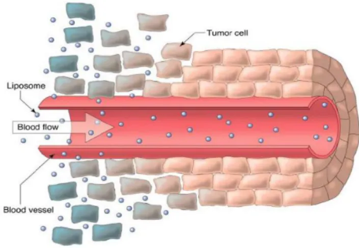
From all above mentioned advantages, the most important strength of these systems is their ability to improve pharmacokinetics (PK) and biodistribution (BD) of the associated therapeutics. Liposomes, depending on their lipid constituents, are able to increase the circulation in the bloodstream of the incorporated drug reducing its clearance. This type of lipid vesicles is designed as long-circulating liposomes (LCL) and they may be constructed by inclusion in the lipid composition of different polymers, covalently linked to phospholipids. Polyethylene glycol (PEG) is the most popular polymer used. (Harris, 2003) The presence of PEG at liposome surface, as shown in Figure 1.8C, reduces the adsorption of plasma proteins. Due to these characteristics, this type of liposomes, after parenteral administration, is able to have a longer circulation time in bloodstream. Regarding BD, alterations can occur via the so called enhanced permeability and retention (EPR) effect, also called passive targeting. (Maeda, 2000, 2001) Several pathological conditions present increased permeability of tissue vasculature. Solid tumors are a good example of compromised vasculature. As tumor tissue starts to grow, the support of nutrients and oxygen will eventually be insufficient for tumor nutritional requirements. To overcome this situation, cytokines and other signaling molecules are released from tumor cells in order to recruit more blood vessels in a process called angiogenesis. Angiogenic blood vessels present gaps of 600 to 800 nm between adjacent endothelial cells (Figure 1.9) allowing the extravasation of liposomes to the interstitial space in a size-dependent manner.Figure 1.9. Extravasation and accumulation of liposomes in tumor tissue due to EPR effect. (Saetern, 2004)
1. Introduction
18
As these lipid systems present sizes substantially lower, they tend to accumulate in the tumor tissue. Thus, to take advantage of this EPR effect, liposomes must present long circulation times in the bloodstream (by the inclusion of PEG in lipid composition, for example) in order to enable, to a higher extent, the extravasation of the drug into the tumor.
In addition to the preferential targeting of liposomes to tumor vasculature, it is important to refer that, in general, tumor tissues lack an effective lymphatic drainage. Therefore, extravasated liposomes are able to accumulate in tumor tissues. (Allen, 2004; Torchilin, 2007; Cruz, 2009) All of these characteristics, combined with the already mentioned biodegradability, make liposomes a suitable and promising carrier for clinical use and, particularly, for cancer therapy.
1. Introduction
19
1.6. Aims and goals
In the present study the first objective was to validate the cytotoxicity of a small library of metallodrugs, already described as AQP3 inhibitors. Based on the cytotoxic screening, one compound was selected to be further incorporated in an adequate delivery system, liposomes. The second objective of the work was the design and development of liposomal formulations able to incorporate the selected compound. The third objective was the in vitro evaluation of the cytotoxic activity of the AQP3 inhibitor previously selected, both in the free and in the liposomal forms, in two cancer cell lines.
In order to fulfill the objectives of the present thesis the work was persecuted according to the following:
1. Validation of cytotoxicity of a small library of metallodrugs already described as AQP3 inhibitors against a human epidermoid carcinoma cell line (A431) overexpressing this aquaglyceroporin;
2. Selection of a single compound to be incorporated in liposomes, based on the cytotoxic activity against A431 cells;
3. Establishment of an adequate methodology for quantification of the selected AQP3 inhibitor;
4. Development and characterization of liposomes incorporating the AQP3 inhibitor. In particular, the influence of lipid composition, mean size and superficial charge on incorporation parameters were studied;
5. Cytotoxic in vitro studies of the selected metallodrug formulations in free and liposomal
forms against two cancer cell lines: the A431 and the murine colon cancer cell line, C26.
It is important to highlight that, due to similarities of the metallodrugs tested, the developed formulation can be seen as a model for further studies involving other AQP3 inhibitors.
21
2. Materials and Methods
23
2.1. Materials
All the metallodrugs were synthesized and gently provided by Dr. Angela Casini (Pharmacokinetics, Toxicology and Targeting, Research Institute of Pharmacy, University of Groningen).
Six mononuclear compounds were included in the present work: [Au(phen)Cl2]Cl
(phen=1,10-phenantroline, Auphen), [Cu(phen)Cl2]Cl (Cuphen) [Au(dien)Cl]Cl2 (dien = diethylentriamine,
Audien), [Au(terpy)Cl]Cl2 (terpy = tripyridine) and two different compounds based on the
following [Au(bipy(R,R’))Cl2]PF6 (bipy = 2,2’-bipyridine, Aubipy), where R=R’= H, or Me.
Aubipy compounds will be referred according to the substituent group:. R=Me, AubipyMe, R=H, Aubipy.
The pure phospholipids, egg phosphatidylcholine (PC), dimyristoyl phosphatidylcholine (DMPC), Dipalmitoyl phosphatidylcholine (DPPC) and distearoyl phosphatidylethanolamine covalently linked to poly(ethylene glycol) 2000 (PEG), used for the preparation of liposomal formulations were purchased from Avanti Polar Lipids (Alabaster, AL).
Deionized water (Milli-Q system; Millipore, Tokio) was used in all experiments. Nuclepore Track-Etch Membranes were purchased from Whatman Ltd, (NY, USA). Culture media and antibiotics were obtained from Invitrogen (Life Technologies Corporation, NY, USA). Reagents for cell proliferation assays were purchased from Promega, (Madison, WI, USA). Octadecylamine (n-Stearylamine (SA)), cholesterol (Chol) and Hoescht 33258 were purchased from Sigma (Sigma-Aldrich, St. Louis, MO, USA). All the remaining chemicals and substrates used were of analytical grade.
2. Materials and Methods
24
2.2. Methods
2.2.1. Preparation of Cuphen liposomal formulations
Liposomes composed of the selected phospholipids were prepared by the dehydration-rehydration method (DRV) (Cruz, 1993; Gaspar, 1996, 2008). Briefly, the selected phospholipids (20µmol/mL) were dissolved in chloroform and the mixture was dried by rotary evaporation (Buchi, Switzerland) of the organic solvent to obtain a thin film in a round-bottom flask. The film was then dispersed in a Cuphen solution, frozen (-70ºC) and lyophilized (Freeze dryer, Edwards, USA) overnight.
The rehydration of the lyophilized powder was performed with a buffer constituted of 10 mM HEPES and 145 mM NaCl, pH 7.4 (HEPES buffer) in two steps, in order to enhance the Cuphen incorporation (Lasch, 2003): first, a 30 minute step where two-tenth of the original dispersion was added, and subsequently, the addition of the remaining volume (up to the starting volume). Here it is important to refer that the hydration steps should always be performed at a temperature above the phase transition temperature (Tc) of the phospholipids. In order to reduce and homogenize the mean size of liposomes, the so formed vesicles were submitted to an extrusion step through polycarbonate membranes of appropriate pore size until the desired vesicle size is reached (1, 0.8, 0.6, 0.4 (3x), 0.2 (3x) and 0.1 (3x) µm) under nitrogen pressure (10-500 lb/in2) with an Extruder device (Lipex: Biomembranes Inc., Vancouver, British Columbia, Canada).
The separation of non incorporated Cuphen was performed by ultracentrifugation at 250,000 g for 120 min at 15ºC in a Beckman LM-80 ultracentrifuge (Beckman Instruments, Inc, USA.) Finally, the pellet was ressuspended in HEPES buffer, according to the final concentration desired.
2.2.2. Characterization of Cuphen liposomal formulations
Liposomes were characterized in terms of lipid composition, lipid (Lip) and Cuphen concentration, mean diameter, zeta potential and by the following incorporation parameters: initial and final Cuphen to lipid ratios [(Cuphen/Lip)i and (Cuphen/Lip)f, respectively] and incorporation efficiency (I.E.) defined as the percentage of [(Cuphen/Lip)f]/[(Cuphen/Lip)i]. The I.E., being a ratio between final to initial (Cuphen/Lip), determines the ability that a particular lipid mixture presents for incorporating Cuphen in liposomal matrix.
2. Materials and Methods
25
2.2.2.1. Cuphen quantification
UV/Vis spectroscopy, due to its simplicity and reliability was the selected technique for Cuphen quantification. Moreover, the destruction of liposomes was performed by addition of absolute ethanol (EtOH).
From a stock solution of 200nmol/mL in EtOH, serial dilutions were performed with the same organic solvent and UV spectra were traced (220nm-800nm) in a UV/Vis spectrophotometer (Shimadzu UV 160A).
After selection of the appropriate wavelength, calibration curves were performed in order to evaluate the relation between the absorbance values and concentration. The selected concentrations ranged from 2 μM up to 20 μM, in order to ensure linearity and enabling the determination of Cuphen content in developed formulations after liposome disruption with EtOH.
2.2.2.2. Phospholipid quantification
The method for phospholipid quantification was based on the colorimetric determination of phosphate (PO4). In the presence of ammonium heptamolybdate ((NH4)6Mo7O24.4H2O) the
inorganic phosphate was converted to phosphomolybdic acid, which was quantitatively converted to a blue color due to reduction of ascorbic acid during heating, using the method described by Rouser and co-workers (Rouser, 1970). Briefly, samples (in triplicate) containing phosphate quantities between 20 and 80 nmol (sample volume below 100 µL) were pipetted into 15 mL glass tubes. In parallel, a calibration curve from a 0.5 mM phosphate solution was prepared: in triplicate, phosphate amounts of 20, 30, 40, 50, 60 and 80 nmol were pipetted into glass tubes. All tubes were heated (180ºC) in a heating block until dryness. After cooling, 0.3 mL of perchloric acid (70-72%) was added to all tubes. In order to avoid volume losses, marbles were placed on the top of all glass tubes. At this point, all tubes were heated in the heating block (180ºC) for 45 min, to convert all the organic lipid phosphate to the inorganic form and until achievement of a clear solution. After cooling samples to room temperature, 1.0 mL of H2O, 0.4
mL of hexa ammonium heptamolybdate solution [1.25% (w/v)] followed by 0.4 mL of ascorbic acid solution [5% (w/v)] were added to all glass tubes. A blue color solution was obtained due to the reduction of ascorbic acid during heating in a boiling water bath for 5 min. After cooling, the absorbance of all samples was recorded (797nm) in a UV-mini 1240 Spectrophotometer (Shimadzu). The amount of phosphate in samples was obtained through the calibration curve with the aid of linear regression. The calibration curve was linear up to absorbance values of around 1.000.
2. Materials and Methods
26
2.2.2.3. Liposome size measurements
Liposome mean diameter was determined by dynamic light scattering based on Brownian motion of the particles in a hydrodynamic sizing system (Zetasizer Nano S (Zen 1600), Malvern Instruments, UK). For viscosity and refractive index, the values of pure water were used. As a measure of particle size distribution of the dispersion, the system reports the polydispersity index, (P.I.). P.I. ranges from 0.0, for an entirely monodisperse sample, up to 1.0 for a polydisperse suspension. To determine the mean diameter and P.I. of liposomal preparations, samples were diluted to a final lipid concentration of 0.2 µmol/mL in HEPES buffer. All the measurements were done in an appropriate polycarbonate cell at a temperature of 25ºC.
All liposomal formulations were prepared in order to obtain a P.I. < 0.30.
To ensure that appropriate mean diameter and P.I. were achieved, these parameters were also determined during the extrusion procedure.
2.2.2.4. Zeta potential determination
Zeta potential of liposomal formulations was measured in a hydrodynamic sizing system (Zetasizer Nano Z (Zen 2600), Malvern Instruments, UK). Zeta potential is defined as an electric potential between the membrane surface and the ionic dispersion medium. Zeta potential was measured using a combination of the measurement techniques: Electrophoresis and Laser Doppler Velocimetry, sometimes called Laser Doppler Electrophoresis. This method measures how fast a particle moves in a liquid when an electrical field is applied. For viscosity and refractive index, the values of pure water were used.
Before determination of the zeta potential of liposomal formulations, an initial check of the apparatus was made with a standard known zeta potential value (standard DTS5050, Malvern Instruments, Ltd., UK). Dilutions of liposomal formulations were made in HEPES buffer, at a final lipid concentration of about 0.2 µmol/mL. Samples were slowly introduced into a clear disposable zeta cell with a syringe to avoid air bubbles. The zeta potential of samples was recorded at a temperature of 25ºC.
2. Materials and Methods
27
2.2.2.5. Stability of Cuphen liposomes
The stability in suspension of Cuphen liposomes was assessed by quantifying Cuphen and lipid contents after storage at 4ºC for 10 days.
After this storage period (t=10), samples were taken (t=0), diluted in HEPES buffer and submitted to a centrifugation step (250,000 g for 120 min at 15ºC). Liposomes were ressuspended in HEPES buffer according to the initial volume. Cuphen and phospholipid contents were quantified. The stability was defined as the ratio in percentage between Cuphen to lipid ratio at t=10 and the initial Cuphen to lipid ratio at zero time (t=0), according to the following formula: [(Cuphen/Lip)t=10/(Cuphen/Lip)t=0] x 100.
In addition, vesicles mean size and zeta potential were also determined.
2.2.3. Cell Culture
Human epidermoid carcinoma cells, A431, gently provided by Prof. Eschevarria (Instituto de Biomedicina de Sevilla (IBIS), Hospital Universitario Virgen del Rocío, Seville, Spain) were seeded in culture flasks and maintained in Dulbecco’s modified Eagle’s medium (DMEM) with high-glucose (4500 mg/L), supplemented with 10% fetal bovine serum (FBS) and 100 μg/mL penicillin/streptomycin. Murine colon cancer cells, C26, obtained from CLS (Cell Lines Service, Life Technologies) were plated in culture flasks and maintained in Roswell Park Memorial Institute (RPMI) 1640 with the supplementation described above. Both cell lines were kept at 37° C under a 5% CO2 atmosphere. Maintenance of cultures was performed every
two/three days, until cells reached a confluence of about 80%. At this point, sub-culturing was performed using a solution of TrypLE (Invitrogen, Life Technologies Corporation, NY, USA). Briefly, after media removal, the cell layer was washed with phosphate buffered saline (PBS) and incubated with TrypLE for 7-10 minutes at 37ºC. After cells detachment, complete growth medium was added. The cells were then centrifuged in a bench centrifuge (Beckman, Izasa, Spain) at 500g for 10 min and the pellet ressuspended in fresh culture medium. Appropriate aliquots of the cell suspension were seeded in new culture flasks.
Cell storage was done in liquid nitrogen, in cryotubes with freezing medium consisting of FBS and 10% dimethylsulfoxide (DMSO).