RESEARCH ARTICLE
Cite this:Med. Chem. Commun.,
2017,8, 1993
Received 19th April 2017, Accepted 6th September 2017
DOI: 10.1039/c7md00196g
rsc.li/medchemcomm
Encapsulation of nor-β-lapachone into poly
IJD
,
L
)-lactide-
co
-glycolide (PLGA) microcapsules: full
characterization, computational details and
cytotoxic activity against human cancer cell lines†
Marcília P. Costa,
aAnderson C. S. Feitosa,
bFátima C. E. Oliveira,
bBruno C. Cavalcanti,
bGleiston G. Dias,
cEwerton W. S. Caetano,
deFrancisco A. M. Sales,
deValder N. Freire,
fStefano Di Fiore,
gRainer Fischer,
ghLuiz O. Ladeira,
iEufrânio N. da Silva Júnior
*
cand Claudia Pessoa*
bjIn this work, we characterize nor-β-lapachone-loaded (NβL-loaded) microcapsules prepared using an emulsification/solvent extraction technique. Features such as surface morphology, particle size distribution, zeta potential, optical absorption, Raman and Fourier transform infrared spectra, thermal analysis data, drug encapsulation efficiency, drug release kinetics andin vitrocytotoxicity were studied. Spherical microcap-sules with a size of 1.03 ± 0.46μm were produced with an encapsulation efficiency of approximately 19%. Quantum DFT calculations were also performed to estimate typical interaction energies between a single nor-β-lapachone molecule and the surface of the microparticles. The NβL-loaded PLGA microcapsules exhibited a pronounced initial burst release. After thein vitrotreatment with NβL-loaded microcapsules, a clear phagocytosis of the spheres was observed in a few minutes. The cytotoxic activity against a set of cancer cell lines was investigated.
1. Introduction
Cancer is a proliferative disease characterized by atypical growth of uncontrolled cells that invade other tissues. Cancer-ous cells, also called malignant cells, move to the blood circu-lation and spread to lymph nodes (metastasis), eventually
de-veloping new tumours in different organs.1 These aberrant
proliferations can be clinically detectable as neoplasia, which literally means new growth.2Many studies have attested that
these mutations can allow the functional activation of some genes (oncogenes) or the inactivation of tumour suppressor genes.1,3Oncogenes contribute to the stimulation of
prolifera-tion or the deactivaprolifera-tion of senescence and/or apoptosis, whereas tumour suppressor genes generally act as check-points to proliferation or cell death.1,4Epigenetic factors
con-nect external (toxic compounds, drugs or diet) and internal factors (cytokines or growth factors) involved in cancer development.5
Numerous plant-derived compounds were reported to pos-sess robust anticancer activity.6 Among the most promising
drugs, quinones constitute a diversified class of compounds, which are widely distributed in nature, classified by the aro-matic moieties existent in their structure and associated with diverse biological activities.7 The National Cancer Institute
(NCI) identified the quinone moiety as an important pharmacophoric group owing to its cytotoxic activity in screening of natural products.8
As reported so far, the cytotoxicity of quinones can be re-lated to a number of mechanisms, in particular redox cycling, which produces reactive oxygen species (ROS), and the alkyl-ation of cellular nucleophiles, prompting covalent binding aPharmacy Course, Federal University of Piauí, 64049-550 Teresina, PI, Brazil
bDepartment of Physiology and Pharmacology, Federal University of Ceará,
60430-270 Fortaleza, CE, Brazil. E-mail: [email protected]
cInstitute of Exact Sciences, Department of Chemistry, Federal University of
Minas Gerais, Belo Horizonte, 31270-901, MG, Brazil. E-mail: [email protected]; Tel: +55 31 34095720
dDepartment of Secondary School and Teachers College, Federal Institute of
Ceará, 60040-531 Fortaleza, CE, Brazil
eFederal Institute of Ceará, 63503-790 Iguatu, CE, Brazil
fDepartment of Physics, Federal University of Ceará, 60455-760 Fortaleza, CE,
Brazil
gFraunhofer Institute for Molecular Biology and Applied Ecology IME, 52074,
Aachen, Germany
hInstitute for Molecular Biotechnology, RWTH Aachen University, 52074 Aachen,
Germany
iInstitute of Exact Sciences, Department of Physics, Federal University of Minas
Gerais, Belo Horizonte, 31270-901, MG, Brazil
jOswaldo Cruz Foundation (Fiocruz), 60180-900 Fortaleza, CE, Brazil
†Electronic supplementary information (ESI) available: All the experimental de-tails. See DOI: 10.1039/c7md00196g
Published on 07 September 2017. Downloaded by Federal University of Ceará on 19/12/2017 11:42:50.
View Article Online
with proteins and/or DNA9and producing irreversible cellular
changes and death.10
Quinoidal compounds are the second largest class of anti-cancer drugs used clinically for anti-cancer chemotherapy.9b,11
Re-lated to this class, lapachol (extracted from the heartwood of Tabebuia sp.) and its derivatives α-lapachone and β-lapachone are among the most important naphthoquin-ones studied due to their biological activities.12β-Lapachone
is one of the most widely studied naphthoquinones currently in phase II clinical trials.13β-Lapachone was shown to be a
selective inducer of cell death in cancer cells,14acting against
a wide variety of multidrug resistant cancer cell lines.15
Fur-thermore,β-lapachone increased the lethality against human cancer cells in association with other drugs (taxol, mitomycin C and paclitaxel) and acted as a co-adjuvant in killing human cancer cells during radiotherapy treatment.14Specifically, this
compound is bioactivated by NADIJP)H:quinone oxidoreduc-tase 1 (NQO1). NQO1 induces futile cycling of the drug that exhausts NAD(P)H in the cell, leading to a considerable quan-tity of ROS that causes DNA damage.16Unfortunately, the low
solubility and non-specific distribution of β-lapachone have limited its suitability for clinical assays.17To overcome these
problems, several delivery release strategies were proposed: β-lapachone complexed with hydroxypropyl-β-cyclodextrin16
or methylated-β-cyclodextrin18 and β-lapachone-containing
poly(ethylene glycol)-block-poly(D,L-lactide) polymer
mi-celles.16 Gold nanoparticles with surface modifications with
polyethylene glycol loaded with β-lapachone in per-6-thio-β -cyclodextrin pockets and complexed with an anti-epidermal growth factor receptor antibody as a targeting ligand were al-ready demonstrated.19 More recently, β-lapachone in
lipo-somes20 and a β-lapachone-poloxamer-cyclodextrin ternary
system17were also put to the test.
Our research group has dedicated effort to the synthesis and antitumor studies of lapachone derivatives.21Recognized
as an important prototype with anti-cancer activity, nor-β -lapachone (NβL) (Fig. 1) was studied by us against several cancer cell lines, exhibiting IC50values in the range of 0.3–
2.5μM.10,22Interestingly, this drug showed low cytotoxic
ac-tivity in non-tumor cells and genotoxic and mutagenic effects only at high concentrations.23,24Compared withβ-lapachone,
nor-β-lapachone also has great advantages in synthetic terms. NβL is prepared from nor-lapachol in a quantitative yield. Nor-lapachol is easily prepared from a commercial quinone, lawsone.21
Strategies for cancer treatment based on controlled deliv-ery systems are the focus of many investigations to overcome liposolubility and/or low bioavailability problems, as well as the instability of the drug in physiological fluids and off-target toxicity of anticancer drugs, optimizing therapeutic ef-ficacy and reducing toxic side effects.25Gradual release of the
drug with constant plasma levels simplifies its uptake by can-cer cells, improving the drug availability at the desired target and consequently increasing its efficacy.25a Besides, it de-creases the drug resistance developed by tumour cells.26
Numerous chemotherapeutic agents have been micro-encapsulated in a polyIJD,L-lactide-co-glycolide) (PLGA) polymer
(Fig. 1).26a,27 PLGA is a biodegradable and biocompatible
polymer approved by the US FDA for human use28 and
ap-plied in pharmaceutical and medical devices which allows the gradual release of entrapped drugs over a long duration.25a PLGA-based drug delivery systems undergo two main release mechanisms, namely, diffusion and degrada-tion/erosion,29 and may present an adjustable degradation
rate based on the amount of the D,L-lactide/glycolide
poly-mers.28All these properties have turned PLGA into an
attrac-tive option for the development of controlled release delivery devices.
For the reasons discussed above, recently we have shown the efficiency of NβL in PLGA microparticles for improving its cytotoxicity against prostate cancer cells.30In this previous
report, we have discussed our first impressions related to this efficient strategy to make this lapachone derivative more po-tent against cancer cells.
In this work, we present a complete study on a PLGA-microencapsulated formulation containing nor-β-lapachone. We evaluated some modifications to increase the drug load. The interaction of NβL and PLGA after encapsulation was then studied by absorbance measurements, luminescence spectroscopy, Raman spectroscopy, Fourier transform infra-red (FT-IR) spectroscopy and differential scanning calorime-try (DSC). The drug loading, encapsulation efficiency, and in vitrodrug release kinetics were measured by spectroscopic analysis, while scanning electron microscopy (SEM) was employed to assess the surface morphology and size distribu-tion of the microparticles. Surface charges were evaluated by zeta potential measurements, and classical molecular dynam-ics, annealing, and adsorption techniques were combined with quantum density functional theory (DFT) calculations to estimate the typical binding energy values of the drug on the PLGA microparticle surface. The in vitro cytotoxic effect of the NβL-loaded microcapsules was investigated by examining their viability on different cancer cell lines in comparison with empty PLGA microcapsules and the free drug. The in vitro cellular internalization of NβL-loaded microcapsules was visualized by using optical microscopy.
2. Results and discussion
Nano/microparticles are important carriers for drug delivery. Microparticles have diameters between 1 and 250μm and are
Fig. 1 Representation of the nor-β-lapachone (NβL) and PLGA mole-cules: (a) planar chemical structure of NβL; (b) 3D chemical structure of NβL; (c) planar chemical structure of polyIJD,L)-lactide-co-glycolide (PLGA);x(lactic acid) andy(glycolic acid) indicate the number of repe-titions of each unit.
prepared through conventional emulsion solvent evaporation techniques or spray drying,28 which are easy to implement.
Microencapsulation methods can form microspheres (mono-lithic matrix) or microcapsules (hollow capsules),31in general
exhibiting a spherical morphology with monodisperse32 or
polydisperse particle size distributions.31a Microparticles, in comparison with nanoparticles, have the advantage of carry-ing higher drug amounts and exhibit better control over sustained release profiles.32
Initially, we have focused our efforts on the preparation and characterization of PLGA microparticles involving NβL. Based on the physicochemical properties of NβL, PLGA microparticle formulations were prepared by the emulsion solvent evaporation method, either with a simple (o/w) or a double (w/o/w) emulsion system. The w/o/w method was not effective for this drug, the simple emulsion method being the most satisfactory for the preparation of NβL microparticles. Evaluation of the role of formulation parameters such as the drug/polymer ratio and PVA solution volume in the micropar-ticles was carried out. Table S1 (ESI†) gives the details of 10 formulations prepared using an o/w emulsion and 9 formula-tions prepared using a w/o/w emulsion. Formulation 7 showed the best results in terms of morphology and encapsu-lation rate and was selected for all subsequent studies. The percentage yield of each adequate encapsulation was deter-mined and was greater than 70% for all formulations.
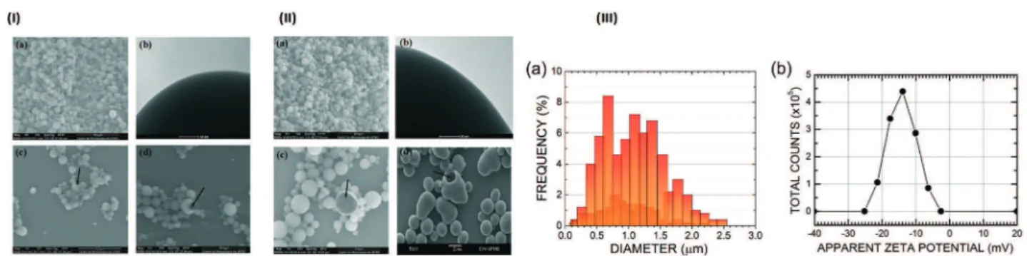
Following our strategy, the microparticle morphology, size distribution and zeta potential were also studied. Scanning electron microscopy (SEM) and transmission electron micros-copy (TEM) were employed for the morphological characteri-zation of the PLGA microparticles with (Fig. 2, IIa–d) and without NβL (Fig. 2, Ia–d). The PLGA microparticles containing NβL showed a regular spherical shape, a heteroge-neous size and the absence of agglomeration (Fig. 2, IIa), looking not very different from the case of pure PLGA micro-particles (Fig. 2, Ia). Their surfaces were smooth and nonporous, and no drug crystal formation was observed
(Fig. 2, IIb). The electron microscopy studies also confirmed that the formulated microparticles had a hollow structure (microcapsules) (Fig. 2, IIc), as in the samples of pure PLGA microparticles (Fig. 2, Ic). Following incubation in distilled water at 37°C for 24 h, we observed changes in morphology and polymer degradation after just one day, as shown in Fig. 2(Id), while for pure PLGA the same level of degradation was observed after five days of degradation in water at the av-erage core human body temperature of 37 °C. PLGA micro-particles containing NβL exhibit a bimodal size distribution with maxima at 0.68μm and 1.23μm, an average diameter of 1.03 ± 0.46 μm (Fig. 2, IIIa), a minimum observed diameter of 0.15μm (0.2% frequency) and a maximum observed diam-eter of 2.47 μm (0.4% frequency). The most common PLGA microparticle diameter found was about 0.68 μm, with 8% frequency, followed by 1.1μm with 7.2%, 1.37μm with 6.8% and 1.23μm with 6%. The surface electrical properties of the microcapsules revealed that empty PLGA microparticles had a zeta potential of −23.4 ± 0.35 mV, while PLGA
microparti-cles containing NβL exhibited a less negative value (−14.0 ±
0.17 mV) (Fig. 2, IIIb), corresponding to 4.4 × 10⁵counts at
−14.0 mV, 3.4×10⁵counts at−17.6 mV and 2.9 ×10⁵counts
at−10.0 mV (the number of counts falls to practically zero
be-low−25.2 mV and above −2.50 mV). Thus, PLGA
microparti-cles loaded with NβL tend to be less stable against coagula-tion than colloidal dispersions of pure PLGA microparticles.
The incorporation of NβL into the microcapsules was also detected through spectroscopic techniques. Fig. 3(I) shows the UV-vis absorption curves of pure NβL, PLGA microparti-cles without the drug and PLGA microcapsules containing NβL molecules. One can see that the NβL molecule has its absorption onset at about 580 nm with two bumps at 470 nm and 285 nm and a pronounced maximum at 256 nm. The PLGA microcapsules, on the other hand, exhibit a sharp ab-sorption onset at 250 nm. When both spectra are added and averaged (μPLGA,NβL, the dashed-dotted gray curve in Fig. 3(I)), the resulting plot resembles a scaled down version
Fig. 2 (I) Microphotographs illustrating the regular spherical form, different sizes and surface features of empty PLGA microparticles obtained by the simple emulsion (o/w) process. (a) Scanning electron micrograph (SEM) at a magnification of×5000; (b) transmission electron microscopy image showing a microparticle surface at a magnification of×300; (c) SEM showing cryofractured microcapsules (black arrow); (d) SEM showing the irregular form and erosion of PLGA microparticles (black arrow) after 5 days of degradation in water at 37°C. (II) Microphotographs illustrating the shapes, different size ranges and the surface structure of PLGA microparticles containing NβL obtained by the simple emulsion (o/w) process. (a) Scanning electron micrographs (SEM) at a magnification of×5000; (b) Transmission electron microscopy image showing a microparticle surface at a magnification of×300; (c) SEM showing cryofractured microparticles (black arrow); (d) SEM showing the irregular form and erosion of PLGA microparticles (black arrow) after 1 day of degradation in water at 37°C. (III) Characterization of PLGA microparticles containing NβL: a) size distribution of 500 particles and b) zeta potential (N= 3).
of the NβL loaded PLGA curve for wavelengths smaller than 305 nm. The NβL loaded PLGA UV-vis absorption spectrum, on the other hand, has a very pronounced maximum at 261 nm related to the NβL spectral peak at 256 nm and a small absorption bump at 580 nm. Other features of the NβL UV spectrum can be seen in Fig. 3(I) as well: absorption shoul-ders at 288, 339, and 453 nm are matched in the pure NβL spectrum with equivalent structures at 285, 343, and 470 nm.
The FT-IR spectrum (Fig. 3, IIa) of the PLGA microcap-sules loaded with NβL follows closely the spectrum of the unloaded microparticles, exhibiting a series of absorption bands between 490 and 780 cm−1(related to CH bending
vi-brations), 980 and 1480 cm−1 (CH
2 bending vibrations and
wagging vibrations for the lowest wavenumbers, backbone CCO stretching vibrations for the highest wavenumbers), a sharp peak at about 1760 cm−1 (C
O stretching vibrations),
and bands centered at 2950 (CH2 stretching vibrations) and
3380 cm−1 (OH stretching motion). The pure NβL curve, in
contrast, has a set of low intensity absorption bands scattered between 500 and 1740 cm−1and high wavenumber
bands at 2920 cm−1 and 3440 cm−1. In the region between
1500 and 1670 cm−1, one can see a set of characteristic peaks
of NβL, which are also present in the NβL-loaded PLGA microcapsules (Fig. 3, IIb), but absent in the empty formula-tion. These peaks occur (for pure NβL) at wavenumbers 1568, 1587, 1611, 1631, and 1643 cm−1 (there is also a small
ab-sorption band at 1694 cm−1 which is barely visible in the
PLGA microparticles with NβL resembling the E2g normal
mode of benzene at 1763 cm−1). The peak at 1568 cm−1can
be assigned to the bending motion of the CH3groups of the
NβL molecule, while the 1587 cm−1 absorption band
corsponds to the bending of two CH bonds in the benzene re-gion of NβL (in the same fashion as the A2gnormal mode of
benzene at 1544 cm−1). At 1611, 1631, and 1643 cm−1, the
vi-brational modes can be depicted as CH3 scissoring motions
and the combination of CH benzene bending (reminiscent of the E1ubenzene normal mode at 1658 cm−1) with a small
de-gree of C–C bond stretching. For the drug loaded PLGA microparticles, the same peaks in the order of increasing wavenumber occur at 1571, 1589, 1614, 1632, 1645, and 1693 cm−1, the first five exhibiting small blue shifts (of the order
of 2 cm−1) with respect to the spectral curve of the isolated
drug.
Fig. 3(III) shows the Raman spectra of the pure NβL,
empty PLGA microcapsules and PLGA microcapsules
containing NβL. The presence of NβL was verified by the ap-pearance of four signature peaks in the 1560–1660 cm−1
wavenumber range, namely at 1571, 1591, 1614, and 1647 cm−1. The maxima at 1571 and 1591 cm−1correspond to the
infrared absorption lines observed at 1568 and 1587 cm−1,
with the wavenumber differences being ascribed to experi-mental uncertainties. These lines involve CH3 and benzene
ring vibrations, as described previously, while the Raman bands at 1614 and 1647 cm−1 are related to infrared
absorp-tion peaks at 1611 and 1643 cm−1(also assigned to benzene
ring normal modes), respectively. These Raman intensities are slightly blue shifted (by about 1 cm−1) for the PLGA
microparticles with drug incorporation; the peak at 1571 cm−1 moves to 1572 cm−1, the peak at 1591 cm−1 shifts to
1594 cm−1, the 1614 cm−1line stays at the same wavenumber,
and the peak at 1647 cm−1shifts to 1648 cm−1.
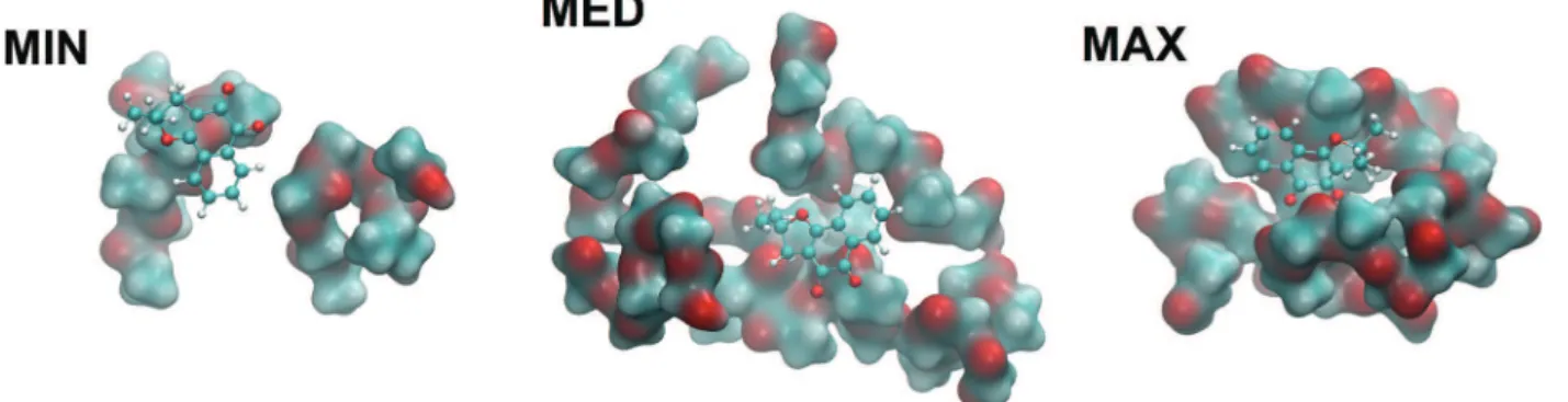
We have also established by computational analysis (Fig. 4) the optimized structures of PLGA chains with mono-mers of lactic acid and glycolic acid in a random sequence interacting with a single NβL molecule. After carrying out classical geometry optimizations and annealing computa-tions, the best geometries were reoptimized using quantum density functional theory (DFT) in order to estimate the bind-ing energy of NβL on the surface of a PLGA microparticle. It was observed that the polymeric structure is highly irregular before and after the classical annealing procedures, giving rise to connected clusters with dissimilar sizes and which are rich in indentations, cavities and clefts suitable for the bind-ing of NβL. Monte Carlo calculations were employed to probe the surface of the annealed polymer, and 50 adsorption sites were found. Their adsorption energies were estimated to be ranging from −9.85 kcal mol−1 (physisorption or weak
bind-ing) to −31.5 kcal mol−1 (chemisorption or strong binding).
Three of these structures were selected, with adsorption ener-gies at the classical levels of −9.85 kcal mol−1 (MIN), −18.5
kcal mol−1 (MED), and
−31.5 kcal mol−1(MAX). Fig. 4 shows
the DFT adsorption geometries obtained from these
Fig. 3 (I) Absorbance spectra of NβL (pure and encapsulated in PLGA microparticles) and PLGA microparticles without the drug (empty). The arrow shows the peak of NβL in the formulation containing the drug (black line). (II) (a) FT-IR spectra of pure NβL (dotted line), empty PLGA micro-particles (dashed line) and PLGA micromicro-particles containing nor-β-lapachone (solid line). (b) The gray rectangle highlights the peaks of NβL in the formulation containing the drug in the 1500–1800 cm−1range. (III) Raman spectra of pure NβL, empty PLGA microparticles and PLGA
microparti-cles containing NβL. The gray rectangle shows the signature peaks of NβL in the formulation containing the drug.
classically optimized structures using a GGA dispersion corrected exchange-correlation functional. In the maximum binding energy configuration, the NβL molecule is docked to a cleft where the effective contact surface with PLGA is the largest in comparison with the MED and MIN adsorption ge-ometries and with a DFT adsorption energy of −40.9 kcal
mol−1, which indicates a quantum correction to the classical
energy of about−9.4 kcal mol−1or, conversely, an increase in
the binding energy of about 30%. For the MED geometry, the quantum correction was −8 kcal mol−1 (increase of 43% in
the binding energy) and for the MIN case, −5.5 kcal mol−1
(56% increase in the binding energy). So it seems that the classical calculations tend to underestimate the strengths of both physisorption and chemisorption processes in the Nβ L-PLGA adsorbate. Adsorption energies calculated at the quan-tum level can be useful in the understanding of the drug loading process and are helpful to describe the first phase of the drug release kinetics (a burst release mechanism due to adsorption was observed in the NβL loaded microparticles). In particular, classical and quantum molecular dynamics simulations can be employed to investigate water solvation effects and the role of pH in the drug release mechanism from PLGA microparticle and nanoparticle surfaces.
Finally, after the complete characterization of nor-β -lapachone-loaded microcapsules, their in vitro
anti-proliferative activity in human tumor cells was evaluated. MTT, a yellow dye that is reduced in living cells to form an insoluble dark blue product, was used to assess the cytotoxic effect of pure NβL, empty PLGA microcapsules and PLGA microcapsules containing NβL on tumor cell lines SF295, OVCAR-8, HCT-116, DU145, PC3M, and PC3. The IC50 data
(μg mL−1 and μM) for the cytotoxic activity after 72 h are
presented in Table 1, along with the data obtained for doxo-rubicin (positive control). PLGA microcapsules containing NβL demonstrated cytotoxic activity against all cell lines,
giv-ing high or moderate activity. PLGA microcapsules
containing NβL showed greater sensitivity for the prostate cancer cell lines (DU145, PC3M and PC3). The IC50values for
the cytotoxic activity against these cells after 24 h and after 72 h are shown in Fig. 6(I). According to Pérez-Sacauet al.,33
compounds are classified according to their activity as highly active (IC50 < 1 μg mL−1), moderately active (1 μg mL−1 <
IC50<10μg mL−1) or inactive (10μg mL−1>IC50).
Encapsu-lated NβL has the best inhibitory activity for the PC3M cell line, in which the PLGA microcapsules containing NβL exhibited a higher activity than the free drug. As can be seen from Fig. 6(I), PC3M cells exhibited the maximum activity of the encapsulated drug after just 24 h. After incubation for 24 h, the IC50values of the free and encapsulated NβL
formula-tions were 1.887 (1.54–2.31) and 2.442 (1.86–3.21) μg mL−1,
Fig. 4 NβL molecule (orange) adsorbed on the PLGA structure in three configurations: MIN, MED, and MAX, which stand for minimum, medium and maximum adsorption energies.
Table 1 Cytotoxic activity expressed as IC50for pure NβL, PLGA microcapsules containing NβL and Dox (positive control) after 72 h of exposure to dif-ferent cancer cell lines
Compounds IC50μg mL−1(μM) (CI 95%)–72 h
Pure NβL SF-295 (glioblastoma)
OVCAR-8 (ovarian)
HCT-116 (colon)
DU145 (prostate)
PC3M (prostate)
PC3 (prostate) PBMCa
0.330(1.447) 0.588(2.579) 0.359(1.575) 0.316(1.386) 2.045(8.969) 0.331(1.453) 0.219–0.496
(0.961–2.175)
0.515–0.672 (2.246–2.947)
0.268–0.481 (1.175–2.110)
0.299–0.335 (1.311–1.469)
1.971–2.122 (8.643–9.307)
0.261–0.420 (1.144–1.844)
>2.28(10.000) PLGA microcapsules
containing NβL
1.688(7.404) 3.132(13.737) 1.706(7.482) 0.543(2.382) 1.046(4.587) 0.423(1.854) 0.950–2.990
(4.167–13.114)
2.674–3.668 (11.728–16.088)
1.246–2.335 (5.465–10.241)
0.495–0.597 (2.171–2.618)
0.818–1.340 (3.593–5.856)
0.370–0.482 (1.624–2.116)
>2.28(10.000) Dox 0.217(0.399) 0.337(0.620) 0.120(0.221) 0.083(0.153) 0.200(0.368) 0.240(0.442) 0.924(1.700)
0.158–0.239 (0.291–0.440)
0.310–0.364 (0.570–0.670)
0.090–0.170 (0.166–0.313)
0.006–0.115 (0.011–0.212)
0.170–0.230 (0.313–0.423)
0.210–0.270 (0.386–0.497)
0.489–1.685 (0.900–3.100)
aThe Alamar Blue assay was performed on human peripheral blood mononuclear cells (PBMC) after 72 h of drug exposure. Doxorubicin (Dox)
was the positive control. CI, confidence interval.
respectively. After 72 h, the corresponding values were 2.045 (1.971–2.122) and 1.046 (0.82–1.34) μg mL−1, and after 96 h
they were 1.787 (1.63–1.96) and 1.401 (1.20–1.64) (data not shown in the graph). The free drug was mostly toxic towards PC3M cells within the first 24 h and no additional cytotoxic-ity was observed until 96 h, probably due to its potent cyto-toxicity. We suggest that in 72 h, the activity of the Nβ L-loaded PLGA microcapsules was increased because of the higher concentration of the drug within the cells. For PC3, no difference in cytotoxicity between free and encapsulated NβL was observed in 24 and 72 h. For DU145, differences were not observed between free and encapsulated NβL in 24 h; however, in 72 h, a superior cytotoxic activity was observed in the free drug. For DU145 and PC3, there was no difference between the cytotoxicity profiles of free and encapsulated NβL in 72 h and 96 h. That is, the maximal cytotoxic activity was achieved in 72 h. Aiming to exclude possible cytotoxic ef-fects of the microcapsules without the drug, a cytotoxicity test was also carried out in empty microcapsules. Empty PLGA microcapsules did not demonstrate proliferative inhibition at the maximum concentration of 10μM (2.28μg mL−1). Alamar
Blue, a non-fluorescent dye that is reduced to a pink-colored fluorescence, was used to evaluate thein vitrocytotoxic activ-ity on primary cell cultures (PBMC). The IC50data (μg mL−1
andμM) are displayed in Table 1. Pure NβL and PLGA micro-capsules containing NβL did not present proliferative inhibi-tion at the maximum concentrainhibi-tion of 2.28μg mL−1(10μM)
on PBMC. Alamar Blue is widely used to investigate the selec-tive cytotoxicity of compounds toward normal proliferating cells because of its low toxicity to normal cells. Optical microscopy experiments revealed that the microcapsules ad-hered to the surface and then were phagocytized (Fig. 6, II). In Fig. 6, IIa, it is possible to see the internalization after 20 min of treatment in PLGA microcapsules containing NβL in DU145 cells. Confocal microscopy imaging showed the PLGA microcapsules around the cell membrane (Fig. 6, IIb) after 60 min of treatment; however, the internalization was not clear. Fig. 6(III) shows the morphological changes in the PC3M prostate cell line caused by free and encapsulated NβL in 24, 48 and 72 h. Changes over time in the cell cytoplasm and death by apoptosis were observed.
The thermograms of the pure NβL, empty PLGA microcap-sules and PLGA microcapmicrocap-sules containing NβL are shown in Fig. 5. The DSC trace of the pure NβL shows a sharp endother-mic peak at 192.04°C, which corresponds to its melting point (Fig. 5, Ia). This peak was not observed in the PLGA microcap-sules containing NβL. The thermogravimetric analysis (TGA) and the differential thermal analysis (DTA) thermograms are shown in Fig. 5(Ib) and (Ic), respectively. The thermal gravimet-ric analysis (TGA) of the PLGA microcapsules containing NβL showed a mass gain due to decomposition (Fig. 5, Ib). Several preparations were made to optimize the drug entrance into the microcapsules. Microcapsules were prepared from PLGA 50 : 50 with different drug/polymer ratios (1 : 10–1 : 30). The 1 : 20 drug/ polymer ratio showed the maximum efficiency in the drug en-capsulation with a total drug loading of 1.19 ± 0.07% and an encapsulation efficiency of 19.36 ± 1.15%. Thein vitrorelease kinetics of NβL-loaded microcapsules was monitored in PBS buffer (pH 7.4) at 37°C. The NβL release from the microcap-sules followed a biphasic profile (Fig. 5II). In the first 6 hours, around 70% of the total amount of drug was released. An ini-tial burst effect was observed with around 90.05 ± 6.76% of the drug released within 24 h followed by a sustained release for an extended time period.
As drug carriers, microparticles have been demonstrated to be very promising for the treatment of several types of cancer.25a,c,34 In this work, the encapsulation of NβL, a
li-pophilic synthetic naphthoquinone, in PLGA microparticles has proven to be a considerable challenge. Numerous en-capsulation protocols have been described in the literature, the selection of the method being dependent on the chemi-cal properties of the compound. For hydrophobic com-pounds, o/w methods are frequently used, in which the polymer and the drug are dissolved together in an organic phase.35This encapsulation method has been used for
anti-cancer drugs such as aclarubicin, lomustine, and
paclitaxel.31a Due to the increased solubility of NβL in the aqueous phase constituted of PVA solution by the o/ w technique, the o/w/o approach has been regarded as a possible alternative to improve the drug entrapment. How-ever, NβL encapsulation was possible in this work only by the simple emulsion (o/w) technique.
Fig. 5 (I) Thermal analysis of pure NβL (dotted lines), empty PLGA microparticles (dashed lines) and PLGA microparticles containing NβL (solid lines). (a) Differential scanning calorimetry (DSC); (b) thermogravimetric analysis (TG); (c) differential thermal analysis (DTA). (II) Release profile of NβL from PLGA microparticles, under physiological conditions, at 37°C in PBS buffer (pH 7.4). Data are expressed as the mean ± S.D. of three experiments.
The microcapsules formulated here showed good morpho-logical characteristics. The NβL loading had no effect on the surface morphology in comparison with empty microcap-sules. The SEM images (Fig. 2, IID), on the other hand,
re-vealed that the microcapsule degradation involves swelling, loss of form and erosion. The less negative value of the zeta potential in the PLGA microcapsules containing NβL clearly points to the adsorption of the drug by the microparticles,
Fig. 6 (I) IC50for free and encapsulated NβL (PLGA micro NβL) after 24 and 72 h of incubation in human prostate cell lines.*With significant difference from the PLGA micro NβL (24 h) group (p< 0.05) according to ANOVA and Tukey's test. (II) Photomicrographs illustrating the interaction of the DU145 cancer cell line with PLGA microparticles containing NβL. (a) Optical microscopy image of DU145 cells phagocyting PLGA microparticles containing NβL after 20 min of treatment, with a magnification of ×400; (b) confocal fluorescence microscopy image of microparticles containing NβL (red particles) in the membrane of DU145 cells after 60 min of treatment, with a magnification×600. (III) Nor-β -lapachone induces morphological changes in PC3M human prostate cell lines. Microscopic analysis of hematoxylin/eosin-stained cells after 24, 48 and 72 h of incubation with free nor-β-lapachone (NβL) at 11.2μM and PLGA microcapsules containing nor-β-lapachone (PLGA Micro NβL) at 5.4 μM. The cells were analyzed by light microscopy (400×). The black arrows show cells in different stages of apoptosis.
while the UV absorption bands and the FT-IR spectra (ben-zene ring infrared absorption bands) of both pure NβL and PLGA microcapsules containing NβL confirmed the drug loading. However, no significant differences in the position of the absorption spectral bands of NβL were observed, indi-cating the absence of strong chemical interactions between NβL and PLGA. Raman spectroscopy, which is a useful method to reveal the solid-state interactions between mole-cules,36validated the presence of drug non-covalently bound
to the microcapsules as well. The adsorption geometry of a single NβL in PLGA was modelled using classical molecular dynamics, annealing and DFT geometry optimization, reveal-ing that the drug is adsorbed with bindreveal-ing energies varyreveal-ing from−6 kcal mol−1(physisorption) to−52 kcal mol−1
(chemi-sorption). At the same time, thermal analysis has established that the incorporation of NβL changes the thermal properties of the microcapsules. The absence of the peak corresponding to the melting point of pure NβL in the PLGA microcapsules containing NβL suggests that the drug was dissolved or mo-lecularly dispersed within the polymer in an amorphous fash-ion, with no drug crystallization during the microencapsula-tion process,27a,f helping the diffusion and dissolution of NβL in the release medium, corroborating the TEM results which showed no drug crystals at the surface of the micropar-ticles. DSC analysis reinforced this picture.
One can assume that the low amount of encapsulated drug (1.19% w/w loading) is due to the drug diffusion to the external aqueous phase. According to Shahani and Panyam,37
many hydrophobic drugs have been encapsulated in PLGA microparticles using o/w emulsion solvent evaporation with loading values of 10–20% (w/w), but it is not always possible to achieve a high drug loading through this technique.
Thein vitro drug release rate of polymeric microparticles can be influenced by several factors, including the type and composition of the polymer, porosity, particle size, propor-tion of non-encapsulated drug and formapropor-tion of drug crystals on the surface of the microcapsules.26a,31aThe release profile of NβL from the PLGA microcapsules in this study occurred in two stages. An initial burst release phase takes place within the first day, followed by a second gradual release phase. According to Wischke and Schwendeman,31a this burst release effect has often been attributed to the drug adsorbed on the surface, which results in greater initial drug diffusivity. The initial burst observed here has often been reported.27a,c,f Indeed, the o/w method is able to transport drug molecules to the microparticle surface due to the flow of the solvent out of the oil phase during solvent evaporation.31a
From Table 1, one can see that PLGA microcapsules containing NβL exhibit cytotoxic activity against all the can-cer cell lines investigated but did not show higher activity than pure NβL for the following cell lines: SF-295 (glioblas-toma), OVCAR-8 (ovarian) and HCT-116 (colon). Encapsulated NβL exhibited the best inhibitory activity against the human prostate cancer cell lines. For DU145 and PC3M, the maxi-mum activity of the drug could already be observed within 24
h (Fig. 5) as thein vitrodrug release was around 90% in this time interval. Empty PLGA microcapsules showed negligible cytotoxicity, with no effect on cell proliferation. These results are satisfactory concerning the PLGA biocompatibility and the presence of possible residues from the formulation pro-cess (organic solvent, PVA solution).38 Simón-Yarza and
co-workers39 mentioned in their work the differences between
drug release under in vitroconditions (PBS at 37 °C, under shaking) and that in tissue, in which the drug release deceler-ates. Thus, the release profile in vivowill probably be differ-ent, with the peak plasma level of the drug going below 90% after 24 h. The microscopy data reveal that microcapsules containing NβL are quickly internalized, causing cell death through apoptosis as can be seen from images of cellular re-duction, chromatin condensation, cell fragmentation and bleb formation.40
3. Conclusions
In this study, a PLGA microparticle-based drug delivery sys-tem was developed for NβL using the emulsion solvent evapo-ration method. Simple o/w and w/o/w emulsions were tested to optimize the formulation, and microcapsules with good morphological features were obtained. The characterization of NβL by UV spectroscopy, Raman spectroscopy and FT-IR spectroscopy techniques confirmed the occurrence of drug loading with vibrational signatures related to the benzene ring normal modes of NβL. Classical molecular dynamics, annealing and quantum DFT calculations carried out to in-vestigate the interaction of a NβL molecule with the PLGA
microparticle surface revealed the occurrence of
physisorption and chemisorption processes. DSC analysis and microscopy data suggest that NβL is incorporated in an amorphous state dispersed into the polymer. In terms of the in vitrodrug release profile, NβL had an initial burst release within the first day, followed by a constant release rate after-wards. Cytotoxicity studies indicated that PLGA microcap-sules containing NβL exhibited cytotoxic activity against vari-ous cancer cell lines, with better activity against prostate cancer cells (DU145, PC3M and PC3). Moreover, the cytotoxic activity of NβL against PC3M cells was more effective when the drug was delivered in PLGA microcapsules in comparison with the free drug, indicating that microcapsules containing NβL could be a promising drug delivery system for prostate anticancer treatment.
Conflicts of interest
These authors declare no competing interests.
Acknowledgements
The authors would like to thank the Brazilian Research Agency CNPq for financial support through projects 550579/ 2012-5 (Edital Jovens Pesquisadores), 305385/2014-3, PVE 401193/2014-4, 402329/2013-9, 470767/2012-0 and Edital Uni-versal MCTI/CNPq No 01/2016. E. W. S. C. received financial
support from CNPq project 307843/2013-0 and FUNCAP (Fundação Cearense de Apoio ao Desenvolvimento Científico e Tecnológico, Brazil). C. P. received financial support from CNPq through projects 555020/2010-0 and 490045/2011-1 (FUNCAP-PRONEX). E. N. S. J. would like to thank the Programa Pesquisador Mineiro PPM-X, FAPEMIG (APQ-02478-14) and INCT-catálise.
Notes and references
1 (a) A. Harlozinska, Anticancer Res., 2005,25, 3327–3333; (b)
P. P. Y. Goon, G. Y. H. Lip, C. J. Boos, P. S. Stonelake and A. D. Blann, Neoplasia, 2006, 8, 79–88; (c) N. A. Lobo, Y.
Shimono, D. Qian and M. F. Clarke, Annu. Rev. Cell Dev. Biol., 2007,23, 675–699.
2 J. S. Bertram,Mol. Aspects Med., 2001,21, 167–223.
3 G. Gasparre, A. M. Porcelli, G. Lenaz and G. Romeo, Cold Spring Harbor Perspect. Biol., 2013,5, a011411.
4 (a) D. Hanahan and R. A. Weinberg,Cell, 2000,100, 57–70;
(b) D. Hanahan and R. A. Weinberg, Cell, 2011, 144,
646–674.
5 (a) A. P. Feinberg and B. Tycko, Nat. Rev. Cancer, 2004, 4,
143–153; (b) L. Vecchio, P. F. S. Etet, M. J. Kipanyula, M. Krampera and A. H. N. Kamdje, Biochim. Biophys. Acta, 2013,1836, 90–104.
6 D. J. Newman and G. M. Cragg, J. Nat. Prod., 2016, 79,
629–661.
7 (a) A. Esteves-Souza, D. V. Figueiredo, A. Esteves, C. A. Câmara, M. D. Vargas, A. C. Pinto and A. Echevarria,Braz. J. Med. Biol. Res., 2007, 40, 1399–1402; (b) E. L. Bonifazi, C.
Ríos-Luci, L. G. León, G. Burton, J. M. Padrón and R. I. Misico,Bioorg. Med. Chem., 2010,18, 2621–2630; (c) S. L. de
Castro, F. S. Emery and E. N. da Silva Júnior, Eur. J. Med. Chem., 2013,69, 678–700; (d) E. N. da Silva Júnior, G. A. M.
Jardim, R. F. S. Menna-Barreto and S. L. de Castro,J. Braz. Chem. Soc., 2014, 25, 1780–1798; (e) B. Hosamani, M. F.
Ribeiro, E. N. da Silva Júnior and I. N. N. Namboothiri,Org. Biomol. Chem., 2016,14, 6913–6931.
8 P. Wei, X. Zhang, S. Tu, S. Yan, H. Ying and P. Ouyang, Bioorg. Med. Chem. Lett., 2009,19, 828–830.
9 (a) J. L. Bolton, M. A. Trush, T. M. Penning, G. Dryhurst and T. J. Monks, Chem. Res. Toxicol., 2000, 13, 135–160; (b) S.
Sagar, M. Kaur, K. P. Minneman and V. B. Bajic,Eur. J. Med. Chem., 2010, 45, 3519–3530; (c) R. Vessecchi, F. S. Emery,
S. E. Galembeck and N. P. Lopes, Rapid Commun. Mass Spectrom., 2010,24, 2101–2108.
10 E. N. da Silva Júnior, M. C. B. V. Souza, A. V. Pinto, M. C. F. R. Pinto, M. O. F. Goulart, F. W. A. Barros, C. Pessoa, L. V. Costa-Lotufo, R. C. Montenegro, M. O. Moraes and V. F. Ferreira,Bioorg. Med. Chem., 2007,15, 7035–7041.
11 (a) J. D. B. Marinho-Filho, D. P. Bezerra, A. J. Araújo, R. C. Montenegro, C. Pessoa, J. C. Diniz, F. A. Viana, O. D. L. Pessoa, E. R. Silveira, M. O. Moraes and L. V. Costa-Lotufo, Chem.-Biol. Interact., 2010, 183, 369–379; (b) E. J. S.
Salustiano, C. D. Netto, R. F. Fernandes, A. J. M. Silva, T. S. Bacelar, C. P. Castro, C. D. Buarque, R. C. Maia, V. M.
Rumjanek and P. R. R. Costa, Invest. New Drugs, 2010, 28,
139–144; (c) D. Siegel, C. Yan and D. Ross, Biochem. Pharmacol., 2012,83, 1033–1040.
12 C. Ríos-Luci, E. L. Bonifazi, L. G. León, J. C. Montero, G. Burton, A. Pandiella, R. I. Misico and J. M. Padrón,Eur. J. Med. Chem., 2012,53, 264–274.
13 https://clinicaltrials.gov/ct2/results?term=lapachone&Search= Search. Accessed April 10, 2017.
14 D. R. Rocha, A. C. G. Souza, J. A. L. C. Resende, W. C. Santos, E. A. Santos, C. Pessoa, M. O. Moraes, L. V. Costa-Lotufo, R. C. Montenegro and V. F. Ferreira, Org. Biomol. Chem., 2011,9, 4315–4322.
15 J. R. G. Castellanos, J. M. Prieto and M. Heinrich, J. Ethnopharmacol., 2009,121, 1–13.
16 E. Blanco, E. A. Bey, Y. Dong, B. D. Weinberg, D. M. Sutton, D. A. Boothman and J. Gao, Cancer Res., 2007, 70,
3896–3904.
17 S. Seoane, P. Díaz-Rodríguez, J. Sendon-Lago, R. Gallego, R. Pérez-Fernández and M. Landin, Eur. J. Pharm. Biopharm., 2013,84, 497–504.
18 S. B. Jang, D. Kim, S. Y. Kim, C. Park, J. H. Jeong, H. J. Kuh and J. Lee,Korean J. Physiol. Pharmacol., 2013,17, 9–13.
19 S. Y. Jeong, S. J. Park, S. M. Yoon, J. Jung, H. N. Woo, S. L. Yi, S. Y. Song, H. J. Park, C. Kim, J. S. Lee and E. K. Choi, J. Controlled Release, 2009,139, 239–245.
20 I. M. F. Cavalcanti, E. A. M. Mendonça, M. C. B. Lira, S. B. Honrato, C. A. Camara, R. V. S. Amorim, J. Mendes Filho, M. M. Rabello, M. Z. Hernandes, A. P. Ayala and N. S. Santos-Magalhães,Eur. J. Pharm. Sci., 2011,44, 332–340.
21 (a) E. H. G. da Cruz, P. H. P. R. Carvalho, J. R. Corrêa, D. A. C. Silva, E. B. T. Diogo, J. D. de Souza Filho, B. C. Cavalcanti, C. Pessoa, H. C. B. de Oliveira, B. C. Guido, D. A. da Silva Filho, B. A. D. Neto and E. N. da Silva Júnior,New J. Chem., 2014, 38, 2569–2580; (b) G. A. J. Jardim, T. T.
Guimarães, M. C. F. R. Pinto, B. C. Cavalcanti, K. M. de Farias, C. Pessoa, C. C. Gatto, D. K. Nair, I. N. N. Namboothiri and E. N. da Silva Júnior, Med. Chem. Commun., 2015, 6, 120–130; (c) S. B. B. B. Bahia, W. J. Reis,
G. A. M. Jardim, F. T. Souto, C. A. de Simone, C. C. Gatto, R. F. S. Menna-Barreto, S. L. de Castro, B. C. Cavalcanti, C. Pessoa, M. H. Araujo and E. N. da Silva Júnior,Med. Chem. Commun., 2016,7, 1555–1563.
22 E. N. da Silva Júnior, C. F. Deus, B. C. Cavalcanti, C. Pessoa, L. V. Costa-Lotufo, R. C. Montenegro, M. O. Moraes, M. C. F. R. Pinto, C. A. Simone, V. F. Ferreira, M. O. F. Goulart, C. K. Z. Andrade and A. V. Pinto, J. Med. Chem., 2010,53, 504–508.
23 B. C. Cavalcanti, F. W. A. Barros, I. O. Cabral, J. R. O. Ferreira, H. I. F. Magalhães, H. V. N. Júnior, E. N. da Silva Júnior, F. C. Abreu, C. O. Costa, M. O. F. Goulart, M. O. Moraes and C. Pessoa, Chem. Res. Toxicol., 2011, 24,
1560–1574.
24 E. R. Almeida, F. R. S. Lucena, C. V. N. S. Silva, W. S. Costa-Junior, J. B. Cavalcanti, G. B. L. Couto, L. L. S. Silva, D. L. Mota, A. B. Silveira, S. D. Sousa Filho and A. C. P. Silva, Phytother. Res., 2009,23, 1276–1280.
25 (a) M. Farazuddin, B. Sharma, A. A. Khan, B. Joshi and M. Owais, Int. J. Nanomed., 2012,7, 35–47; (b) E. Fattal and G.
Barratt, Br. J. Pharmacol., 2009, 157, 179–194; (c) Z. Wang,
W. K. Chui and P. C. Ho,Pharm. Res., 2011,28, 585–596; (d)
I. Kim, H. J. Byeon, T. H. Kim, E. S. Lee, K. T. Oh, B. S. Shin, K. C. Lee and Y. S. Youn,Biomaterials, 2013,33, 5574–5583.
26 (a) R. Lin, L. S. Ng and C. H. Wang,Biomaterials, 2005,26,
4476–4485; (b) Z. Chen,Trends Mol. Med., 2010,16, 594–602;
(c) H. Sah, L. A. Thoma, H. R. Desu, E. Sah and G. C. Wood, Int. J. Nanomed., 2013,8, 747–765.
27 (a) A. S. Zidan, O. A. Sammour, M. A. Hammad, N. A. Megrab, M. D. Hussain, M. A. Khan and M. J. Habib,AAPS PharmSciTech, 2006,7, E38–E46; (b) J. K. Jackson, T. Hung,
K. Letchford and H. M. Burt,Int. J. Pharm., 2007,342, 6–17;
(c) C. Wischke, Y. Zhang, S. Mittal and S. P. Schwendeman, Pharm. Res., 2010,27, 2063–2074; (d) Y. H. Zhang, Z. J. Yue,
H. Zhang, G. S. Tang, Y. Wanga and J. M. Liu,Eur. J. Pharm. Biopharm., 2010,76, 371–375; (e) Y. H. Zhang, H. Zhang and
J. M. Liu,Med. Oncol., 2011,28, 901–906; (f) D. H. P. Ossa,
M. Lorente, M. E. Gil-Alegre, S. Torres, E. García-Taboada, M. R. Aberturas, J. Molpeceres, G. Velasco and A. I. Torres-Suárez,PLoS One, 2013,8, 1–8.
28 R. C. Mundargi, R. V. Babu, V. Rangaswamy, P. Patel and T. M. Aminabhavi,J. Controlled Release, 2008,125, 193–209.
29 S. Fredenberg, M. Wahlgren, M. Reslow and A. Axelsson,Int. J. Pharm., 2011,415, 34–52.
30 M. P. Costa, A. C. S. Feitosa, F. C. E. Oliveira, B. C. Cavalcanti, E. N. da Silva Jr., G. G. Dias, F. A. M. Sales, B. L.
Sousa, I. L. Barroso-Neto, C. Pessoa, E. W. S. Caetano, S. D. Fiore, R. Fischer, L. O. Ladeira and V. N. Freire, Molecules, 2016,21, 873.
31 (a) C. Wischke and S. P. Schwendeman, Int. J. Pharm., 2008,364, 298–327; (b) S. Fujii, M. Okada, T. Nishimura, H.
Maeda, T. Sugimoto, H. Hamasaki, T. Furuzono and Y. Nakamura,J. Colloid Interface Sci., 2012,374, 1–8.
32 D. D. Ateh, V. H. Leinster, S. R. Lambert, A. Shah, A. Khan, H. J. Walklin, J. V. Johnstone, N. I. Ibrahim, M. M. Kadam, Z. Malik, M. Gironès, G. J. Veldhuis, G. Warnes, S. Marino, I. A. McNeish and J. E. Martin, Biomaterials, 2011, 32,
8538–8547.
33 E. Pérez-Sacau, R. G. Díaz-Peñate, A. Estévez-Braun, A. G. Ravelo, J. M. García-Castellano, L. Pardo and M. J. Campillo, J. Med. Chem., 2007,50, 696–706.
34 D. S. Dhanda, P. Tyagi, S. S. Mirvish and U. B. Kompella, J. Controlled Release, 2013,168, 239–250.
35 M. Pellach and S. Margel,Chem. Cent. J., 2011,5, 1–6.
36 L. S. Taylor, F. W. Langkilde and G. Zografi, J. Pharm. Sci., 2001,90, 888–901.
37 K. Shahani and J. Panyam, J. Pharm. Sci., 2011, 100,
2599–2609.
38 Y. Kang, J. Wu, G. Yin, Z. Huang, Y. Yao, X. Liao, A. Chen, X. Pu and L. Liao,Eur. J. Pharm. Biopharm., 2008,70, 85–97.
39 T. Simón-Yarza, F. R. Formiga, E. Tamayo, B. Pelacho, F. Prosper and M. J. Blanco-Prieto, Int. J. Pharm., 2013, 440,
13–18.
40 S. Elmore,Toxicol. Pathol., 2007,35, 495–516.