IN ST IT U T O D E C IÊ N C IA S B IO M ÉD IC A S A B EL S A LA Z A R Jo ão A n d ra d e M o u ta . C H A R A C T E R IZ A T IO N O F H O X B 7 A N D H O X B 9 E X P R E S S IO N I N B R E A S T CA NC E R P A T IE N TS
C
HAR
A
C
T
E
R
IZ
A
T
ION
O
F
H
O
X
B
7
A
N
D H
O
X
B
9 E
XP
R
E
S
S
IO
N I
N
BR
E
A
S
T
C
A
N
C
E
R
P
A
T
IE
N
T
S
JO
Ã
O
A
N
DR
A
D
E MO
U
TA
CHARACTERIZATION OF HOXB7
AND HOXB9 EXPRESSION IN
BREAST CANCER PATIENTS
JOÃO ANDRADE MOUTA
M
2019M
.ICB
AS
2019
MESTRADO EM ONCOLOGIA ONCOLOGIA LABORATORIALJoão Andrade Mouta
Characterization of HOXB7 and HOXB9 expression in
breast cancer patients
Dissertação de Candidatura ao grau de
Mestre em Oncologia submetida ao
Instituto de Ciências Biomédicas de
Abel Salazar da Universidade do Porto
Orientadora Doutora Carla Renata Gonçalves de
Freitas
Categoria Investigadora Auxiliar e Professora
Auxiliar Convidada.
Afiliação
Grupo “Cell Growth and Differentiation”
Instituto de Biologia Molecular e Celular
Instituto de Investigação e Inovação em
Saúde- Universidade do Porto.
Instituto de Ciências Biomédicas Abel
Salazar - Universidade do Porto
Co-orientador Doutora Carmen de Lurdes Fonseca
Jerónimo
Categoria Investigadora Auxiliar e Professora
Catedrática convidada
Afiliação Grupo de Epigenética e Biologia do
Cancro
CI-IPO-Instituto
Português
de
Oncologia
Instituto de Ciências Biomédicas Abel
Salazar - Universidade do Porto
Table of Contents
Acknowledgements
... iv
Resumo
... v
Abstract
... vi
List of Abbreviations
... vii
Figures Index
... ix
Tables Index
... xii
1. Introduction
... 1
1.1
Breast Cancer
... 2
1.1.1 Epidemiology
... 2
1.1.2 Risk Factors
... 4
1.1.3 Man Breast Cancer
... 7
1.1.4 Screening
... 7
1.1.5 Breast anatomy
... 8
1.1.6 Histopathological types
... 9
1.1.7 Molecular subtypes
... 11
1.1.8 Biological Markers
... 13
1.2 Hox genes and cancer
... 15
1.2.1 Homeobox Genes and homeodomain
... 15
1.2.2 Altered HOX gene expression in cancer
... 16
1.2.3 Altered HOX genes in breast cancer
... 17
1.2.4 HOXB7-9 genes in breast cancer
... 19
2. Preliminary Results
... 22
3. Aims
... 25
4. Material and Methods
... 27
4.1 Patients sample
... 28
4.2 cDNA synthesis
... 29
4.3 Immunohistochemistry
... 30
4.4 Western-Blot
... 31
5. Results
... 32
5.1 HOXB9 in BrCa
... 33
5.2 Basal protein profile of HOXB9 in BrCa cell lines
... 35
5.3 HOXB9 protein expression
... 36
5.4 HOXB7 protein expression
... 39
6. Discussion
... 43
6.1 HOXB genes and BrCa
... 44
6.2 Role of HOXB9 in BrCa tissue
... 44
6.3 HOXB9 Protein profile in BrCa biopsies
... 45
6.4 Basal protein profile of HOXB9 in BrCa cell lines
... 46
6.5 HOXB7 protein expression
... 47
7. Conclusions
... 48
Acknowledgements
The journey to accomplish this master’s thesis was not easy, and every day I have learnt something new and grown at professional level as well as personal, however this was only possible with the contribution of several people.
First, I would like to thank to Professor Carmen Jerónimo of the Master Course in Oncology for accepting my application in the course and for collaborating in this project as co-supervisor, encouraging me to always give my best.
To Dr. Renata Freitas, for welcoming me into the project and for always having the patience to guide me. You have given me strength and motivation throughout this course, as well as numerous tools to achieve my objective and to be successful in my professional career. For that, I will always be grateful.
To the Cancer Biology and Epigenetics group at IPO-Porto, for receiving me as one of your own and for helping me whenever I needed, without exception. To Sofia Salta and Claúdia Lima for introducing me to immunohistochemistry, both of your help was fundamental for this work.
I would also like to show my gratitude to all Cell Growth and Differentiation group, for always helping me see the bright side. To Simone Bessa, for all your calmness and consideration and for helping me in so many different ways. To Mafalda Pereira, one of my dearest friends, for keeping me on tracks and for always make me laugh during the hardest times. I sincerely hope that you achieve everything that you want.
To my family, you were the ones who make this achievement possible, without your support any of this would be possible. Thank you for everything, and I am sorry for all the mood swings and no responses during this time. To Luís, Diogo and Marta, thank you all for your comprehension, making sure that I was doing fine and for never letting me walk alone, for that I am lucky to call you my friends.
Last but not least, I would like to thank my girlfriend for never letting me quit, for your motivation lectures and for every kind gesture that you made during this time, without your support this would not be the same. For your encouragement, toleration and inspiration I will always be grateful.
This master’s thesis does not only belong to me, but for everyone who helped me in every single way.
Resumo
Os genes HOX codificam fatores de transcrição cruciais para o desenvolvimento embrionário, que afetam a proliferação celular, diferenciação, migração e apoptose. No entanto alguns destes genes são também expressos no tecido adulto e frequentemente desregulados em processos cancerígenos, como é o caso do cancro de mama, a principal causa de morte nas mulheres, devido a doença oncológica. Resultados preliminares do nosso laboratório, demonstraram que o gene HOXB7 é sobre-expresso em linhas celulares de cancro da mama representativas dos distintos subtipos molecular usados na clínica. No entanto estes resultados não foram replicáveis num grupo de pacientes com cancro de mama do IPO-Porto.
O principal objetivo desta dissertação foi aprofundar a caracterização da expressão dos genes HOXB7 e HOXB9 nestes pacientes. Com esta finalidade, realizaram-se análises de qPCR para avaliar os níveis de mRNA do HOXB9 em biopsias de tecido normal e cancerígeno. De seguida, foi otimizado o protocolo para analisar a expressão proteica do HOXB9 por western blot. O nível proteico do HOXB7 e HOXB9 em pacientes, foi explorado também por imunohistoquímica, o que envolveu igualmente otimização de protocolos.
A analise quantitativa dos níveis de mRNA e de proteína revelaram níveis de expressão mais altos nos tecidos normais do que nos tecidos cancerígenos, o que contraria os resultados preliminares realizados em linhas celulares no nosso laboratório e também outros estudos publicados.
Este trabalho permitiu a otimização de protocolos visando a caracterização da expressão do HOXB7 e HOXB9 em cancros de mama, tendo em conta distintos subtipos moleculares, e enalteceu a necessidade de aumentar o número de casos estudados e de incorporar controlos visando uma menor heterogeneidade genética. Adicionalmente, os dados obtidos por imunohistoquímica permitiram uma caracterização das células e das estruturas mamárias que demonstram aberrante expressão destes genes durante os desenvolvimentos do cancro da mama, tendo em conta os diferentes subtipos moleculares, o que é relevante para a consolidação do estudo da sua função nestes contextos oncológicos.
Abstract
HOX genes encode transcription factors crucial for embryonic development that affect cell proliferation, differentiation, migration, and apoptosis. However, some of these genes are also expressed in adult tissue and are often deregulated in cancerous processes, such as breast cancer, the leading cause of death in women due to cancer. Preliminary results from our laboratory demonstrated that the HOXB7 gene is overexpressed in breast cancer cell lines representing the distinct molecular subtypes used in the clinic. However, these results were not replicable in a group of IPO-Porto breast cancer patients.
The main objective of this dissertation was to further characterize the expression of HOXB7 and also HOXB9 in these patients. To this end, qPCR analyzes were performed to evaluate HOXB9 mRNA levels in normal and carcinogenic tissues. Then, the protocol to analyze HOXB9 protein levels by western blot was optimized. The protein level of HOXB7 and HOXB9 in patients was also explored by immunohistochemistry, which also involved protocol optimization.
Quantitative analysis of mRNA and protein levels revealed higher expression levels in normal tissues than in cancerous tissues, which contradicts preliminary results performed on cell lines in our laboratory and also other published studies.
This work allowed the optimization of protocols aiming to characterize HOXB7 and HOXB9 expression in breast cancers, taking into account different molecular subtypes, and emphasized the need to increase the number of cases studied and to incorporate controls aiming at less genetic heterogeneity. Additionally, data obtained by immunohistochemistry will allow characterization of breast cells and structures showing aberrant expression of these genes during the development of breast cancer, considering the distinct molecular subtypes, which is relevant to consolidate study addressing their function in these oncological contexts.
List of Abbreviations
ATM – Ataxia telangiectasia mutated AUC – Area under the curve
bFGF – Basic fibroblast growth factor BrCa – Breast cancer
CDH1 – E-cadherin gene
ChIP – Chromatin immunoprecipitation
COMMD7 – Copper Metabolism MURR1 Domain-containing 7 CRC – Colo rectal cancer
DCIS – Ductal carcinoma in situ DNA – Deoxyribonucleic acid
DNTM3B – DNA methyltransferase 3 beta EMT – Epithelial-mesenchymal transition ER – Estrogen receptor
FW - Forward
GAPDH – Glyceraldehyde-3-phosphate dehydrogenate GC – Gastric cancer
GF – Growth factors GH – Growth hormone
HER2 – Human epidermal growth factor 2 receptor HMW – High molecular weight
HOX – Homeobox HOXB7 – Homeobox B7 HOXB8 – Homeobox B8 HOXB9 – Homeobox B9
HRT – Hormonal replacement therapy IDC – Invasive ductal carcinoma
IGF1 – Insulin-like growth factor 1 IHC – Immunohistochemistry ILC – Invasive lobular carcinoma IMD – Intratumoral microvessel density LCIS – Lobular carcinoma in situ MRI – Magnetic resonance imaging NBr – Normal breast
NRG2 – Neuregulin 2
PCR – Polymerase Chain Reaction PgR – Progesterone receptor qPCR – Quantitative real-time PCR RNA – Ribonucleic acid
ROC – Receiver Operating Characteristic RV – Reverse
SNPs – Single nucleotide polymorphisms TDLU – Terminal duct lobular unit
TGF-β – Transforming growth factor beta TNBC – Triple negative breast cancer TP53 – Tumour protein 53
Figures Index
Figure 1 - Main causes of death in Europe (Last Eurostat data from 2015, EU),
standardized rates per 100 000 inhabitants. Note higher mortality for the women population, in comparison with the men population, detected only for malignant neoplasm of breast (arrow). ... 2
Figure 2 - BrCa incidence in Portugal 1998–2011 (overall and by region) [11].
Age-standardized rates from women aged 30-84 years and in specific age-groups (30–44, 45–69, 70–84 years). ... 4
Figure 3 - Breast anatomy. All mammals have mammary glands that produce breast
milk to feed their offspring [44]. ... 9
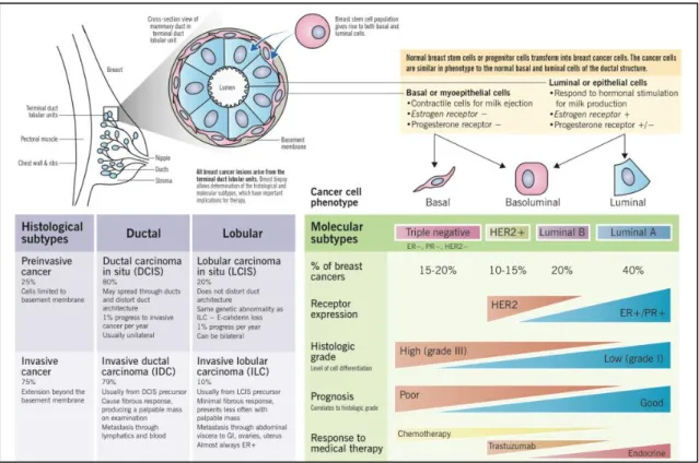
Figure 4 - BrCa pathogenesis and histologic versus molecular subtypes, by Eric Wong
and Joana Rebelo. ... 11
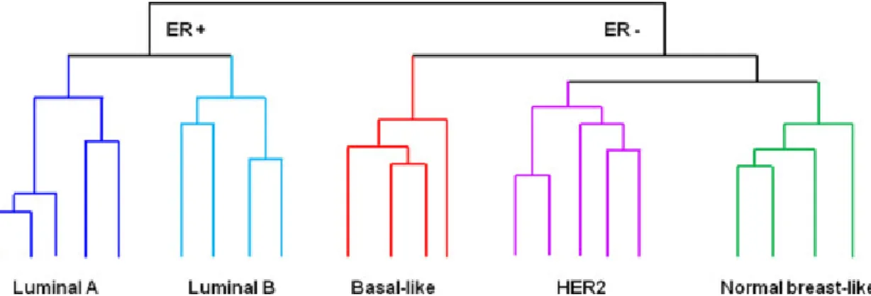
Figure 5 - Schematic illustrations of the five major clusters that represent the molecular
subtypes of BrCa based on Perou and colleagues work carried out a cDNA microarray analysis of 38 invasive BrCa, 1 ductal carcinoma in situ, 1 fibroadenoma and 3 normal breast samples, and a number of biological replicates of tumours from the same patients [52]. Hierarchical clustering led to identify four subtypes: luminal, normal breast-like, human epidermal growth factor receptor 2 (HER2), and basal like. Later, it was shown that similar molecular subtypes can be identified in multiple cohorts and that luminal cancers can be subclassified into luminal A and B [53]. ... 12
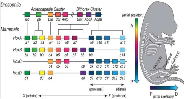
Figure 6 - Localization of the Hox gene within the clusters and their expression in mouse
embryos. In the left, a coloured box represents each Hox gene and the line represents the non-coding regions linking them [70]. Color‐coding highlights conserved relationships between Drosophila and mammalian Hox genes, and the paralogous relationships within the mammalian cluster. In the right, Hox expression along the antero-posterior and proximo-distal axes reflects the formation of distinct skeleton elements along these axes. ... 16
Figure 7 - Hox gene expression and function during mammary gland (MG) development
in mice (in article submitted [86]). A. MG embryonic development. Gene expression is colour-coded in the image. ep: epithelium; me: mammary ectoderm; mm: mammary mesenchyme. Note ventrolateral surface ectoderm (milk line) marked by the expression of HoxC8 at E.10.5 and then restriction of several Hox genes to mammary placodes or surrounding mesoderm in subsequent stages. B. MG development after birth and detected Hox gene expression. TEB: Terminal embryonic buds: AB: Alveolar buds; MA: mature milk alveoli. ... 18
Figure 8 - HOXB7 (A) and HOXB9 (B) basal mRNA expression levels, analyzed by
qPCR [108]. Expression levels of BrCa cells (MCF-7, BT-474, SKBR3, MDA-MB-231, MDA-MB-468) compared to normal breast cells (MCF10A). The * denotes p-value < 0,05, the ** denotes p-value < 0,01 by unpaired T test with Welch’s correction. ... 24
Figure 9 - Box-plots of mRNA expression levels of HOXB9 in normal (NBr=11) and
breast cancer tissues (BrCan=117) analyzed by qPCR. Y-axis as the 2-∆CT values of
HOXB9 expression. Outliers were identified by the SPSS software and indicated with ˚ when considered as “Mild Values” and with * when considered as “Extreme Values”. A. Comparison between NBr and BrCa tissues reveals no significant differences. B. Comparison between distinct molecular subtypes reveals that BrCa Triple negative and HER2+ seem to have lower expression than NBr tissue. Luminal A (n=33), Luminal B (n=43), Triple negative (n=32) and HER2+ (n=9). C. A group of 28 patients shown higher HOXB9 expression in comparison with NBr (n=11). D. That group was analysed considering their molecular subtype. Luminal A (n=9), Luminal B (n=13), Triple negative (n=6) and HER2+ (n=1). ... 34
Figure 10 - Box-plots of mRNA expression levels of HOXB9 in human NBr and BrCa
tissues in comparison with normal breast cells (PCS-600-010, MCF10A). HOXB9 is significant upregulated in NBr, when compared to the cell lines. Outliers were identified by the SPSS software and indicated with ˚ when considered as “Mild Values” and with *
when considered as “Extreme Values”. Y-axis as the 2-∆CT values of HOXB9 expression.
... 35
Figure 11 - Box-plots of mRNA expression levels of HOXB9 in the differents tumour
grades. Outliers were identified by the SPSS software and indicated with ˚ when considered as “Mild Values” and with * when considered as “Extreme Values”. Y-axis as
the 2-∆CT values of HOXB9 expression. ... 35
Figure 12 - HOXB9 basal protein expression level in MDA-MB-231 cell line by western bloting. ... 36
Figure 13 - HOXB9 expression in different tissues by immunohistochemistry. (A) HOXB9
expression in colon cancer (positive control), magnification of 10x; (B) HOXB9 stained positive in NBr, magnification of 10x; (C) HOXB9 stained negative in BrCa tissue, magnification of 10x; (D) HOXB9 stained positive in BrCa tissue, magnification of 10x; (E) Closer magnification (40x) of (D), HOXB9 stains positive in the nucleus of breast cancer cells. ... 37
Figure 14 - HOXB9 protein expression in BrCa tissue. (A) Box-plots of HOXB9 H-score
in human NBr and BrCa tissues analyzed through immunohistochemistry. (B) ROC curve for HOXB9 and differences between NBr and BrCa. ... 38
Figure 15 - Kaplan-Meier plots of overall survival in positive and
HOXB9-negative patients with Breast Cancer. ... 38
Figure 16 - Box-plots of hoxb9 H-score in different molecular subtypes of BrCa. Through
Kruskal-Wallis analysis, we can observe that there are not any significant differences (p value ˃ 0,05) between BrCa molecular subtypes. ... 39
Figure 17 - Box-plots of HOXB9 H-score in different tumour grades. Through
Kruskal-Wallis analysis, we can observe that there are not any significant differences (p value ˃ 0,05) between different tumour grades. ... 39
Figure 18 - HOXB7 expression in different tissues by immunohistochemistry. (A) HOXB7
expression in bladder cancer (positive control), magnification of 10x; (B) HOXB7 stained positive in NBr, magnification of 10x; (C) HOXB7 stained negative in BrCa, magnification of 10x; (D) HOXB7 stained positive in BrCa, magnification of 10x; (E) Closer magnification (40x) of (D), HOXB7 shows positive nuclear staining in breast cancer cells. ... 40
Figure 19 - HOXB7 protein expression in BrCa tissue. (A) Box-plots of HOXB9 H-score
in human NBr and BrCa tissues analyzed through immunohistochemistry. (B) ROC curve for HOXB7 and differences between NBr and BrCa. ... 41
Figure 20 - Kaplan-Meier plots of overall survival in positive and
HOXB7-negative patients with Breast Cancer. ... 41
Figure 21 - Box-plots of HOXB9 protein expression in different molecular subtypes of
BrCa. Through Kurskal-Wallis analysis, we can observe that there are not any significant differences (p value ˃ 0,05) between BrCa molecular subtypes Outliers were identified by the SPSS software and indicated with ˚ when considered as “Mild Values” and with * when considered as “Extreme Values”. ... 42
Tables Index
Table 1 - Top 25 countries with highest rates of breast cancer worldwide. Three first
columns show data from Continuous Update Project for 2018 [5] and last column indicates the economic status of each country/region, according to the report on World
Economic Situation and prospect in 2014 (United Nations) [10]. ... 3
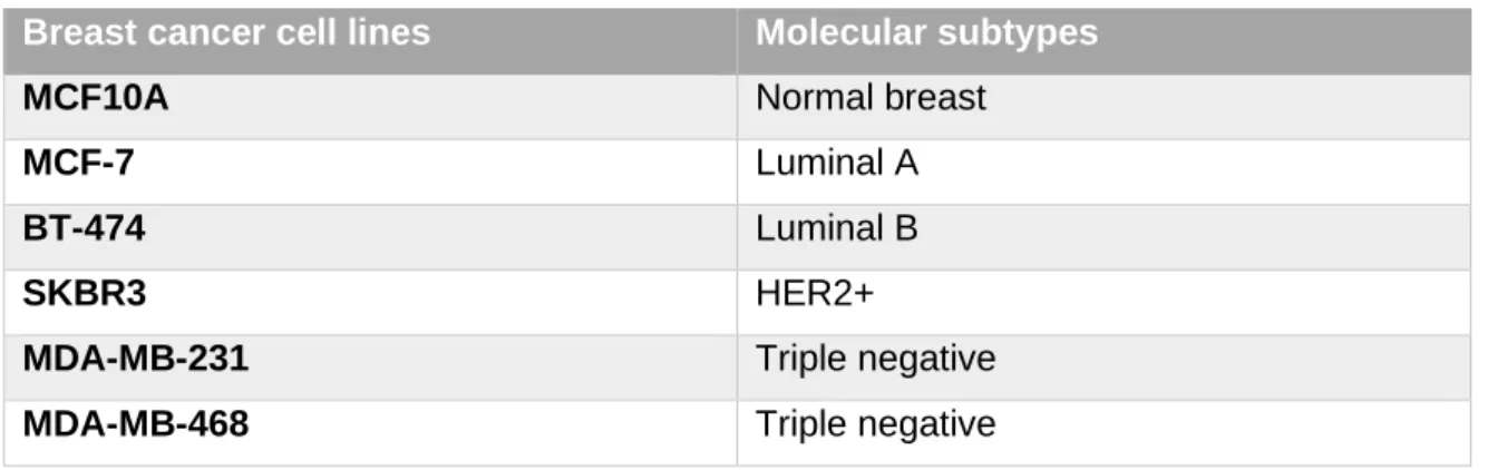
Table 2 - BrCa cell lines that were used, represents different BrCa molecular subtypes. ... 24
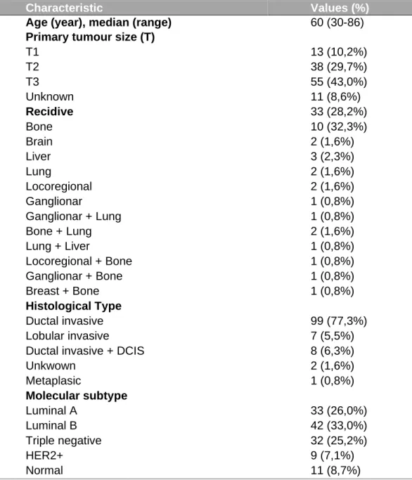
Table 3 - Clinicopathological characteristics of the cohort group from IPO-Porto. ... 28
Table 4 - cDNA synthesis. ... 29
Table 5 - Components of the PCR reaction to validate cDNA synthesis. ... 29
Table 6 - PCR amplification protocol for GAPDH. Primer Forward: ACTGGCGTCTTCACCACCAT; Primer Reverse: TCTTGAGGCTGTTGTCATACTTC; Product size: 142 bp. ... 30
Table 7 - HOXB9 and GAPDH primers sequence, product length and annealing temperature. ... 30
Table 8 - HOXB7 and HOXB9 Antibodies characteristics for immunohistochemistry assays. ... 31
1.1 Breast Cancer
1.1.1 Epidemiology
Oncological diseases are extremely common in Europe, affecting 1 in 4 people throughout their lifetime. In addition, these diseases figure among the most common causes of death in Europe and, as for other main causes of death, affect most significantly the men population (Fig.1) [1]. The only exception, in which a main cause of death affects more women than men, is breast cancer (BrCa). After lung cancer, which has the highest number of new cases and deaths per year, BrCa is the second most common cancer type, affecting women in the world [2, 3]. In 2018, 2 million new cases of this oncological disease have been identified worldwide and it was also the most incident type of cancer among women, and also the deadliest [4, 5]. However, men also develop BrCa but in a much lower percentage, which represents about 1% of the diagnosed cases [6].
Figure 1 - Main causes of death in Europe (Last Eurostat data from 2015, EU), standardized rates per 100 000 inhabitants. Note higher mortality for the women population, in comparison with the men population, detected only for malignant neoplasm of breast (arrow).
Within the first 25 countries with the highest rate of BrCa, 19 are from Europe and 24 are from countries classified as having “upper middle” to “high” per capita gross national income
(Tab.1). This has been related with risk factors more prevalent in these countries such as aging of the population, low parity, increasing age of the first pregnancy, use of birth control and post-menopausal medication, obesity, physical inactivity, among others [7]. In countries with high income, BrCa incidence and mortality are expected to decrease, due to advances in early diagnosis and therapy, while in underdeveloped countries the opposite tendency is expected [8]. Recent studies suggest that the increasing incidence of BrCa in Northern and Eastern Europe is associated to mammography screening and reduction of post-menopausal hormonal replacement therapy [2]. However, these countries have available the best early detection programs, neo-adjuvant therapies and health care in general, which is contributing to decrease BrCa mortality. In contrast, in underdeveloped countries the number of BrCa cases is expected to increase, associated to risk factors such as obesity, physical inactivity, late first-pregnancy or smoking but also due to lack of routine screenings and poor health care [2]. In these countries, the number of new BrCa cases is expected to ascends to 2.4 million per year worldwide in 2030 [9].
Table 1 - Top 25 countries with highest rates of breast cancer worldwide. Three first columns show data from Continuous Update Project for 2018 [5] and last column indicates the economic status of each country/region, according to the report on World Economic Situation and prospect in 2014 (United Nations) [10].
In Portugal, BrCa incidence rates have been rising for years (Fig.2) [11, 12]. Also in Portugal, this oncological disease is the most common cancer type and the principal cause of cancer mortality in women, accounting for 30% of all cancer cases and 16% of all cancer
deaths in 2012 [7]. Two recent studies addressed the incidence in distinct country regions and found a higher number of cases for most age groups in Lisbon area [11, 12]. In general, the South presents the highest age-standardized rate (155.8/100 000 inhabitants), while the North presents the quickest rate of increase (3.6%/year). Therefore, these studies suggest that the Northern region could be number one among all regions in a few years. For now, younger women from the North have lower risk to develop BrCa in comparison to women with the same age gap from South and Centre. However, this risk is reversed in older women. The authors of these studies suggest that differences found in distinct regions of the country might be due to a combination of different diagnostic practices and/or exposure to different risk factors. The heterogeneity of the disease in younger and older women may also explain partially the differences in age-specific rates [12]. The number of diagnosed cases in Azores and Madeira was much lower than in the continental region, representing only 4,2% of the cases occurring in the time period from 1998 through 2011 [11].
Figure 2 -BrCa incidence in Portugal 1998–2011 (overall and by region) [11]. Age-standardized rates from women aged 30-84 years and in specific age-groups (30–44, 45–69, 70–84 years).
1.1.2 Risk Factors
The identification of risk factors associated to the development of BrCa is a critical step for the prevention of this disease. There are 5 major categories of risk factors that have been associated to BrCa 1) aging, 2) family history, 3) genetics, 4) estrogen levels and 5) life habits.
1) Aging: It is the major risk factor for BrCa, which has an increasing incidence throughout a woman´s lifetime [13]. For example, 2 in 3 women with an Invasive BrCa are over 55 years old and post-menopausal. This is explained by accumulation of genetic damage (mutations) throughout life that eventually promotes oncological processes. However, it is also true that BrCa that occurs in younger ages, prior to 40 years old, are more difficult to treat, have higher risk of relapse and cause more death [14]. The first screening age, ideal according to several studies, is between 40-45 years, in order to detect BrCa with worse prognosis [14]. However, when women have family history of BrCa, they are advised to start the screening much earlier, at the age of 25. Thus, the genetic background alters the age of BrCa risk. An intriguing example was found in African-American women, which seems more prone to develop aggressive BrCa types before the age of 40, contrarily to Caucasian-American women [13].
2) Family history: It is strongly relevant for the risk to develop BrCa. As above mentioned, certain types of BrCa (5% to 10%) are related with hereditary genetic background and those are the ones affecting younger women and responsible for higher mortality rate. Thus, the risk for BrCa directly relates with the number of first-degree relatives with this disease. Moreover, even in older women the genetic background is relevant for the outcome of the disease. Thus, post-menopausal women with familiar history of BrCa have much higher risk to develop this disease. In certain cases, a strong family history of BrCa is linked to mutated genes, such as BRCA1, BRCA2 gene or CHEK2 gene, which play a central role in the development of different types of BrCa [15].
3) Genetics: DNA damage causes abnormal cell growth and function and is strongly associated to oncogenesis. It can be inherited or appear de novo over the course of life, resulting from natural aging or exposure to chemicals in the environment. Most inherited cases of BrCa are associated to mutations in BRCA1 and BRCA2 genes, involved in cell damage repair [16]. Women with BRCA1 or BRCA2 mutations (or both) have up to a 72% risk of being diagnosed with BrCa, which occurs particularly in younger women. They are tumour suppressors crucial for the repair of double-strand DNA breaks. BRCA1 is also necessary for cell replication and DNA synthesis, but due to epigenetic inactivation or mutations, increases BrCa risk [17-19]. Single nucleotide polymorphisms (SNPs) seem to increase BrCa risk in women with or without BRCA1 mutation. Several other gene mutations have been associated to high risk of BrCa: ATM (DNA repair); BARD1
(DNA repair); BRIP1 (DNA repair); CDH1 (cell adhesion); CHEK2 (cell growth regulation); MRE11A (DNA repair); MSH6 (DNA repair); NBN (DNA repair); PALB2 (DNA repair); PMS2 (DNA repair); PTEN (cell growth regulation); RAD50 (DNA repair); RAD51C (DNA repair); STK11 (cell growth regulation); TP53 (cell growth regulation); HER2 (epidermal growth factor receptor, which activates cell proliferation, survival and adhesion) [20-24].
4) Estrogen levels: Early menarche and late menopause increase BrCa risk, given the increased exposition to estrogens [25]. The breast cells from women that did not have a full-term pregnancy remain very active and responsive to estrogen. Thus, these women have a higher BrCa risk [25, 26]. Due to an increased exposure to estrogen, women who have their first child after age of 30 also have higher BrCa risk compared to women who gave birth before that. Pregnancy also reduces the total number of lifetime menstrual cycles, which may explain its protective effect against BrCa. Lactation also decreases BrCa risk, especially if it lasts longer than 1 year. Women who started menstruating younger than age 12 have higher BrCa risk later in life, as well as women who go through menopause older than 55. The earlier breasts form, the sooner they interact with internal or external hormones and with chemicals in hormone disruptor products, which increases BrCa risk. In addition, the hormonal replacement therapy (HRT) used by post-menopausal women have been strongly associated to BrCa diagnosis, increasing the risk up to 75% [27].
5) Life habits: Diets rich in carbohydrates and fat, combined with lack of exercise, frequently conduct to overweight and obesity, which increases the risk of BrCa, especially after menopause. In addition, overweight and obesity also increase the risk of recurrence after treatment. This relation between body weight and BrCa risk is due to the capacity of the adipocytes to produce estrogen, which may trigger BrCa develop and grow. The higher is the exposition to extra estrogen over time, the higher is the risk to develop BrCa. Several studies also show an association between exercising regularly (4 to 7 hours/week) to a lower BrCa risk. Regular exercise seems to lower the levels of insulin growth factor in the blood, hormone that can affect BrCa development. Moreover, people that exercise regularly tends to maintain a healthy weight, therefore, having less body fat than inactive people. Regarding diet, it is also known that women with an abundant processed meat or fast food intake have higher BrCa risk than women with a diet rich in fruits and vegetables [28]. Alcohol consumption is associated with BrCa risk in women, by increasing the levels of estrogen and other hormones associated with
hormone-receptor-positive BrCa or by damaging the DNA. The consumption of three alcoholic drinks per week seems to be enough to increase the risk of BrCa 15% and the risk goes up another 10% for each additional drink women regularly have each day. There are also some evidences that alcohol consumption, even a few beverages per week, increase the risk of relapse after BrCa treatment. Importantly, girls from 9 to 15 years old that consume 3 to 5 drinks per week have 3 times more probability to develop benign breast lumps, which later in life can become carcinogenic. In addition, smoking is linked to a higher risk of BrCa in younger, premenopausal women and very heavy second-hand smoke exposure increases BrCa risk in postmenopausal women. Moreover, Catsburg and colleagues, found a positive association between duration and intensity of cigarette smoking, as well as being a smoker prior to first pregnancy, with the risk to develop BrCa [29].
1.1.3 Man Breast Cancer
Male BrCa is rare and it can affect 1 in 100,000 men, accounting for 1% of all BrCa cases diagnosed [30]. It has some similarities with females BrCa, however, upon its discovery, it tends to present a more advanced stage, and consequently a worse prognosis [31]. In all male BrCa cases, the most common type of invasive carcinoma is the ductal, being the in situ and invasive lobular carcinomas the most rare cases in males [32]. Incidence of male BrCa cancers increases with age (>60) and is influenced by the genetic background, particularly associated to the BRCA1 and BRCA2 mutations [6]. About of 20% male BrCa cases have a first-degree family member with BrCa [32, 33]. The Klinefelter syndrome, gynecomastia, prostate, testicular or liver diseases, obesity, or alcohol consume, can have an impact on the incidence of man BrCa [34]. In addition, occupational exposures to radiation or polycyclic aromatic hydrocarbons could have an implication on the appearance of these cancers in males [35]. Decrease in mortality in male breast cancer is particularly associated to adjuvant systemic treatments, since there is no screening implemented for men [36]. Due to the lack of mammary tissue in males, skin and chest wall involvement is not rare and mastectomy is the standard surgical treatment [32].
1.1.4 Screening
BrCa screening is done by evaluating the breast before any signs or symptoms of the disease and is a practice implemented in several countries [37]. These trials have proven that screen-detected, non-palpable, preclinical tumours of ≤ 15 mm have a better prognosis.
Therefore, BrCa screening is helping to diminish the death associated to this disease. However, incidence of breast cancer is still increasing, in part due to a more efficient detection using periodical screening, along with an increase of the risk factors [38].
Currently, there are four reliable methods for BrCa detection: mammography, breast magnetic resonance (MRI), clinical breast exam and breast self-awareness [9]. The most valid and reliable method is mammography, since it can detect BrCa at an early stage, when treatment is more likely to have a positive outcome [39]. This is a multidisciplinary method and accuracy rate can be from 85% to 90%, which will lead to a reduction in mortality rate from 30% to 50% [9, 40]. However, risk of over-diagnosing or false-positive screening has to be taken into account of this method [37]. In families with BRCA mutation, screening is performed annually with magnetic resonance imaging and mammography combined, to detect the cancer at less invasive stage [37]. Clinical exam is also important for early stage detection and surveillance, spatially in women that have a greater risk for BrCa. In this group are included women with radiographically dense breasts, breast implants, women who have been submitted to radiotherapy or patients without access to an optimal health care [9]. This examination consists in a manual palpation of the breasts and local regional lymph nodes [41]. In countries with low income and underdeveloped health services, breast self-exam is an alternative approach that should not be, however, neglected even when there are other resources [9].
In order to positively confirm BrCa, a sample of the primary tumour and cytology/histology of axillary nodes, if involvement is suspected, needs to be analyzed by a pathologist [37]. The sample should be taken with a core needle biopsy, and must be retracted before any treatment for pathological diagnosis, who should be done according to the World Health Organization classification and the tumour-node-metastasis staging system [37]. The exam should analyze histologic type and grade, and determination of estrogen receptor (ER), progesterone receptor (PgR) and HER2 receptor status by IHC or FISH/CISH assay [41].
1.1.5 Breast anatomy
The breast, or mammary gland, is a structure able to produce milk in females. It is composed mainly by adipose tissue and extends from the collarbone down to the underarm and across
to the middle of the ribcage (Fig.3). It can present different anatomies and physiologies,
depending on age, hormonal status, genetic background and daily life habits [42] and, as glandular structures, they are very perceptive to hormonal changes in the body [43]. They have lobes, which are divided into smaller lobules connected via milk ducts [43]. The adipose tissue of the breast is supplied by a network of nerves, blood vessels, lymph
vessels, lymph nodes, and is also composed by fibrous connective tissue and ligaments [43].
Figure 3 - Breast anatomy. All mammals have mammary glands that produce breast milk to feed their offspring [44].
1.1.6 Histopathological types
BrCa is a highly heterogeneous pathology that has different clinical outcomes and
responsiveness to treatments [45, 46]. In fact, several lines of evidence suggest that BrCa
is, in fact, a collection of different diseases with distinct risk factors, clinical presentation, pathological characteristics, response to therapy and outcomes, which originate in the same anatomical structure (the terminal duct-lobular unit) [47]. Most BrCa are adenocarcinomas (95%) and their phenotypic range can be correlated with diversity in gene expression patterns [46]. Bearing in mind the cell morphology, growth and architecture patterns, there are more than 21 histological subtypes described [48]. However, BrCa can be divided into in situ and invasive carcinomas, which are classified as ductal or lobular according to the origin site [49, 50].
1) Ductal carcinoma in situ (DCIS) is non-invasive potentially malignant intraductal
proliferation of epithelial cells, characterized by cellular or nuclear atypia, that can evolve to invasive carcinoma [49]. Classification of DCIS depends on the degree of nuclear atypia, intraluminal necrosis, miotic activity and calcification, then it can be divided into three types, Low-Grade, Intermediate-Grade and High-grade, which are correlated to the risk of becoming invasive [49]. Historically, DCIS has been classified into five architectural subtypes: comedo, solid, cibriform, papillary and micropapillary. DCIS is consistently positive for E-cadherin and β-catenin.
2) Lobular carcinoma in situ (LCIS) is characterized by intra-lobular proliferation of small, and loosely cohesive cells, originated in the terminal duct lobular unit (TDLU). It constitutes a risk factor but a nonobligatory precursor for subsequent development of invasive carcinoma [49]. There are two immunohistochemical markers to distinguish LCIS, the lack of E-cadherin and β-catenin expressions and the positivity for high molecular weight (HMW) keratin [51].
3) Invasive ductal carcinoma (IDC) is a malignant ductal proliferation along with stromal invasion in the presence or absence of DCIS and can be classified according to cell type, number, type and location of secretion, architectural features and immunohistochemical profile [49]. It is the most frequent invasive breast cancer and accounts for 55% of breast cancer incidence upon diagnosis.
4) Invasive lobular carcinoma (ILC) is a malignant abnormal proliferation of neoplastic cells in the lobules, which invaded the stroma. ILC represents 5-15% of all breast cancer cases, and its incidence appears to be increasing, affecting postmenopausal women, and it could be related to hormone replacement treatment. ILC has 5 distinct histological groups, classic type, pleomorphic, histiocytoid, signet ring and tubolobular [49].
Figure 4 - BrCa pathogenesis and histologic versus molecular subtypes, by Eric Wong and Joana Rebelo.
1.1.7 Molecular subtypes
Human BrCa heterogeneity correlates with different gene expression patterns [52], which can be used as a prognostic marker [53]. Using hierarchical clustering, Perou and colleagues realized that part of the BrCa produce estrogen receptor (ER+) and the remaining one do not produce this hormone receptor (Fig.4,5) [52]. The ER+ tumours fall into luminal subtypes (A and B), given that the luminal cells have the most enriched levels of ER. The ER- tumours (low to absent) fall into Basal, HER2-enriched, Normal Breast-Like subtypes and Triple Negative [53, 54].
Figure 5 - Schematic illustrations of the five major clusters that represent the molecular subtypes of BrCa based on Perou and colleagues work carried out a cDNA microarray analysis of 38 invasive BrCa, 1 ductal carcinoma in situ, 1 fibroadenoma and 3 normal breast samples, and a number of biological replicates of tumours from the same patients [52]. Hierarchical clustering led to identify four subtypes: luminal, normal breast-like, human epidermal growth factor receptor 2 (HER2), and basal like. Later, it was shown that similar molecular subtypes can be identified in multiple cohorts and that luminal cancers can be subclassified into luminal A and B [53].
Thus, there are five molecular subtypes of BrCa according to their gene expression profiles belonging to five distinct groups:
1) Luminal tumours are the most common subtype of BrCa cases, and they can be divided into two subtypes, Luminal A and B, being the luminal A the most common. These subtypes express hormone receptors for estrogen (ER) and progesterone
(PR). However, while Luminal A does not express the human epidermal growth
factor receptor 2 (HER2) and have low levels of Ki-67 protein, which is involved in
tumour growth regulation, the luminal B subtype may express or not HER2, always have high levels of the protein Ki-67 [50, 54]. Luminal B tumours have a higher expression of genes involved in cell proliferation when compared with luminal A subtypes [55]. In general, luminal tumours have a good prognosis, but luminal A subtype have a significant better prognosis than luminal B subtype, being a low-grade subtype, while luminal B usually is a higher-low-grade, having a lower survival rate [50, 54].
2) The HER2+ enriched subtype, are not only overexpressing this gene but are also amplifying other genes in the HER2 amplicon, 40 to 80% of tumours from this molecular subtype have a mutation related to p53 gene, subsequently being associated with a poor prognosis [50, 54, 56]. This group also has a low expression of basal-related genes such as keratin 5 [55].
3) Basal tumours have an increased expression of basal markers such as keratins 5, 6, 14 and 17, and EGFR. This molecular subtype is also known to have increased expression of genes related with proliferation and to higher probability to present
low BRCA1 expression, and higher frequency of p53 mutation, which controls proliferation and maintains the integrity and stability of the genome, this gene is actually mutated in approximately in 80% of the cases [54, 57-59]. Most cases of basal tumours are triple negative breast cancer cases (TNBC), which account for 60-90%, of these tumours. TNBCs do not express ER and PR, and HER2 gene is not amplified or overexpressed [54, 60]. Overall, TNBC presents an aggressive phenotype, being histologically classified as high-grade, invasive, ductal carcinomas of no special type with basal-like features, thus it would not express biomarkers such as ER, PR and HER2, but would express cytokeratins, EGFR, vimentin and p-cadherin, which are characteristic of high-grade invasive breast carcinomas [60, 61]. 4) Recent studies have defined a new molecular subtype of BrCa, claudin-low, which is typically triple negative. This subtype is characterized by downregulation of tight junction proteins, such as E-cadherin, and by overexpression of genes associated with epithelial-mesenchymal transition and BrCa cell phenotype [50, 59, 62]. 5) Another subtype of BrCa is the normal-like subtype, which is a controversial group
since some authors think that this subtype represents normal cell contamination on samples rather than a real subtype, also this group expresses genes associated with adipose tissue and other stromal cell types [50]. This subtype accounts for 7,8% of all breast cancer cases, and its gene expression is characterized by a high expression of genes of basal epithelial cells and adipose cells [52, 54].
1.1.8 Biological Markers
After BrCa diagnosis, there is the need to determine patient prognosis and identification of the most appropriate adjuvant systemic treatment. For a more accurate prognosis, some characteristics can be reviewed such as clinicopathological prognostic factors and predictive biological markers. Clinicopathological characteristics include lymph nodes with metastasis, tumour size and tumour grade. Although there are several new molecular tests, these factors are still very important for aiding therapy in newly diagnosed cases [63]. However, these markers are insufficient for proper patient treatment, especially in a time where personalized care is becoming a fundamental aim in biomedical research. Thus, with that in mind there was a need to discover additional molecular biomarkers that could correlate with clinical outcomes and treatment response. ER, PR biomarkers are currently used to predict response to endocrine therapy and HER2 to predict the benefit of trastuzumab treatment [64].
ER stimulates BrCa cell growth by binding to regulatory elements in the genome. Thus, when ER is activated in BrCa patients they can benefit from anti-estrogenic therapy [64] and currently, ER detection is used to identify patients for adjuvant treatments targeting ER and its downstream targets, preventing BrCa proliferation [64]. PR is used as a biomarker alongside with ER. Indeed, it was shown that ER could stimulate PR expression and that PR in the presence of progesterone could modify the location at which ER binds to chromatin. Thus, this alteration could lead to a switch in the regulation of genes that take part in cell proliferation, apoptosis and differentiation and so, when PR is present in ER+ BrCa patients the prognostic is better [64].
The third most used predictive biological marker is HER2. Its measurement is required on cases of invasive BrCa. HER2, when overexpressed, can activate MAPK and PI3K/AKT that leads to tumour growth, enhancing cell proliferation, invasion and metastization [20, 64]. HER2 is overexpressed in 15-20% of BrCa cases and, when ER is overexpressed, in nearly 80% of BrCa cases [64, 65]. This biomarker is used for predicting response to anti-HER2 therapy [64].
1.2 Hox genes and cancer
1.2.1 Homeobox Genes and homeodomain
HOX genes were primarily discovered in Drosophila and found to have a common sequence of 183bp, the homeobox, which encodes a 61-amino acid DNA-binding homeodomain (Fig.6) [66, 67]. This sequence was found to be expressed in all three kingdoms of multicellular organisms [66]. The conservation that is observed between all organisms, extends past the gene sequences into their relative positioning along the chromosome [68]. During vertebrate evolution, HOX genes underwent gene duplications followed by specific gene loss events, which resulted in 39 HOX genes divided in four clusters in mammals [69, 70]. Holland and colleagues divided the HOX genes into several groups: group 1 and 2: (HOX1 and HOX2, set to be the “anterior” genes), group 3 (HOX3), group 4 (HOX4-8 genes, set to be the “central” genes), group 5 (HOX9-13 genes, set to be the “posterior genes” [71]. In humans, HOX clusters are located in four distinct chromosomal loci: HOXA Chr 7p15.3; HOXB Chr 17q21.3; HOXC Chr 12q13.3 and HOXD Chr 2q31, and each can contain between 9 to 11 genes [72, 73].
These genes encode a family of transcription factors that regulate early morphogenesis, for example controlling the anterior-posterior and proximo-distal differentiation during development by interfering with the cell fate within these axes [68, 72, 74, 75]. In a certain way, HOX genes act as a selector gene, since their combinatorial expression can play a major role in identity of an individual body segment [76]. In addition, these genes were found to regulate cell proliferation, specification and cell death and are required for non-transcriptional functions such as DNA replication and repair, and mRNA translation [77]. Interestingly, their position within the cluster reflects the relative positions of the structures that they specify along this axis. Thus, if a gene is at the 3’ end of a cluster it is expressed in anterior regions of the embryo, while if it is located closer to the 5’ it is expressed in more posterior regions of the embryo. Akam and colleagues, suggested that during early Drosophila development HOX expression domains are determined by three regulatory inputs: transcriptional regulation from earlier segmentation genes, cellular memory system based on the action of Polycomb/tithorax group proteins and cross-regulatory interactions among the HOX genes themselves [78].
Figure 6 - Localization of the Hox gene within the clusters and their expression in mouse embryos. In the left, a coloured box represents each Hox gene and the line represents the non-coding regions linking them [70]. Color‐coding highlights conserved relationships between Drosophila and mammalian Hox genes, and the paralogous relationships within the mammalian cluster. In the right, Hox expression along the antero-posterior and proximo-distal axes reflects the formation of distinct skeleton elements along these axes.
1.2.2 Altered HOX gene expression in cancer
As explained previously, these genes have a key role in development. However, they also are implicated in tumour initiation and progression during adulthood. In fact, atypical expression of homeobox genes have been described in tumours, and such can be due to re-expression of HOX genes that were only activated during embryonic development, and then gained expression in neoplastic cells [5, 79]. In some cases, there are homeobox genes that are expressed in neoplastic cells derived from tissues where that particular gene was not expressed in embryonic stages, or in other cases the homeobox genes can be downregulated in malignant cells that are derived from a tissue where a particular gene is expressed in the fully differentiated state [79]. The role of HOX genes in cancer seems to be highly tissue-specific, given that they can be up or downregulated, depending of the cancer tissue. For example, HOXA9 is silenced in lung cancer cases through epigenetic mechanisms but is upregulated in acute lymphocystic leukemia [80]. In contrast, HOXB13 is upregulated in BrCa, but it is silenced in prostate cancer [80]. These genes can also have two distinct roles in tumours, acting as oncogenes by promoting cell growth and invasion, or as tumour suppressors by modifying cell survival and morphogenesis [81, 82].
Actually, HOX genes are often deregulated in both solid tumours and liquid tumours. This family of genes are expressed during hematopoiesis and each cluster have a specific pattern of lineage-restricted expression, where HOXA genes are expressed in myeloid cells, HOXB genes in erythroid cells, HOXC genes in lymphoid cells and HOXD genes are not expressed in this process. HOXA9 is one of the HOX genes that is widely studied, being a key regulator of hematopoiesis and as an oncogenic role in leukemia, enhancing the expression of proliferative genes and inhibiting apoptosis [83].
HOX genes expression can be regulated through epigenetic control, as described for example for HOXA9 in lung cancer [80]. These genes have CpG islands in the promoters, and when silenced these islands can be found methylated. In addition, HOX gene expression can be affected by histone modifications [81]. In consequence of these mechanisms, among others, abnormal activity of HOX genes can affect pathways that promote tumourigenesis, or as a result of their altered expression they can maintain a more embryonic state through the activation of anti-apoptotic pathways or suppression of differentiation [81]. HOX genes can act as transcriptional activators in some types of cancer, but in other cases they have a role as transcriptional repressors, so upregulation and downregulation of these genes is crucial to promote tumourigenesis [84].
1.2.3 Altered HOX genes in breast cancer
The mammary glands start to develop during embryonic development with the formation of the mammary line, or milk line, and differentiation of placodes within this line (Fig.7A). At birth, these placodes already descend into the underlying mesenchyme forming the rudimentary ductal structure of the mammary glands. Then, subsequent stages of development are observed during pubertal, pregnancy, lactation, and involution states. During puberty, branching morphogenesis initiates under the influence of growth hormone (GH), estrogen and insulin-like growth factor 1 (IGF1). Upon pregnancy, progesterone and prolactin combined action generates alveoli able to secrete milk during lactation. The involution state initiates when lactation stops and involves remodelling of the gland back to the pre-pregnancy state. These processes require a variety of signalling pathways that regulate specialized subpopulation of mammary stem cells potentiating drastic changes in the gland occurring with each pregnancy. Particular HOX genes have been implicated in all these development states of the mammary glands, participating in their embryonic development, branching morphogenesis, alveoli formation and involution processes [82, 85] (Fig.7B).
Several studies report abnormal HOX gene expression in BrCa: HOXA6, HOXA13, HOXB2, HOXB4, HOXB5, HOXB6, HOXB7, HOXB8, HOXB9, HOXC5, HOXC13, HOXD1, and HOXD8 [84]. Hur and colleagues quantified the expression levels of the HOX genes in BrCa using reverse transcriptase PCR showing that 14 out of 25 HOX genes, present statistically significant differences between malignant and non-malignant breast cancer tissues. Other studies regarding invasiveness and migration of BrCa cells, have demonstrated that 11 HOX genes were up-regulated in BrCa in comparison with normal breast tissues [67]. However, there are HOX genes that are downregulated in BrCa. HOXA5, for example, presents decreased expression in 60% of BrCa, which has been associated to apoptosis modulation in BrCa [84]. Similarly, HOXD10 appears to have less expression in epithelial cells, as malignancy progresses. Indeed, when HOXD10 expression is restored in cell lines, cell migration seems to diminish [81].
Figure 7 - Hox gene expression and function during mammary gland (MG) development in mice (in article submitted [86]). A. MG embryonic development. Gene expression is colour-coded in the image. ep: epithelium; me: mammary ectoderm; mm: mammary mesenchyme. Note ventrolateral surface ectoderm (milk line) marked by the expression of HoxC8 at E.10.5 and then restriction of several Hox genes to mammary placodes or surrounding mesoderm in subsequent stages. B. MG development after birth and detected Hox gene expression. TEB: Terminal embryonic buds: AB: Alveolar buds; MA: mature milk alveoli.
1.2.4 HOXB7-9 genes in breast cancer
HOXB7 has been associated with tissue development of the mammary gland and with BrCa development [85]. Microarray analyses suggest that HOXB7 is significantly overexpressed in both the primary cancer and distant metastasis [87, 88]. Moreover, in cell lines, overexpression of this gene is directly or indirectly responsible for regulating the expression of vascular endothelial growth factor (VEGF) and basic fibroblast growth factor (bFGF), which promote BrCa progression [89]. Indeed, HOXB7 overexpression in BrCa has been associated to BrCa progression and to the promotion of the epithelial-to-mesenchymal transition (EMT), through up-regulation of bFGF and activation of the MAPK pathway [81, 88, 90, 91]. Importantly, some studies suggest that HOXB7 overexpression increases resistance to tamoxifen through activation of receptor tyrosine kinase pathways.
In other solid tumours, HOXB7 function has been associated to cancer progression and poor clinical outcome and suggested as a potential prognostic factor and therapeutic target [90]. Moreover, there are studies that reveal that this gene is overexpressed in nearly 60% of BrCa cases. This was seen either in cell lines (231, 468, MDA-MB-453, MCF-7, SkBr-3) or BrCa specimens [90, 91]. Interestingly, HOXB7 protein seems to have an important role in DNA repair. Rubin and colleagues have shown that BrCa cell lines expressing HOXB7 present an enhanced survival after irradiation, so this resistance could allow these cells to accumulate mutations that might lead to tumourigenesis [90].
HOXB8, which is localized immediately after HOXB7, within the HOXB cluster, is known to be overexpressed in colorectal cancer (CRC), when compared to normal colon tissue [92]. A study performed by Wang and colleagues, revealed that when HOXB8 is silenced can lead to a decreased proliferation, migration and invasion of CRC cells [93]. In BrCa, HOXB8 does not seem to be deregulated in cell lines or breast cancer tissues, taking in consideration gene expression detection performed using reverse transcriptase PCR [67]. HOXB9, located after HOXB7 and HOXB8 in the cluster, and potentially sharing regulatory mechanisms with them, encode a transcription factor with major roles in mammary gland development [85]. Apparently, the deregulation of this gene is strongly associated to cancer progression, being involved in increment of cell migration, invasion, angiogenesis, and in the formation of lung metastasis derived from primary BrCa [94-96]. The relevance of its deregulation for cancer progression makes it an important prognostic factor in different cancer types [94, 95, 97].
HOXB9 expression and function, has been studied mainly in the gastric cancer (GC) context, where it has been associated with a poor survival rate, when overexpressed,
inducing tumour progression, invasion and occurrence of metastasis. Kato and colleagues revealed that HOXB9 expression increased in 51,7% of GC patients, and its expression was localized in the nucleus of the GC cells. Also, patients with “positive” HOXB9 expression, seemed to exhibit a poorer prognosis when compared to those with a lower expression, being highly associated with lymphatic invasion and lymph node metastasis [98]. However, another study suggests that overexpression of HOXB9 in GC cells leads to an increase in the apoptotic rate, thus, reducing the metastatic ability [99]. In endometrial cancer, HOXB9 overexpression correlates with cell migration and to the upregulation of an oncogenic transcription factor (E2F3), which controls cell cycle, differentiation, apoptosis and response to stress [100]. The overexpression of HOXB9 was also linked to colon cancer, where it seems to contribute for the formation of metastasis and a poor survival rate of patients [101]. This transcription factor was studied in BrCa, being upregulated when compared to normal breast tissues and it was associated with lower overall survival, relapse-free survival, distant metastasis-free survival and post-progression survival for patients diagnosed with BrCa [102].
Seki and colleagues studied HOXB9 expression in BrCa through immunohistochemical staining [103]. Regarding those results, it was notable that 69 (48,9%) of a total of 141 tumour specimens stained positive and were associated with a higher nuclear grade [103]. Hayashida and colleagues, found HOXB9 to be overexpressed in 42% of all BrCa cases analyzed, especially on those with high histological grade. HOXB9 abnormal expression in BrCa was proposed to be a tumour progression factor rather than a tumour-initiating event, since its expression has oncogenic effects, such as inducing growth factors that can alter tumour-specific cell fates, angiogenic factors and also leading to the formation of metastasis [103, 104]. Indeed, HOXB9 levels of expression relate to an increased expression of VEGFs that contributes to tumour progression and influence intratumoural microvessel density (IMD), a measure of neovascularization surrounding the tumour tissue [105]. Thus, higher HOXB9 expression can increase IMD and, therefore, contribute to a worse prognostic in BrCa patients. Shrestha and colleagues also demonstrated that HOXB9 is upregulated in BrCa tissues, being its expression also higher in various tumour cells including ER+ BrCa cells, MCF7 [106]. Thus, HOXB9 overexpression in BrCa can be due to a higher estrogenic activity present in BrCa tissues [106].
Other studies attempted to apply ionizing radiation to HOXB-overexpressing breast cells showing that they become more resistant due to an enhanced DNA damage response, due to an increase of baseline ataxia telangiectasia mutated (ATM) phosphorylation [107]. So, in non-irradiated cells HOXB9 can induce detectable DNA damage, such as double-strand DNA breaks, that will increase ATM phosphorylation, leading to DNA damage response.
Thus upon radiation, BrCa cells that are overexpressing HOXB9, have their ATM hyperactivated in the early stages of the double-stranded DNA break response, leading to a more rapid response. In fact, production of growth factors (GF) and cytokines can alter tumour microenvironment and modulates cell fate, resulting on an increase of tumour aggressiveness and chemotherapy resistance. Thus, HOXB9 overexpression triggers the production of TGF-β, which is GF, and it can alter cell fate by inducing EMT [107].
In summary, HOXB9 overexpression can lead to an increased production of 1) TGF-β controlling cell cycle, inducing cell differentiation and promoting apoptosis; 2) ErbB ligands inducing epithelial to mesenchymal transition and invasion; 3) bFGF supporting differentiation 4) VEGFs, promoting angiogenesis; 5) NRG2 influencing growth, differentiation and survival of different cell types [104, 106]. Also, HOXB9 influences BrCa progression, triggering ErbB ligands and TGF-β production, which causes phosphorylation and posterior activation of HER2/3 and TGF-β pathways, respectively. Although, activation of HER2/3 promotes cell motility, the TGF-β pathway promotes EMT [107].
Our lab has been investigating the role of HOXB genes in BrCa, particularly HOXB7 and HOXB8. Primarily, the HOX expression profile was characterized through qPCR and western blot in MDA-MB-231 and MDA-MB-468 BrCa cell lines, followed by HOX functional analysis through knockdown of HOXB7 using specific siRNAs. These assays made us suggest that HOXB7 Tf may regulate CDH1. However, our lab then performed aggregation assays, after successfully knocking down HOXB7 in MDA-MB-231 cell line, using as control cells that form compact aggregation as spheroids (MCF-7). The results demonstrated that silencing of HOXB7 in MDA-MB-231 cells does not affect the rate of aggregate formation in the experimental condition, in comparison with the control. Thus, the silencing of HOXB7 seems to be insufficient to modify the function of CDH1 proteins.
In order to further explore the role of HOXB7 in BrCa, we went on to characterize its expression in distinct BrCa molecular subtypes, with the aim to update these analyses using the qPCR approach and also to facilitate the selection of the most suitable cellular models for future functional assays in vitro and in vivo. To this end, the lab expanded the analyses of gene/protein expression analyses to cell lines MCF-7, BT-474, SKBR3, MDA-MB-231 and MDA-MB-468 (Fig.8A). Human tissues were also analyzed, belonging to a cohort of BrCa patients from IPO-Porto. The evaluation of the cell lines leads us to conclude that HOXB7 was overexpressed in all of them, in comparison with normal cells (MCF10A). However, the expression was particularly high in MCF-7 and BT-474, in accordance with previous studies by Hur and colleagues using RT-PCR instead of qPCR [67]. Complementary Western blot analyses further confirmed these results. Contradictory results emerged, however, when analyzing the expression levels of HOXB7 in the cohort of BrCa patients. Where, HOXB7 appeared to be more significantly expressed in normal breast (NBr) than in BrCa tissues. In addition, no significant differences were found in the expression levels of distinct BrCa molecular subtypes. Analyzing CDH1 expression in cell lines and tissues, we also noticed lack of differences between tissues belonging to distinct molecular subtypes, which were very clearly identified in the cell lines. Thus, these results suggested the need to increase the cohort of patients analyzed and to test methods of protein detection that might be more efficient to evaluate differential expression levels in NBr and BrCa tissues (immunohistochemistry).
HOXB9 expression was also re-analyzed in our lab using qPCR and aiming to fulfill the same aims above described for HOXB7. The results showed that this gene is overexpressed in cell lines MCF-7, BT-474 and MDA-MB-231, when compared to MCF10A normal breast cells (Fig.8B). However, the expression levels of this gene were still uncharacterized in the cohort of BrCa patients from IPO-Porto previous to the initiation of this master thesis.
Figure 8 - HOXB7 (A) and HOXB9 (B) basal mRNA expression levels, analyzed by qPCR [108]. Expression levels of BrCa cells (MCF-7, BT-474, SKBR3, MDA-MB-231, MDA-MB-468) compared to normal breast cells (MCF10A). The * denotes p-value < 0,05, the ** denotes p-value < 0,01 by unpaired T test with Welch’s correction.
Table 2 - BrCa cell lines that were used, represents different BrCa molecular subtypes.
Breast cancer cell lines Molecular subtypes
MCF10A Normal breast
MCF-7 Luminal A
BT-474 Luminal B
SKBR3 HER2+
MDA-MB-231 Triple negative
Overall, this project aims to overcome limitations from the literature regarding the characterization of HOXB7 and HOXB9 expression, at a transcriptional and protein level and considering BrCa cell lines and patients’ biopsies from distinct molecular subtypes. This work will be important for the planning of future experiments aiming to further comprehend the function of these genes in BrCa.
Our specific aims were:
Aim 1 – Characterize basal expression profiles of HOXB9 in BrCa tissues from a cohort of patients from IPO-Porto (n=128) representatives of the distinct molecular subtypes, using qPCR and characterize protein expression profile of HOXB9 using cell lines representing all recognized molecular subtypes of BrCa by western blot;
Aim 2 – Characterize HOXB7 and HOXB9 protein level in patients’ biopsies, through
4.1 Patients sample
All human breast cancer and normal RNA samples were received from IPO-Porto. The BrCa samples were used to quantify HOXB9 protein and mRNA expression and HOXB7 protein expression. mRNA expression was analyzed through qPCR and protein expression through immunohistochemistry assay. This cohort group consisted of 127 patients, being 11 patient’s cancer free and 117 patients with BrCa (table 8).
Table 3 - Clinicopathological characteristics of the cohort group from IPO-Porto.
Characteristic Values (%)
Age (year), median (range) 60 (30-86)
Primary tumour size (T)
T1 13 (10,2%) T2 38 (29,7%) T3 55 (43,0%) Unknown 11 (8,6%) Recidive 33 (28,2%) Bone 10 (32,3%) Brain 2 (1,6%) Liver 3 (2,3%) Lung 2 (1,6%) Locoregional 2 (1,6%) Ganglionar Menungeal 1 (0,8%) 1 (0,8%) Ganglionar + Lung 1 (0,8%) Bone + Lung 2 (1,6%) Lung + Liver 1 (0,8%) Locoregional + Bone 1 (0,8%) Ganglionar + Bone 1 (0,8%) Breast + Bone Pleural + Bone Bone + Lung + Brain Bone + Lung + Liver
1 (0,8%) 1 (0,8%) 1 (0,8%) 1 (0,8%) Histological Type Ductal invasive 99 (77,3%) Lobular invasive 7 (5,5%)
Ductal invasive + DCIS 8 (6,3%)
Unkwown 2 (1,6%) Metaplasic Normal Breast 1 (0,8%) 11 (8,6%) Molecular subtype Luminal A 33 (26,0%) Luminal B 42 (33,0%) Triple negative 32 (25,2%) HER2+ Normal Breast 9 (7,1%) 11 (8,6%) Normal 11 (8,7%)

![Table 1 - Top 25 countries with highest rates of breast cancer worldwide. Three first columns show data from Continuous Update Project for 2018 [5] and last column indicates the economic status of each country/region, according to the report on Wo](https://thumb-eu.123doks.com/thumbv2/123dok_br/15145955.1012345/16.892.228.640.630.1013/countries-highest-worldwide-continuous-project-indicates-economic-according.webp)
![Figure 2 -BrCa incidence in Portugal 1998–2011 (overall and by region) [11]. Age-standardized rates from women aged 30-84 years and in specific age-groups (30–44, 45–69, 70–84 years)](https://thumb-eu.123doks.com/thumbv2/123dok_br/15145955.1012345/17.892.135.768.472.858/figure-incidence-portugal-overall-region-standardized-specific-groups.webp)
![Figure 3 - Breast anatomy. All mammals have mammary glands that produce breast milk to feed their offspring [44]](https://thumb-eu.123doks.com/thumbv2/123dok_br/15145955.1012345/22.892.182.689.172.468/figure-breast-anatomy-mammals-mammary-glands-produce-offspring.webp)
![Figure 7 - Hox gene expression and function during mammary gland (MG) development in mice (in article submitted [86])](https://thumb-eu.123doks.com/thumbv2/123dok_br/15145955.1012345/31.892.127.727.475.994/figure-expression-function-mammary-gland-development-article-submitted.webp)