Universidade de Trás-os-Montes e Alto Douro
miR-371a-3p as a tool for subtyping Testicular Germ Cell
Tumors
Dissertação de Mestrado em Genética Molecular Comparativa e Tecnológica
Bárbara Denise Vilela Salgueiro
ORIENTADOR: Professora Doutora Carmen de Lurdes Fonseca Jerónimo
CO-ORIENTADOR: Professora Doutora Raquel Maria Chaves
Universidade de Trás-os-Montes e Alto Douro
miR-371a-3p as a tool for subtyping Testicular Germ Cell
Tumors
Dissertação de Mestrado em Genética Molecular Comparativa e Tecnológica
Bárbara Denise Vilela Salgueiro
ORIENTADOR: Professora Doutora Carmen de Lurdes Fonseca Jerónimo
Professora Associada convidada com Agregação
Departamento de Patologia e Imunologia Molecular
Instituto de Ciências Biomédicas Abel Salazar Universidade do Porto
Investigadora Auxiliar e coordenadora do Grupo de Epigenética e Biologia do Cancro
Centro de Investigação
Instituto Português de Oncologia do Porto Francisco Gentil, EPE
CO-ORIENTADOR: Professora Raquel Maria Chaves
Professora do Departamento de Genética e Biotecnologia
Universidade de Trás-os-Montes e Alto Douro
Composição do Júri:
______________________________________________
______________________________________________
“I would never die for my belief, because I might be wrong.”
This original research was developed to achieve the Master Degree in Molecular Comparative and Technological Genetics “DR, 2ª série – Nº 133 – Regulamento nº 658/2016 de 13 de julho de 2016”.
ACKNOWLEDGEMENTS
Chegou ao fim mais uma etapa da minha vida, etapa essa que desde criança sonhei em concretizá-la, mas que não seria possível fazê-lo sem a ajuda e o apoio de diversas pessoas, às quais expresso os meus agradecimentos.
Antes de mais gostaria de agradecer à minha orientadora, Professora Doutora Carmen Jerónimo por me ter aceite no Grupo de Epigenética e Biologia do Cancro e pela confiança que depositou em mim desde o início do projeto. Agradeço acima de tudo pela partilha de conhecimentos, pela disponibilidade em ajudar em qualquer situação, e por ter contribuído para o meu desenvolvimento profissional na área da investigação Oncológica.
À minha co-orientadora, Professora Doutora Raquel Chaves pelo apoio incondicional ao longo destes últimos dois anos e por todos os conselhos construtivos que sempre deu aos seus alunos.
Ao Professor Doutor Rui Henrique, pela ajuda e pelo acompanhamento prestado durante o desenvolvimento deste projeto.
Ao Professor Doutor Manuel Teixeira, Diretor do Departamento de Genética e do Centro de Investigação do Instituto Português de Oncologia (IPO) do Porto, agradeço por me ter permitido realizar este projeto no Centro de Investigação.
À Professora Doutora Raquel Chaves, na qualidade de Diretora do Mestrado em Genética Molecular Comparativa e Tecnológica e à sua antecessora, Professora Doutora Paula Lopes, pela aceitação da minha candidatura no referido ciclo.
Ao Serviço de Anatomia Patológica do IPO do Porto, em particular ao Dr. João Lobo, por todas as horas em que ficou até mais tarde a trabalhar para este projeto, pelos sábios conselhos e acima de tudo pela prontidão que sempre demonstrou na resolução de qualquer eventualidade. Às Técnicas Mariana Cantante e Rita Guimarães pela realização dos cortes histológicos e à Técnica Paula Lopes, encarregue da imunohistoquímica.
À Ana Laura, o meu braço direito do laboratório, que me acolheu de braços abertos desde o primeiro dia, a pessoa que esteve sempre lá nos bons e nos maus momentos e que sem ela eu sei que este trabalho teria sido muito mais difícil de realizar. Obrigada do fundo do coração, por tudo, mas acima de tudo pela amizade que criei contigo.
À Daniela Silva, obrigada por toda a ajuda e por toda a paciência que tiveste comigo, por teres tornado aqueles programas de estatística bem mais fáceis de perceber e pelo bom gosto que sempre tiveste e colocaste em tudo o que me ajudaste a fazer.
À Catarina Barbosa por estar sempre lá cheia de carinho para dar, pela calma e otimismo que sempre me transmitiu.
À Sofia Salta, pela pessoa incrível que revelou ser, pelas horas perdidas a rever o meu manuscrito e pelas horas ganhas em frente ao mar com conversas profundas. O meu muito obrigada por me teres permitido conhecer-te melhor.
Aos restantes membros do Laboratório: Ângela, Lameirinhas, Maria, David, Inês, Sara e Vera por todas as horas de trabalho partilhado e de gargalhadas soltas. Sem vocês, nada teria sido igual com certeza.
À Sara Reis, a minha parceira de licenciatura, mestrado e da vida, pelos desabafos ao telemóvel, por estares sempre lá para mim e por nunca me teres deixado desistir de tudo nos momentos mais complicados. Continuo a dizer, não és de sempre mas és para sempre!
Às minhas “besties” Alexandra Torre e Patrícia Silva por me aturarem ao fim de semana com os assuntos do laboratório que, apesar de ser chinês para os ouvidos delas, mesmo assim me deixavam falar horas e horas. Pelas noites e jantares que me faziam ganhar força para a semana que se avizinhava e por toda a amizade que me deu coragem e força para continuar.
À minha Tia Graça, por todo o carinho e hospitalidade durante o meu ano de estágio. Foste sem dúvida a minha segunda avó e nunca me vou esquecer daquilo que fizeste por mim.
Ao Paulo Gomes, obrigada pela amizade, pelo apoio que sempre me deu durante a vida e no meu percurso académico.
À minha irmã, por nada em particular, simplesmente por ser minha irmã. Agradeço a Deus todos os dias por me ter dado uma melhor amiga de sangue. Já não sei o que seria a minha vida sem ti!
À minha avó materna, a minha segunda mãe, o meu porto de abrigo. Obrigada pelo amor de todos os dias e por me incentivares a dar o melhor de mim em tudo aquilo que faço.
“Last but not the least” agradeço ao meu maior pilar, a minha Mãe, por ser um exemplo quer a nível profissional quer pessoal. Obrigada por acreditares em mim, no meu sucesso e nas minhas capacidades. Por me teres criado sozinha desde que o pai morreu e por NUNCA, lê bem, NUNCA teres falhado como mãe. És o meu maior orgulho, o melhor de mim! Amo-te!
Espero que esta dissertação chegue ao céu porque é aos meus anjos que a dedico, ao meu PAI e ao meu AVÔ materno.
This study was funded by a grant of the Research Center of Portuguese Oncology Institute of Porto (CI-IPOP 27)
x
RESUMO
Os tumores de células germinativas testiculares (TGCTs) constituem um grupo heterogéneo de neoplasmas, afetando na maioria homens em idade jovem. As taxas de cura são elevadas e o tratamento adequado depende de uma avaliação clínica e patológica que seja cuidadosa e precisa. De fato, a subtipagem histopatológica dos TGCTs é um ponto crítico para uma decisão terapêutica adequada. Considerando a limitação dos biomarcadores de soro atualmente disponíveis, foram propostos novos candidatos, principalmente o miR-371a-3p, que superou os marcadores de soro clássicos, contudo não existe informação detalhada relativamente ao perfil de expressão nos vários subtipos de TGCTs. Deste modo, avaliamos os níveis dos níveis de expressão do miR-371a-3p nos vários subtipos de TGCT usando uma coorte consecutiva de amostras de tecido testicular. Após o isolamento do RNA e síntese de cDNA, os níveis de expressão do miR-371a-3p foram avaliados em 154 amostras de tecido de TGCT e em 15 tecidos não-tumorais por PCR quantitativo em tempo real (RT-qPCR). A fim de gerar uma curva padrão para a quantificação relativa e verificar a eficiência do PCR, o cDNA do RNA total de referência humano (Agilent, EUA) foi utilizado e, para a normalização, o RNU48 foi utilizado como um gene de referência. O miR-371a-3p discriminou tecidos tumorais de tecidos controlo com alta sensibilidade e especificidade (AUC = 0,99). Além disso, os seminomas apresentaram níveis mais elevados de expressão do miR-371a-3p em comparação com amostras de TGCT não-seminomatoso, revelando também diferenças significativas entre eles. Contudo, os TGCTs pré-púberes apresentaram níveis mais baixos de expressão do miR-371a-3p do que TGCTs pós-púberes. Globalmente, os níveis de expressão do miR-371a-3p diminuíram em paralelo com a diferenciação celular progressiva. Concluímos que o miR-371a-3p é específico para TGCT e pode ser clinicamente útil para a deteção precoce e monitorização desta doença oncológica.
Palavras-chave: Tumores de células germinativas testiculares, microRNAs, biomarcadores,
xi
ABSTRACT
Testicular germ cell tumors (TGCT) are heterogeneous group of neoplasms, mostly affecting young men. Curability rates are high and adequate treatment relies in careful and accurate pathological and clinical assessment. Indeed, TGCT histopathological subtyping is critical for adequate therapeutic decision. Considering limitation of currently available serum biomarkers, novel candidates have been proposed, most notably miR-371a-3p, which outperformed classical serum markers, but no detailed information concerning TGCT subtype was available. Thus, we carried out evaluation of miR-371a-3p expression levels among TGCT subtypes using a consecutive cohort of tissue samples. After RNA isolation and cDNA synthesis, miR-371a-3p expression levels were measured in 154 tissue samples of TGCT and in 15 non-tumoral tissues by real-time quantitative PCR (RT-qPCR). To allowed generation of a standard curve for relative quantification and ascertain PCR efficiency cDNA from total Human Reference RNA (Agilent, USA) was used and for normalization RNU48 was used as a reference gene. MiR-371a-3p discriminated TGCT from control tissues with high sensitivity and specificity (AUC=0.99). Furthermore, seminomas displayed higher miR-371a-3p expression levels compared to nonseminomatous TGCT, which also disclosed significant differences among them. Nonetheless, prepubertal TGCT depicted lower miR-371a-3p expression levels than postpubertal TGCT. Globally, miR-371a-3p expression levels decreased in parallel with progressive cell differentiation. We concluded that miR-371a-3p is TGCT-specific and it might be clinically useful for early detection and disease monitoring.
Keywords: Testicular germ cell tumor, microRNAs, biomarker, miR-371a-3p, postpubertal;
xiii
TABLE OF CONTENTS
I.
INTRODUCTION ... 1
1
Testicular germ Cell Tumors ... 2
1.1
The neoplasia ... 2
1.2
Epidemiology and Risk Factors ... 3
1.3
TGCT classification ... 5
Germ Cell Neoplasia in situ ... 5
1.3.1
Testicular Germ Cell Tumors related to GCNIS ... 6
1.3.2
Testicular Germ Cell Tumors unrelated to GCNIS ... 8
1.4
Diagnosis, Staging, Prognosis and Treatment ... 9
1.4.1
Diagnosis ... 9
1.4.2
Staging and Prognosis ... 9
1.4.3
Treatment ... 12
2
Epigenetics ... 13
2.1
DNA Methylation ... 14
2.2
Histones: Post-translational modifications and variants ... 15
2.3
Chromatin remodelling ... 15
2.4
Non-coding RNAs ... 16
2.4.1
MicroRNAs: Biogenesis and Function ... 16
xiv
2.4.3
miR-371-373 cluster in TGCT ... 18
II.
AIMS ... 20
III.
MATERIAL AND METHODS ... 22
1.
Patients and samples collection ... 23
2.
RNA Extraction ... 23
3.
MicroRNA cDNA Synthesis ... 24
4.
MicroRNAs Expression: Individual Assays ... 24
5.
Statistical Analysis ... 25
IV.
RESULTS ... 26
Clinical characterization and association between miR-371a-3p expression levels and
standard clinicopathological parameters ... 27
Assessment of miR-371a-3p expression levels in TGCT and controls ... 28
Differential expression of miR-371a-3p among TGCT subtypes and discriminative
power ... 29
V.
DISCUSSION... 34
VI.
CONCLUSIONS AND FUTURE PERSPECTIVES ... 34
xv
FIGURES INDEX
Figure 1 Global incidence of Testicular Germ Cell Tumors. Adapted from [9]. ... 3
Figure 2 A diagrammatic illustration of Testicular germ cell tumors subtypes
pathogenesis. TGCTs- testicular germ cell tumors; GCNIS- germ cell neoplasia in situ;
ESC- embryonic stem cell; PGC- primordial germ cell; EmbrCa- embryonal carcinoma;
TE- postpubertal-type TE; YST- postpubertal-type yolk-sac tumor; CH-
choriocarcinoma; MGCT- mixed germ cell tumor. Adapted from [3]. ... 5
Figure 3 Four distinct mechanisms of epigenetic regulation. M – 5-methylcytosine; the
symbols hexagon and triangle represent different translational modifications in
N-terminal tails of histones proteins. (CBEG, IPO Porto). ... 14
Figure 4
MicroRNAs biogenesis. Kindly provided by [107]. ... 17
Figure 5 Box-plots (left panel) of miR-371a-3p expression levels in Normal Tissue
Testis (Controls) and TGCT (Tumors) samples (A) and respective Receiver Operating
Characteristic Curve (right panel) (B). Abbreviations: AUC – Area Under the Curve. 29
Figure 6 Box-plot of miR-371a-3p expression levels in Normal Testis Tissue (Controls),
Pure Seminomas (Pure SE) and Nonseminomatous tumors (NST) samples. ... 29
Figure 7 Box-plot of miR-371a-3p expression levels between the different subtypes of
TGCT. ... 30
Figure 8 Box-plot (left panel) (A) of miR-371a-3p expression levels in Pure Seminoma
(Pure SE) and SE in mixed germ cell tumors samples and respective Receiver
Operating Characteristic Curve (right panel) (B). Abbreviations: AUC – Area Under the
Curve. ... 30
Figure 9 Box-plot (left panel) (A) of miR-371a-3p expression levels in Controls and
Postpubertal Teratoma samples and respective Receiver Operating Characteristic
Curve (right panel) (B). Abbreviations: AUC – Area Under the Curve. ... 31
xvi
Figure 10 Box-plot of miR-371a-3p expression levels in GCNIS-unrelated TGCTs samples
and GCNIS-related TGCT samples. ... 31
Figure 11 Box-plot of miR-371a-3p expression levels in Prepubertal-type TE and
Postpubertal-type TE as component of mixed TGCT samples (left panel) and box-plot
of miR-371a-3p expression levels in Prepubertal-type YST and Postpubertal-type YST
as component of mixed TGCT samples (right panel). ... 32
Figure 12 Box-plot of miR-371a-3p expression levels in Controls and Prepubertal
TGCTs samples and respective Receiver Operating Characteristic Curve.
Abbreviations: AUC – Area Under the Curve. ... 32
xvii
TABLES INDEX
Table 1 Main risk factors for TGCT... 4
Table 2 Clinical and pathological TNM classification of testicular germ cell tumors
(Adapted from [3, 73]). ... 10
Table 3 Tumor stage information of testicular germ cell tumors (Adapted from [3, 73]).
... 11
Table 4 Prognostic-based staging system for metastatic disease (International Germ
Cell Cancer Collaborative Group) (Adapted from [73, 75]). ... 12
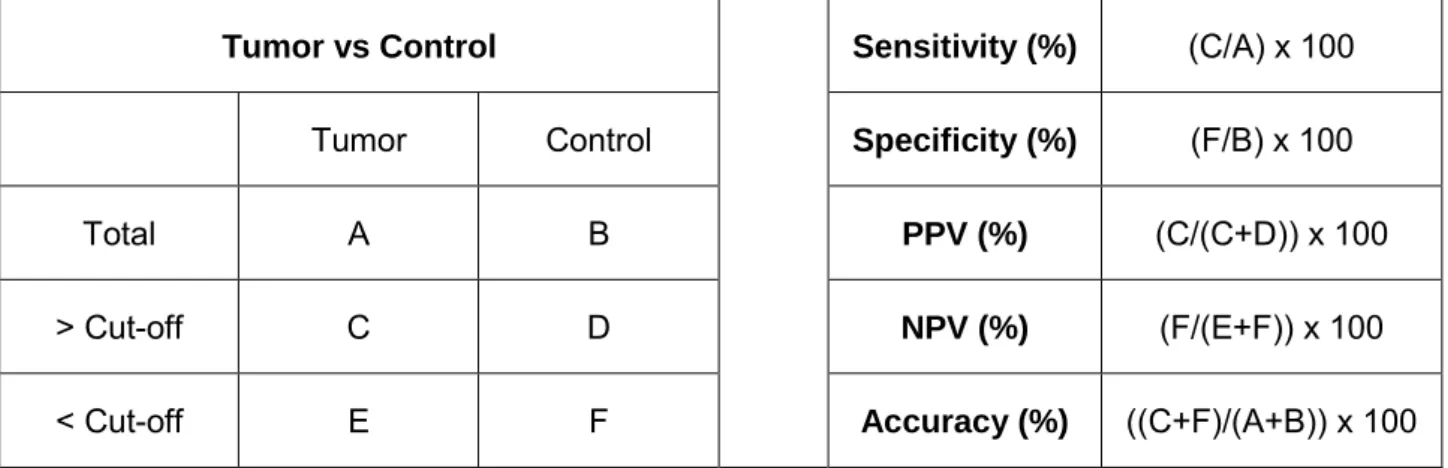
Table 5 Formulas used for biomarker parameters calculation. ... 25
Table 6 Clinicopathological information of TGCT patients and controls. ... 27
Table 7 Serum tumor markers levels in testicular germ cell tumors cases. ... 28
Table 8 miR-371a-3p expression levels in controls and testicular germ cell tumors. 28
Table 9 Performance of miR-371a-3p expression levels as biomarker for different
detections... 33
xviii
LIST OF ABREVIATIONS5mC – 5-methylcytosine
AFP – α-fetoprotein
Ago 2 – Argonaute protein 2
AJCC – American Joint Committee on Cancer
AUC – Area Under the Curve
BEP – Bleomycin, Etoposide and Cisplatin
β-hCG – β-subunit of human chorionic gonadotropin CDK – Cyclin-dependent kinases
CH – Choriocarcinoma CIS – Carcinoma in situ
CpG – Cytosine-phosphate-Guanine
DNA – Deoxyribonucleic Acid
DNMT – DNA methyltransferase EmbrCa – Embryonal Carcinoma
EP – Etoposide and cisplatin
FFPE – Formalin-fixed paraffin-embedded
GCNIS – Germ Cell Neoplasia in situ
HCC – Hepatocellular carcinoma IQR – Interquartile Range
IU/L – International unit per litre
JEB – Carboplatin, etoposide, bleomycin
LATS2 – Large Tumor Suppressor Kinase 2 LDH – Lactate dehydrogenase
miRNA – microRNA mRNA – Messenger RNA
NcRNA - Non-coding RNA ng – Nanograms
NPV – Negative Predictive Value
NST – Nonseminomatous tumors PCR – Polymerase chain reaction
PPV – Positive Predictive Value
Pre-miRNA – Percursor microRNA
Pre-TE – Prepubertal-type Teratoma Pre-YST – Prepubertal-type Yolk sac tumor
xix
Pri-miRNA – Primary microRNApTNM – Pathological TNM
RISC – RNA induced silencing complex
RNA – Ribonucleic Acid
ROC – Receiver Operating Characteristic
RPLND – Retroperitoneal Lymph Node Dissection RT – Reverse Transcription
RT-qPCR – Quantitative real-time Polymerase chain reaction SAM – S-adenosymethionine
SE – Seminoma TE – Teratoma
TGCT – Testicular Germ Cell Tumors TIP – Paclitaxel, Ifosfamide and Cisplatin
TP53 – Tumor protein p53
TSmiR – Target Serum microRNA test
TVB – Testicular Vein Blood
UICC – Union for International Cancer Control
VIP – Etoposide, Ifosfamide and Cisplatin
WHO – World Health Organization YST – Yolk sac tumor
INTRODUCTION
2
1 Testicular germ Cell Tumors
1.1 The neoplasia
Testicular germ cell tumors (TGCTs) are a diversified group of neoplasms, with a wide variety of histological types, derived from a primitive germ cell [1] These tumors are of utmost importance mainly for three major reasons: firstly because they are the most common tumors diagnosed in men aged 20–35 years; secondly because they represent roughly 95% of malignant testicular tumors; and thirdly because incidence of these tumors has increased over the last five decades [2].
According to the 4th edition of the World Health Organization (WHO) Classification of Tumors
of The Urinary System and Male Genital Organs published in 2016, TGCTs are classified into those that are derived from Germ Cell Neoplasia in Situ (GCNIS) and those that are not [3]. The term “GCNIS” is a fusion of the terms “carcinoma in situ” and “intratubular germ cell neoplasia, unclassified” and refers to neoplastic, embryonic-type germ cells confined to the spermatogonial stem cell niche.
TGCT unrelated to GCNIS are very rare and, thus, the overall epidemiological data reported for TGCT in fact is generally concerning to TGCTs related to GCNIS. The GCNIS-derived tumors have several typical characteristics such as postpubertal occurrence and malignant behavior. They can be subdivided into two major groups: seminomas (SE) and nonseminomatous tumors (NST). These two subgroups are clinically distinct regarding therapy, management and prognosis. The NST group includes yolk sac tumor, postpubertal-type (YST), embryonal carcinoma (EC), choriocarcinoma (CH) and teratoma, postpubertal-type (TE). Moreover, in NST group include tumors that present more than one tumor subtype designated as mixed TGCT which constitute the most common NST subtype [4].
Treatment and management protocols of TGCTs may conduct to aggressive long-term side effects such as infertility, vascular and heart damage, respiratory disease and secondary neoplasms. Therefore, an accurate diagnosis is imperative in order to prevent both overtreatment of low risk patients and undertreatment of advanced disease patients and, finally improve the treatment of TGCTs [2].
TGCTs belongs to a group of tumors that present the highest cure rates, displaying 5-year survival rates over 95%, persisting high (over 80%) for patients with metastatic disease [2]. Nevertheless, TGCTs remains the focus of research for several reasons. Despite outstanding cure rates, about 15-20% of patients with disseminated disease frequently relapse. Moreover, many tumors are resistant to cisplatin, and genetic and epigenetic mechanisms underlying this resistance requires more investigation. Moreover, the efforts to decrease morbidity is imperative, since
INTRODUCTION
3
treatment entail important side effects. Furthermore, no validated molecular markers are currently used for screening, diagnostic or prognostic assessment [5-7].
1.2 Epidemiology and Risk Factors
TGCTs comprise 1% of all male tumors worldwide, despite their rareness. These neoplasms are very common in caucasian men, and show a characteristic age distribution with two main peaks, one during childhood and the other after puberty [3]. Furthermore, this malignancy constitute more than 60% of all neoplasias diagnosed in men with 20 to 40 years-old. [8]
As mentioned above, TGCT incidence has increased over the last years. In 2030, 65.827 new cases are predicted worldwide, more 10.5 thousands cases than in 2012 [8].
The biggest discrepancies in incidences of TGCTs are seen in caucasian populations with an industrialized lifestyle, where incidence rates are higher and in african populations living in developing countries, with lower incidence rates. These differences among populations supports the hypothesis that tumor incidence is associated with ethnic background and lifestyle factors (Figure 1) [3].
Figure 1 Global incidence of Testicular Germ Cell Tumors. Adapted from [9].
Research into risk factors has been of increasing interest, due to the rapidly rising incidence of TGCT with multiple hypotheses being explored. The Table 1 presents some of the most studied risk factors.
INTRODUCTION
4
Table 1 Main risk factors for TGCT
Risk factor Comments References
Personal History
Approximately 3% of testis tumor survivors have a higher risk (12-fold) of developing another primary tumor.
[10, 11]
Cryptorchidism
The risk of developing a testis tumor increases with history of an undescended testicle or cryptorchidism. Males with cryptorchidism are several times more likely to develop testicular cancer than those with normally descended testicles.
[12]
Family History
Having a family history of testicular cancer increases the risk for development of the disease. Evidences suggest that brothers and sons of men with testis cancer have as high as a 7 to 10-fold increased risk of developing testis cancer themselves.
[13]
Germ Cell Neoplasia in
situ
GCNIS is a well-established risk factor for testis cancer. It is often an associated finding with cryptorchid testicles, and contralateral testicles in men with a history of testis cancer.
[14]
Genetic Alterations
Down syndrome, Klinefelter syndrome and somatic gains of both chromosomes 21 and X are frequently observed in TGCTs and, for this reason, authors believe that these genetic alterations are associated with a higher risk of developing TGCTs.
[15-17]
Environmental Exposures
The main substances with potential association with testicular cancer include organochlorine pesticides, polychlorinated biphenyls, polyvinyl chlorides, phthalates, marijuana, and tobacco.
[18, 19]
Infertility
Symptomatic infertility probably precedes the development of occult testicular cancer. Men with primary infertility also have higher rates of testicular cancer compared with men who have undergone a vasectomy. This link suggests a possible common etiology between testicular cancer and certain causes of infertility, such as testicular dysgenesis syndrome, Hiwi protein and chromosome 12 aneuploidy, DNA mismatch repair, and Y-chromosome instability.
INTRODUCTION
5
1.3 TGCT classification
Germ Cell Neoplasia in situ
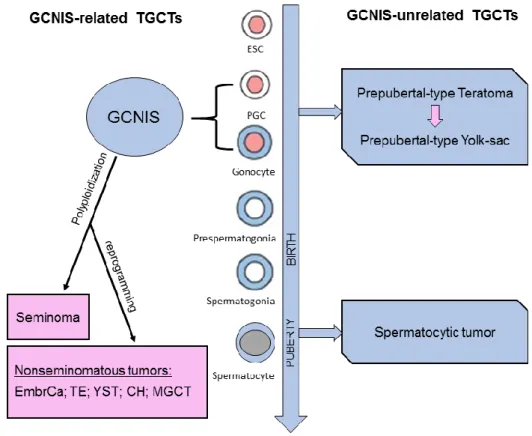
GCNIS derives from the maturation blockage of primordial germ cell or gonocyte to a pre-spermatogonia [22]. Polyploidization is an initial step in the formation of GCNIS results in altered germ cell that remains dormant until puberty. After puberty, it evolves either to SE or to one of the NST subtypes, which may also comprise a SE component (Figure 2). Most of patients with GCNIS have no known genetic abnormalities and these tumors are specially related with disorders of sex development, in which prevalence of GCNIS can be as high as 70% [23]. Unfortunately, until now, it is not possible to predict whether GCNIS will progress to SE or NST, although this progression has been associated to the gain and loss of specific chromosomal regions [3].
Figure 2 A diagrammatic illustration of Testicular germ cell tumors subtypes pathogenesis. TGCTs- testicular germ cell tumors; GCNIS- germ cell neoplasia in situ; ESC- embryonic stem cell; PGC- primordial germ cell; EmbrCa- embryonal carcinoma; TE- postpubertal-type TE; YST- postpubertal-type yolk-sac tumor; CH- choriocarcinoma; MGCT- mixed germ cell tumor. Adapted from [3].
INTRODUCTION
6
Regarding epigenetic profile, GCNIS are characterized by DNA hypomethylation, presence of permissive histone modifications and high specific microRNAs expression levels, such as the miR-371-3 cluster [24-28].
1.3.1 Testicular Germ Cell Tumors related to GCNIS
1.3.1.1 Seminoma (SE)
SE is defined as a “malignant germ cell tumor whose cells are considered the neoplastic counterparts of the primordial germ cells/gonocytes present during early embryonic development” [3]. About 50% of TGCTs are seminomas in its pure form, thus, constituting the most common subtype. Seminoma is rarely seen in men older than 70 years and is only seen in prepubertal children who have a disorder of sex development [29]. Patients with cryptorchidism or immunodeficiency disorders have higher likelihood of developing seminoma than the general population [30, 31]. The presence of syncytiotrophoblastic cells in seminoma might explain the diminutive elevation of β -subunit of human chorionic gonadotropin (beta-hCG) levels in serum and sometimes lead to potential diagnostic confusion with choriocarcinoma [32].
This tumor type is less aggressive than NST and highly sensitive to cisplatin-based chemotherapy and to radiotherapy. Being, these features associated with a good prognosis [3].
1.3.1.2 Nonseminomatous tumors
1.3.1.2.1 Embryonal Carcinoma (EmbrCa)
EmbrCa is “malignant germ cell tumor composed of tumor cells resembling embryonic stem cells” [3]. Forty per cent of all TGCTs and about 87% of all NST are EmbrCa constituting the second most prevalent TGCT subtype [33, 34].
EmbrCa pure form occurs primarily in adult males with a mean age in 3th decade, and rarely
in childhood, except for those with sex development disorders [33, 35].
The prognosis is related to clinical and pathological stage and to the amount of EmbrCa in mixed tumors. Importantly, pure EmbrCa behaves more aggressively than when present in mixed TGCTs. Lymphovascular invasion is a common finding in EmbrCa, which increases the risk of metastatic spread [36, 37].
1.3.1.2.2 Postpubertal-type Yolk sac tumor (YST)
Postpubertal-type YST is a “malignant germ cell tumor that differentiates to resemble extraembryonic structures, including the yolk sac, allantois and extraembryonic mesenchyme” [3]. The majority of cases occur in adult males as a one component of a mixed TGCT (44% of NST), with the pure form being exquisitely rare, accountings for only 0,6% of the cases [38].
INTRODUCTION
7
YST histology is very complex and at diagnosis, serum tumor markers assessment represents an important tool. About 98% of the cases have a strong association between histological identification of YST elements and elevated serum levels of α-fetoprotein (AFP). This NST subtype is associated with chemorresistance and late recurrences [39].
1.3.1.2.3 Choriocarcinoma (CH)
CH is a “malignant germ cell tumor that differentiates to resemble the trophoblastic cells of the extraembryonic chorion, including cytotrophoblastic, intermediate trophoblastic and syncytiotrophoblastic cells” [3]. TGCT subtype that differentiates to resemble the trophoblastic cells of the extraembryonic chorion. Patients with CH are generally in the third or fourth decades of life and, similarly to YST, it appears commonly in the context of a mixed TGCT, with the pure form being extremely rare (0.3% of the cases) [33, 40-42]. In this TGCT subtype beta-hCG is invariably elevated and this elevation reach often values over 50000 IU/L [42].
Unlike others TGCT subtypes, CH patients more often present symptoms related to metastatic disease. [43, 44]. This is the most aggressive TGCT subtype with unfavorable prognosis due to early haematogenous spread, high stage at diagnosis, and hemorrhagic complications [42, 45-47].
1.3.1.2.4 Postpubertal-type Teratoma (TE)
Postpubertal-type TE is a “malignant germ cell tumor composed of several types of tissue representing one or more of the germinal layers (endoderm, mesoderm and ectoderm)” [3]. It is thought to occur through a pathway of differentiation of seminoma, possibly GCNIS, to other types (YST, EC, CH) and subsequent differentiation into Teratoma. It is composed of numerous types of tissues representing one or more of the germinal layers (endoderm, mesoderm and ectoderm) [3].
Most of the cases occurs in young adults, being pure form uncommon and, as a component, occur in 47-50% of mixed TGCTs. Differentiating these tumors from YST is difficult, however serum α-fetoprotein levels may have an important role in diagnosis. [48, 49].
Teratoma constitutes the most chemoresistant component of TGCT, being the most common component present on disease relapses. Nonetheless, this component associates with more favorable prognosis [50].
1.3.1.2.5 Mixed tumors
Mixed germ cell tumors contain two or more TGCT components. They represent more than 69% of all NST and are clinically classified as NST, regardless of the presence or absence of a SE component [33, 51].
Average patient’s age at presentation is 30 years [33, 51]. They can be seen in any combination and more than two components can be present. However, the most frequent
INTRODUCTION
8
combinations includes the EmbrCa with other component: (1) EmbrCa with TE; (2) EmbrCa with SE or (3) EmbrCa with YST [52].
Serum markers are important at diagnosis once they reflect the tumor components present. Elevation of AFP might indicate the presence of YST, whereas beta-hCG elevation might indicate the presence of throphoblastic components. The components of mixed TGCT and the percentage of which component present prognostic implications for the patient being mandatory its report at diagnosis [53]. Several associations have been reported between the presence and proportion of TGCT subtypes and patients’ prognosis. The presence and proportion of EmbrCa has been associated with higher metastastic risk whereas the presence of YST and TE components has been associated with lower metastasis incidence [54, 55]. Moreover, the CH component predominance in mixed TGCT tumor is correlated with high stage at presentation and aggressive behavior [42].
1.3.2 Testicular Germ Cell Tumors unrelated to GCNIS
1.3.2.1 Prepubertal-type Teratoma
As its postpubertal counterpart, this tumor type is “composed of elements resembling somatic tissues derived from one or more of the germinal layers” [3].
Contrary to adult teratomas, this subtype lacks association with GCNIS and have a organoid architecture and commonly lacks 12p amplification [3].
It has been suggested that some teratomas present in adult life might have been present since childhood, although not diagnosed. Importantly, these neoplasms do not recur or metastasize [56].
1.3.2.2 Prepubertal-type Yolk sac tumor
As in adults, this subtype’s cells are similar to extraembryonic structures, such as the yolk sac, allantois and extraembryonic mesenchyme [3]. The incidence of this type of tumor is very low, being about 2-3 per year per 1 million children through age 6 years [57].
Differences between postpubertal counterpart include: (1) no association with cryptorchidism; (2) no association with GCNIS; (3) pure form is more common than as a component of a mixed TGCT [3].
Because of advanced-stage disease’s low incidence and the effective chemotherapy available for those with metastases, survival is almost 100% [58-60]. Evidence suggest that specific microRNA elevation after orchiectomy may predict metastasis [1, 61].
1.3.2.3 Spermatocytic tumor
The reclassification of Spermatocytic seminoma as Spermatocytic tumor by the update of WHO Classification allowed accentuate its nonaggressive behaviour and the lack of common
INTRODUCTION
9
features with the usual SE. This subtype is defined as a “germ cell tumor derived from postpubertal-type germ cells, whose tumor cells resemble spermatogenic cells, most commonly spermatogonia or early primary spermatocytes” [3]. This tumor accounts for about 1% of TGCTs and it occurs at a mean age of 52-59 years.
The behavior of this subtype is nonaggressive with an excellent prognosis and metastization occurs rarely [62-64].
1.4 Diagnosis, Staging, Prognosis and Treatment
1.4.1 Diagnosis
The first initial approach of TGCT diagnosis is the complete examination of testicles and abdomen. This examination often includes imaging tests, ultrasounds and serum tumor markers measurement. These markers might be indicative of the presence of a specific subtype. As previously mentioned, high levels of AFP and HCG are usually associated with the presence of YST and CH, respectively [65, 66]. However, specificity and sensitivity of serum AFP, β-subunit of human chorionic gonadotropin (β-hCG) and lactate dehydrogenase (LDH) is fair, at the most, particularly for seminoma’s patients in which AFP and β-hCG serum levels are either within normal range or display much lower levels than those exhibited by NST [67]. Furthermore, these serum levels might also be deregulated and associated with other type of benign and malignant diseases [68].
Furthermore, GCNIS may be detected accidentally during a biopsy made for other reasons such as fertility problems [69]. Histopathological examination is the current standard diagnostic technique being more sensitive than immunocytochemistry of semen smears. Nevertheless, it can provide false negative result, since the GCNIS cells may not all be located in a testis area [70].
1.4.2 Staging and Prognosis
Patients diagnosed with TGCT are staged for treatment decision making and patient’s stratification. There are two different types of staging published by the American Joint Committee on Cancer (AJCC)/ Union for International Cancer Control (UICC) [71]. The first one is the TNM system, determined using information prior to surgery and combines four categories to describe the tumor: The extent of the primary tumor (T), the regional lymph node (N), spread to the distant metastatic sites (M) and the levels of serum tumor markers (S) (Table 2).
The other type of staging, namely Pathological TNM classification, is more accurate than previous and is performed after surgery by histopathological analysis of the ressected tissue (Table 2) [3, 72].
INTRODUCTION
10
Table 2 Clinical and pathological TNM classification of testicular germ cell tumors (Adapted from [3, 73]).
IGD: in greatest dimension; LDH: lactate dehydrogenase; hCG: human chorionic gonadotropin; AFP: α-fetoprotein; UNL: upper limit of normal range.
INTRODUCTION
11
TNM system is used to assign the stage of the cancer. In Testicular Germ Cell Tumors, T stage is only determined after surgery which means that this stage is always a pathological stage and never a clinical stage. The “p” before the T stage indicates that it is a pathological stage [72]. Tumor stage information is represented in Table 3.
Table 3 Tumor stage information of testicular germ cell tumors (Adapted from [3, 73]).
Additionally, International Germ Cancer Collaborative Group (IGCCG) established a prognostic-based staging for advanced disease based on various parameters as is shown in Table 4 [73-75], in order to determine the best treatment for each patient. Thus, TGCT patients are also classified into a good-risk, intermediate-risk, or poor-risk group.
INTRODUCTION
12
Table 4 Prognostic-based staging system for metastatic disease (International Germ Cell Cancer Collaborative Group) (Adapted from [73, 75]).
PFS- progression free survival; NPVM- nonpulmonary visceral metastases; AFP- α-fetoprotein; LDH- lactate dehydrogenase; β-hCG- human chorionic gonadotropin subunit beta; ULN-upper limit of normal range.
1.4.3 Treatment
Notwithstanding TGCTs have high survival rates, there is a need for more effective treatment strategies for patients with more advanced stages and for those who present disease relapse. Treatment of TGCTs includes one or more therapeutic strategies like surgery, radiotherapy and chemotherapy [74].
INTRODUCTION
13
1.4.3.1 Surgery
Orchiectomy is the primary treatment for TGCT patients and usually is the basis of diagnosis is the histopathological assessment of the surgery specimen. This procedure can reach cure rates of 80% and 70% to SE and NST, respectively [74].
Retroperitoneal Lymph Node Dissection (RPLND) is other type of surgery and is recommended when tumor is already invades abdomen’s lymph nodes. This type of surgery consists on the removal of the lymph nodes and does not affect fertility, decreases the risk of the cancer spreading and preserves sexual function [74, 76].
1.4.3.2 Radiation Therapy
Radiation therapy is the least therapeutic approach due to the side effects in normal germ cells that can compromise androgen production [74]. Furthermore, this type of treatment can increase the risk of other diseases and malignancies. The possibility to administrate lower doses of irradiation can preserve Leydig cell function but may not be so effective in terms of cure rates [77].
1.4.3.3 Chemotherapy
BEP (Bleomycin, Etoposide and Cisplatin) is the standard first-line combination regimen and is given as three or four cycles, depending on the patient’s risk category. Patients with a good prognosis are eligible to received 3 cycles of BEP or, when BEP is contraindicated, 4 cycles of the second-line regimens VIP (Etoposide, Ifosfamide and Cisplatin). Moreover, patients with poor or intermediate prognosis are eligible to receive 4 cycles of BEP. The salvage chemotherapy is based on TIP scheme including Paclitaxel, Ifosfamide and Cisplatin [74].
Several attempts have been made to reduce the toxicity caused by BEP, such as the replacement of carboplatin with cisplatin, unfortunately without success. Furthermore, alternation of several active drugs or dose intensification of etoposide and ifosfamide have been made to increase survival rates in patients with poor prognosis [74].
Metastatic TGCT have been highly curable since the introduction of cisplatin-based chemotherapy which shows a high sensitivity of TGCTs to this form of treatment [78]. However, about 15-20% of these patients develops resistance to treatment and will relapse after first-line chemotherapy. Nonetheless, the mechanisms underlying this sensitivity and resistance are not well understood [78].
2 Epigenetics
The term “ Epigenetics” was defined in 1942 by Conrad Waddington as the “casual interactions by which the genes of the genotype bring about phenotypic effects” [79].Over the years,
INTRODUCTION
14
this definition has been changing and nowadays epigenetic is less focused on genotype and more associated to the processes that controls gene expression, being described as “the study of heritable changes in gene function without alter the DNA sequence” [80].
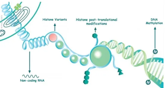
Epigenetic mechanisms may be grouped into four distinct types of mechanisms: DNA methylation, histone post-translational modifications, histone variants and non-coding RNAs (Figure 3) [81]. These four mechanisms have an impact in chromatin dynamics and consequently in the gene regulation and expression [82].Therefore, when this mechanisms are deregulated contribute to the development of several diseases, including cancer [83].
Figure 3 Four distinct mechanisms of epigenetic regulation. M – 5-methylcytosine; the symbols hexagon and triangle represent different translational modifications in N-terminal tails of histones proteins. (CBEG, IPO Porto).
2.1 DNA Methylation
In humans, DNA methylation is the best studied of epigenetic mechanisms and consists in the addition of a methyl group (CH3) at the 5’ position of a cytosine nucleotide that is followed by a
guanine, so called CpG dinucleotide, originating 5-methylcytosine (5mC). This process is catalyzed by DNA methyltransferases (DNMTs) that use S-adenosylmethionine as a methyl donor group [84]. The CpG dinucleotides tend to cluster in regions that are designed as “CpG islands”. The promoters regions present the majority of CpG islands (approximately 60% of human gene promoters) [85] which in normal cells are constantly unmethylated although, some of them, can be specifically methylated during development and tissue differentiation [86]. DNA methylation is associated with gene expression inhibition and with chromatin repressive states once it avoids the binding of transcription factors as well as promotes the recruitment of chromatin remodeling complexes [86, 87].
INTRODUCTION
15
There are five DNMTs in human cells: DNMT1, DNMT2, DNMT3A, DNMT3B and DNMT3L. DNMT1 usually methylates hemi-methylated DNA and it is classified into maintenance DNMT. The DNMT3A and DNMT3B are responsible for de novo methylation playing a critical role during embryogenesis and gametogenesis. DNMT3-L does not have a methyltransferase activity and DNMT2 just appears to be involved in RNA methylation [86, 88].
A cancer cell is characterized by alterations on DNA methylation patterns, namely by global hypomethylation, which can benefits chromosomal instability, and by hypermethylation at specific CpG islands mainly in tumor suppressor genes promoters [89].
2.2 Histones: Post-translational modifications and variants
In Eukaryotic cells, DNA is packaged by positively-charged proteins, the histones, that bind with the negatively-charge DNA [90]. The nucleosomes are constituted by eight histones, one pair of each H2A, H2B, H3 and H4 that with DNA, and they stand basic unit of chromatin fiber. The contact of histones with DNA is facilitated by their structure which is characterized by a globular C-terminal domain and an unstructured N-C-terminal tail [91].
Post-translation modifications that occur in histones N-terminal tails (such as acetylation, methylation and phosphorylation, ubiquitylation and sumoylation) promote the establishment and maintenance of gene activity and consequently conditions chromatin conformation [92]. This post-translational modifications plays an important role in several processes like DNA replication, transcriptional regulation, DNA repair, and others, by unpacking nucleosomal DNA or “slidding” nucleosomes along it [93, 94].
Thus far, histone variants have been described for all canonical histones, excepting for H4. Unlike canonical histones, variants are tissue and temporal-specific, replacing the others during development and differentiation, establishing cell identity [95-97]. Additionally, they promote diversification of chromatin states through its structural features [98].
2.3 Chromatin remodelling
Nowadays, it is known that chromatin remodeling proteins plays a major role in the regulation of gene expression by transiently exposes DNA to binding proteins. Proteins involved in this process utilize ATP to alter the structure of chromatin through disruption of the histone/DNA contacts. Remodeler activity can result in nucleosome sliding, eviction, assembly, spacing or replacement of canonical histones with specific histone variants [99].
Chromatin remodeling enzymes can be divided into four distinct families with different functions: (1) SWI/SNF (SWitching-Defective/Sucrose-NonFermenting) family functions in the sliding and eviction of nucleosomes, (2) ISWI (Imitation-SWItch ) remodelers function in nucleosome assembly and spacing and is also proposed to have functions in higher-level chromatin organization,
INTRODUCTION
16
(3) CHD (chromodomain helicase DNA binding Protein) remodelers are associated with nucleosome sliding, eviction, spacing, and nucleosome assembly and (4) INO80 family is specialized for restructuring the nucleosome [100].
Chromatin remodelers act as an integrator of the extracellular and cytoplasmic signals to the nucleus for precise regulation of target gene expression. Recently, disruption of chromatin remodelers was implicated in cancer development, progression and therapeutic resistance [100].
It is important to note that epigenetic silencing (through nucleosome remodeling, histones and DNA methylation) can contribute to the inactivation or loss of function of chromatin remodelers in human cancer [99].
2.4 Non-coding RNAs
Non-coding RNAs (ncRNAs) are a class of RNA sequences that are transcribed but do not codify proteins. Over the years these RNAs have been considered as non-functional molecules [101]. However, in the last decade several studies showed that ncRNAs are biologically active, functional and essential to a right development [101]. It is, currently, known that gene expression can be controlled by these molecules. Moreover, 90% of our genome is transcribed into RNA molecules although only 2% encodes for proteins, supporting this hypothesis [102, 103]. Indeed, ncRNAs are involved in chromosome dynamic control, RNA editing, translation inhibition and mRNA degradation [104].
This class of molecules can be divided according they size into microRNAs (~ 20 nucleotides), small ncRNAs (30-300 nucleotides) and long ncRNAs (>300 nucleotides) [102]. From these, particularly in cancer, microRNAs (miRNAs) have been the most studied ncRNAs over the last years.
2.4.1 MicroRNAs: Biogenesis and Function
The biological importance of miRNAs was recognized in the early 1990s, when several groups reported the regulation of developmental processes by small non-coding RNA molecules [105]. MiRNAs are small RNA molecules that are involved in several essential biological processes, such as cell cycle, proliferation, differentiation, apoptosis and tumor development [106]. More than one-third of human protein-coding genes are controlled by miRNAs [105].
MiRNAs are transcript from DNA sequences by RNA Polimerase II thought activation of several transcription factors. The transcript is a primary miRNA (pri-miRNA) which forms a typically hairpin loop structure. It would become the final miRNA with regulatory functions of the several steps in processing [107]. This pri-miRNA is recognized by the protein DGCR8 and the ribonuclease (RNase) III enzyme Drosha associated with DGCR8 to form a complex which is able to cut the RNA
INTRODUCTION
17
into a small precursor (pre-miRNA) [108]. The hairpin structure of pre-miRNA is exported into the cytoplasm through the nuclear pore by Exportin5 complex (with Ran-GTPase activity). In the cytoplasm it is recognized by Dicer which cleaves the hairpin loop and produce a short double strand miRNA molecule, with 19 to 22 nucleotides [105]. In the cytoplasm, an Argonaute protein (Ago 2) interacts with Dicer to binds the miRNA. The miRNA is unwinded and one strand is released. The remaining strand interacts with Ago 2 and some additional proteins to form the RISC (RNA Induced Silencing Complex). Then, mature miRNAs interact with complementary target mRNAs within the RISC to regulate mRNA stability or protein translation Figure 4 [105, 107].
Figure 4 MicroRNAs biogenesis. Kindly provided by [109].
These molecules have the potential to qualify as biomarkers in various malignancies because some of them are abundantly expressed in cancer tissuesand most of them reveal high stability in body fluids [106].
Not only miRNA expression profile is altered in many diseases, including urological cancer, but miRNAs’ patterns differ between malignant and benign tissues as well as among different tumor entities, highlighting their potential as biomarkers in various malignancies [110]. These molecules can either act as tumor suppressors or oncogenic drivers, in respect to cancer initiation and progression, depending on the regulated genes, the cellular context, and the cancer type [105].
Circulating miRNAs, especially those present in the serum, are of high diagnostic significance, and have become biomarkers for numerous conditions, including cancer,
INTRODUCTION
18
cardiovascular diseases, metabolic disorders and tissue damage. Furthermore, these molecules present an important role in forensic medicine, prenatal diagnosis and pregnancy monitoring [2].
Several circulating miRNAs have been associated with various types of cancer, such as prostate cancer, lung cancer, hepatocellular carcinoma, gastric cancer, pancreatic cancer, colorectal cancer, breast cancer and cervical cancer [111].
2.4.2 The importance of miRNAs in TGCT
Several approaches have been used for the diagnosis and follow-up of TGCTs such as abdominal ultrasound, chest and abdominal computed tomography with retroperitoneal examination and serum markers assessment. Nevertheless, the specificity and sensitivity of these markers is limited, being altered only in about 60% of TGCT cases [2]. Hence there is an ongoing search for novel biomarkers.
Gillis et al developed a robust protocol for the analysis of miR-371-3 and miR-302 cluster in serum, called the Target Serum miRNA test (TSmiR), which showed a distinctly higher diagnostic sensitivity (98%, in a cohort of 80 patients with testicular cancer and 47 controls) than the standard markers AFP and hCG (36% and 57%, respectively). Additionally, high serum levels of the tested miRNAs in patients with metastasis were identified, as well as normalization of miRNA levels after surgical removal of primary disease [1].
2.4.3 miR-371-373 cluster in TGCT
Increased miR-371-373 cluster expression seems to be a consistent miRNA signature of testicular germ cell neoplasms [112]. It was previously shown that miR-371-373 cluster has an oncogenic potential in TGCT, due to its ability to mask the effect of mutated p53, by inhibiting Ras-induced oncogenic senescence in TGCTs [113]. Subsequently, it was demonstrated that this cluster was significantly upregulated in TGCTs [114]. The mechanisms underlying this upregulation have been elucidated and a feedback loop between the miR-371-373 cluster and Wnt/beta-catenin signaling pathway was disclosed. Remarkably, stem cells showed higher miR-371-373 cluster expression than differentiated cells [115] and since Wnt signaling plays a critical role in regulating cell stemness, miR-371-373 cluster appears to contribute to maintenance of stem cell status [116].
Previous studies showed that serum levels of all three miRNAs from miR-371-373 cluster (miR-371a-3p, miR-372 and miR-373-3p) were significantly higher in TGCT patients. Among them, miR-371a-3p displayed higher expression in SE and NST, disclosing the largest post-operative decrease and outperforming classical TGCT serum biomarkers [106, 112]. Furthermore, significantly higher miR-371a-3p levels were found in testicular vein blood (TVB) than in the peripheral blood, indicating that the primary source for miR-371a-3p upregulation was indeed testicular tumor cells [117]. Hence, miR-371a-3p has been recognized as a promising candidate biomarker for patients
INTRODUCTION
19
with TGCTs [118]. Nevertheless, data on differential expression of miR-371a-3p among TGCT subtypes is lacking and it may impact on its performance as biomarker.
AIMS
21
AIMS
Notwithstanding TGCTs is among the most curable solid tumors, displaying 5-year survival rates over 95%, which remain high (over 80%) for patients with metastatic disease, there is an ongoing search for novel detection tests able to diagnose and characterize more efficiently the disease.
MicroRNAs have emerged as potential diagnostic and prognostic biomarkers replacing serum tumor markers which have limited specificity and sensitivity for the disease.
Thus, the main goal of this dissertation is to evaluate the potential of miR-371a-3p for discriminating TGCTs subtypes.
More specifically, the major aims are:
• Validate miR-371a-3p expression in a retrospective cohort of TGCT tissue samples;
• Assess whether miR-371a-3p expression levels associates with different histological TGCT subtypes and may serve as diagnostic and/or prognostic biomarker, especially for tumors with Stage I-S;
• Evaluate the association of the candidate miRNA expression levels with standard clinicopathological parameters.
MATERIAL AND METHODS
23
1. Patients and samples collection
Archival, formalin-fixed, paraffin-embedded tissue from patients with TGCT [103 GCNIS-related - 68 pure SE and 35 NST (five pure EmbrCa, two pure postpubertal-type TE, and 28 mixed germ cell tumors) – and 16 GCNIS-unrelated - four prepubertal-type YST and 12 prepubertal-type TE], diagnosed and treated with orchiectomy at Portuguese Oncology Institute of Porto, Portugal, between 2005 and 2016 were selected for this study. Additionally, control samples deriving from 15 patients subjected to orchiectomy due to inflammatory or benign testicular pathology were included. All histological slides were retrieved, reviewed by an experienced uro-pathologist, re-classified according to 2016 World Health Organization recommendations [119] and staged according to TNM classification 7th edition [71]. Moreover, the International Germ Cell Consensus Classification [a prognosis factor-based staging system developed by the International Germ Cell Cancer Collaborative Group (IGCCCG)] was applied to all malignant TGCT [75]. Representative areas of all tumors, including different components of mixed TGCT, were identified and selectively collected for subsequent RNA extraction. These areas were then individually considered for the purposes of comparisons among TGCT subtypes. Thus, analyzed tissue samples comprised 138 GCNIS-related TGCT – 68 pure SE and 70 NST (five pure EmbrCa and two pure postpubertal-type TE, as well as 12 SE, 17 EmbrCa, 13 postpubertal-type YST, six CH, and 14 postpubertal-type TE as components of mixed germ cell tumors), four prepubertal-type YST and 12 prepubertal-type TE. This study was approved by institutional ethics review board (Comissão de Ética para a Saúde – CES-IPO-12-017) of Portuguese Oncology Institute of Porto, Portugal.
2. RNA Extraction
Total RNA from FFPE tissue samples was extracted through the Norgen’s FFPE RNA/DNA Purification Plus Kit (Norgen, Canada), according to manufacturer instructions. This Kit provides conditions that allow for the partial reversing of formalin modifications, resulting in a high quality and yield of nucleic acids.
The concentration, purity ratios and quality of each sample were determined using a Nanodrop ND-1000 (ThermoScientific, Wilmington, DE, USA) and RNA samples were then stored at -80ºC until further use.
MATERIAL AND METHODS
24
3. MicroRNA cDNA Synthesis
cDNA was synthesized from total extracted RNA. For all samples, reverse transcription (RT) was carried out using the TaqmanTM MicroRNA Reverse Transcription Kit (Applied Biosystems,
Foster City, CA, USA). 10 ng total RNA from each sample was used and the reactions had a final volume of 15 µL.
The reaction was carried out for miR-371a-3p using stem-loop-primers from the relevant TaqMan microRNA assays (miR-371a-3p, Assay ID 002124, Applied Biosystems).
Reverse transcription was performed in a Veriti® Thermal Cycler (Applied Biosystems®). Thermal-cycling conditions consisted in: 30 minutes at 16ºC, 30 minutes at 42ºC and 5 minutes at 85ºC. Samples were then stored at -20ºC.
4. MicroRNAs Expression: Individual Assays
Quantitative real-time PCR (RT-qPCR) was performed in a 7500 Real-Time PCR system (Applied Biosystems, Foster City, CA, USA), according to the recommended protocol. Briefly, to each well of a 96-well was added: 5 μL of NZYSpeedy qPCR Probe Master Mix (2x) (NZYTech®,
Portugal), 0.5 μL of TaqMan® Small RNA Assay (20X) (Applied Biosystems®, Life TechnologiesTM)
and 4,5 μL of reverse transcription product previously diluted 15x.
For each sample the reactions were run in triplicate, and, according to the manufacturer instructions, the running method consisted in a holding stage of 2 minutes at 50oC, followed by
another holding stage at 95oC for 10 minutes, during which enzyme activation occurs, and 40 cycles
composed of a denaturation stage at 95oC that lasts for 15 seconds and an annealing/extending
stage at 60oC for 60 seconds. Each plate also contained 2 negative controls (only reaction mix) and
five serial dilutions (5x dilution factor) of cDNA from total Human Reference RNA (Agilent, USA) previously synthesized similarly to patient samples for a standard curve generation to relative quantification and PCR efficiency assessement. RNU48 was used as a reference gene for normalization (RNU48, Assay ID 001006) and relative expression of targets tested in each sample was obtained using the formula:
Relative Expression =
𝑻𝒂𝒓𝒈𝒆𝒕 𝒈𝒆𝒏𝒆 𝒎𝒆𝒂𝒏 𝒒𝒖𝒂𝒏𝒕𝒊𝒕𝒚𝑹𝑵𝑼𝟒𝟖 𝒎𝒆𝒂𝒏 𝒒𝒖𝒂𝒏𝒕𝒊𝒕𝒚
MATERIAL AND METHODS
25
5. Statistical Analysis
Differences in miR-371a-3p expression levels among TGCT subtypes were assessed using non-parametric tests, i.e., the Kruskal-Wallis test, followed by Mann-Whitney U-test for pairwise comparisons, when appropriate. P-values inferior to 0.05 were considered statistically significant and Bonferroni’s correction was applied to pairwise comparisons.
To assess the discriminative power of miR-371a-3p expression levels among TGCT subtypes, ROC curves were constructed and biomarker performance parameters [sensitivity, specificity, positive predictive value (PPV), negative predictive value (NPV) and accuracy] were calculated as shown Table 5. Cut-off was established based on the highest value obtained in ROC curve analysis.
Statistical analyses were performed using SPSS Statistics 24 (IBM-SPSS, IL, USA) and graphics were constructed using GraphPad 6 Prism (GraphPad Software, USA).
Table 5 Formulas used for biomarker parameters calculation.
Tumor vs Control Sensitivity (%) (C/A) x 100
Tumor Control Specificity (%) (F/B) x 100
Total A B PPV (%) (C/(C+D)) x 100
> Cut-off C D NPV (%) (F/(E+F)) x 100
RESULTS
27
Clinical characterization and association between miR-371a-3p
expression levels and standard clinicopathological parameters
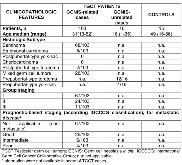
Overall, 134 patients were included in this study. The median age of patients at diagnosis for GCNIS-related TGCTs, GCNIS-unrelated TGCTs and controls was 31, 16 and 49 years, respectively. Post of the patients (65%) with GCNIS-related TGCT presented with clinical stage I (67/103), whereas only 10.7% (11/103) were at stage III. According to prognostic-based staging, 25.2% (26/103) of the cases were classified as good prognosis, 5.8% (6/103) as intermediate prognosis and 3.9% (4/103) as poor prognosis. Relevant clinical and pathological data are presented in Table 6. Serum tumor markers’ levels were available for most patients both pre- and post-operatively Table 7.
MiR-371a-3p expression levels did not associate with serum marker levels or patients’ age (data not shown). No association was disclosed between miR-371a-3p expression levels and TNM staging or prognostic-based staging.
Table 6 Clinicopathological information of TGCT patients and controls.
CLINICOPATHOLOGIC FEATURES TGCT PATIENTS CONTROLS GCNIS-related cases GCNIS-unrelated cases Patients, n 103 16 15
Age median (range) 31(13-52) 16 (1-35) 49 (16-86)
Histologic Subtype
Seminoma 68/103 n.a. n.a.
Embryonal carcinoma 5/103 n.a. n.a.
Postpubertal-type yolk-sac 0 n.a. n.a.
Choriocarcinoma 0 n.a. n.a.
Postpubertal-type teratoma 2/103 n.a. n.a.
Mixed germ cell tumors 28/103 n.a. n.a.
Prepubertal-type teratoma n.a. 12/16 n.a.
Prepubertal-type yolk-sac n.a. 4/16 n.a.
Group staging
I 67/103 n.a. n.a.
II 24/103 n.a. n.a.
III 11/103 n.a. n.a.
Prognostic-based staging (according IGCCCG classification), for metastatic disease*
Not applicable
(non-metastatic) 67/103 n.a. n.a.
Good 26/103 n.a. n.a.
Intermediate 6/103 n.a. n.a.
Poor 4/103 n.a. n.a.
TGCT:Testicular germ cell tumors; GCNIS: Germ cell neoplasia in situ; IGCCCG: International Germ Cell Cancer Collaborative Group; n.a: not applicable.
RESULTS
28
Table 7 Serum tumor markers levels in testicular germ cell tumors cases. SERUM TUMOR MARKERS
GCNIS-related cases Pre-surgery Post surgery
AFP elevated 20/97 (21%) 12/80 (15%) β-hCG elevated 41/95 (43%) 16/80 (20%) LDH elevated 32/82 (39%) 15/67 (22%) GCNIS-unrelated cases AFP elevated 4/5 (80%) 3/3 (100%) β-hCG elevated 2/13 (15%) 0/3 (0%) LDH elevated 4/11 (36%) 1/4 (25%)
AFP: α-fetoprotein; β-hCG: β-human chorionic gonadotropin; LDH: Lactate dehydrogenase; GCNIS: Germ cell neoplasia in situ.
Assessment of miR-371a-3p expression levels in TGCT and
controls
To verify whether miR-371a-3p expression was TGCT-specific, expression levels in normal and neoplastic tissue samples were evaluated (Table 8) and these levels were significantly higher in TGCT tissues compared to controls (p<0.0001).
Table 8 miR-371a-3p expression levels in controls and testicular germ cell tumors. Controls median
(IQR)
Pure SE median
(IQR) NST median (IQR)
0.01
(0-0.03) (5.32-33.01) 11.33 (2.42-11.94) 5.48
NST subtypes median (IQR)
Mixed EmbrCa Mixed YST Mixed CH Mixed TE Mixed SE 8.72
(4.33-69.68) (2.86-9.19) 4.16 (3.25-9.27) 6.61 (0.24-2.36) 0.87 (3.04-7.38) 4.96
Prepubertal TGCT median (IQR)
0.06 (0.02-3.09)
SE: Seminoma; NST: Nonseminomatous tumors; IQR: Interquartile range; EmbrCa: Embryonal carcinoma; YST: Postpubertal-type yolk-sac tumor; CH: Choriocarcinoma; TE: Postpubertal-type teratoma; Mixed: TGCT subtypes as componentes of mixed germ cell tumors.
Subsequently, ROC curve analysis was performed (Figure 5, right panel), disclosing an area under the curve (AUC) of 0.99. Using the cutoff value derived from this analysis (0.0875), miR-371a-3p expression distinguished GCNIS-related TGCTs from normal testicular tissue with 92.2% sensitivity, 93.3% specificity and 92.3% accuracy (Table 9).
RESULTS
29
Figure 5 Box-plots (left panel) of miR-371a-3p expression levels in Normal Tissue Testis (Controls) and TGCT (Tumors) samples (A) and respective Receiver Operating Characteristic Curve (right panel) (B). Abbreviations: AUC – Area Under the Curve.
Differential expression of miR-371a-3p among TGCT subtypes and
discriminative power
To ascertain miRNA-371a-3p differential expression in tissue samples, comparative analyses between TGCT and normal testicular tissues, as well as among TGCT subtypes were carried out.
Both pure SE and NST displayed higher miR-371a-3p expression levels than controls (p<0.001) (Figure 6).
Figure 6 Box-plot of miR-371a-3p expression levels in Normal Testis Tissue (Controls), Pure Seminomas (Pure SE) and Nonseminomatous tumors (NST) samples.
Pure SE was the most common TGCT in this series (n=68), displaying the highest miRNA-371a-3p expression levels, which were significantly higher than those of NST (p<0.0001) (Figure 6 and Figure 7).
RESULTS
30
Figure 7 Box-plot of miR-371a-3p expression levels between the different subtypes of TGCT.
Among NST, EmbrCa displayed the highest miR-371a-3p expression levels, whereas those of postpubertal TE were the lowest, significantly differing from the remainder NST subtypes (Figure 7).
Interestingly, differences in miR-371a-3p expression levels between pure SE and SE in mixed TGCT (p<0.01) were depicted (Figure 4, left panel). Using the calculated cut-off value (7.869), ROC curve analysis disclosed an AUC of approximately 0.75 (Figure 8, right panel). The discriminatory performance was rather modest, with only 30% PPV (Table 9).
Figure 8 Box-plot (left panel) (A) of miR-371a-3p expression levels in Pure Seminoma (Pure SE) and SE in mixed germ cell tumors samples and respective Receiver Operating Characteristic Curve (right panel) (B). Abbreviations: AUC – Area Under the Curve
.
RESULTS
31
Among postpubertal TGCT, TE displayed the lowest miR-371a-3p expression levels but these were still significantly higher than those of normal testicular tissue (p<0.0001). ROC curve analysis disclosed an AUC close to 0.93 (Figure 9, right panel) and using a cutoff value of 0.0875, miR-371a-3p accurately distinguished postpubertal TE from controls (Table 9).
Figure 9 Box-plot (left panel) (A) of miR-371a-3p expression levels in Controls and Postpubertal Teratoma samples and respective Receiver Operating Characteristic Curve (right panel) (B). Abbreviations: AUC – Area Under the Curve.
Globally, miR-371a-3p expression levels were significantly higher in GCNIS-related TGCTs compared to GCNIS-unrelated TGCTs (p=0.0066) (Figure 10).
Figure 10 Box-plot of miR-371a-3p expression levels in GCNIS-unrelated TGCTs samples and GCNIS-related TGCT samples.
Considering the specific subtypes, miR-371a-3p expression was significantly higher in postpubertal vs. prepubertal TE (p=0.0008) (Figure 7), whereas no significant differences were apparent between postpubertal and prepubertal YST (Figure 11).
![Figure 1 Global incidence of Testicular Germ Cell Tumors. Adapted from [9].](https://thumb-eu.123doks.com/thumbv2/123dok_br/15952379.1097730/20.892.131.774.570.837/figure-global-incidence-testicular-germ-cell-tumors-adapted.webp)
![Table 2 Clinical and pathological TNM classification of testicular germ cell tumors (Adapted from [3, 73])](https://thumb-eu.123doks.com/thumbv2/123dok_br/15952379.1097730/27.892.145.753.227.1053/table-clinical-pathological-tnm-classification-testicular-tumors-adapted.webp)
![Table 3 Tumor stage information of testicular germ cell tumors (Adapted from [3, 73])](https://thumb-eu.123doks.com/thumbv2/123dok_br/15952379.1097730/28.892.82.812.275.844/table-tumor-stage-information-testicular-germ-tumors-adapted.webp)
![Table 4 Prognostic-based staging system for metastatic disease (International Germ Cell Cancer Collaborative Group) (Adapted from [73, 75])](https://thumb-eu.123doks.com/thumbv2/123dok_br/15952379.1097730/29.892.98.811.144.901/prognostic-staging-metastatic-disease-international-cancer-collaborative-adapted.webp)
![Figure 4 MicroRNAs biogenesis. Kindly provided by [109].](https://thumb-eu.123doks.com/thumbv2/123dok_br/15952379.1097730/34.892.155.743.336.747/figure-micrornas-biogenesis-kindly-provided.webp)