Sara Raquel Nascimento Santos Pereira da Silva
Graduated in BiochemistryMsmX as model for functional studies of
Multitask ATPases from pathogenic bacteria
Dissertation presented to obtain the Master degree in Biochemistry
Supervisor: Isabel M. G. de Sá-Nogueira, Associate
Professor w. Habilitation (Agregação), FCT/UNL
Júri:
Presidente: Prof. Doutor Pedro António de Brito Tavares
Arguente: Prof. Doutor Luís Jaime Gomes Ferreira da Silva Mota Vogal: Prof. Doutora Isabel Maria Godinho de Sá Nogueira
October 2016
MsmX as model for functional studies of Multitask ATPases from pathogenic bacteria Copyright © reserved to Sara Pereira da Silva, FCT/UNL, UNL
The Faculty of Science and Technology and the New University of Lisbon have the perpetual right, and without geographical limits, to archive and publish this dissertation through press copies in paper or digital form, or by other known form or any other that will be invented, and to divulgate it through scientific repositories and to admit its copy and distribution with educational or research objectives, non-commercial, as long as it is given credit to the author and editor.
A Faculdade de Ciências e Tecnologia e a Universidade Nova de Lisboa têm o direito, perpétuo e sem limites geográficos, de arquivar e publicar esta dissertação através de exemplares impressos reproduzidos em papel ou de forma digital, ou por qualquer outro meio conhecido ou que venha a ser inventado, e de a divulgar através de repositórios científicos e de admitir a sua cópia e distribuição com objectivos educacionais ou de investigação, não comerciais, desde que seja dado crédito ao autor e editor.
Acknowledgments
First of all I would like to profoundly acknowledge my supervisor, Isabel de Sá Nogueira, for all the support, advice and knowledge provided throughout this master thesis, which allowed me to growth not only as a young scientist but also as a person. Having arrived in a time where my colleagues were physically absent from the laboratory, the support and dedication of my supervisor made all the difference during this astonishing scientific journey.
To Research Unit on Applied Molecular Biosciences (UCIBIO), which is financed by national funds from FCT/MEC (UID/Multi/04378/2013) and co-financed by the ERDF under the PT2020 Partnership Agreement (POCI-01-0145-FEDER-007728), for partially supporting this work.
I thank my lab colleagues for the support they were able to give me in spite of being absent from the lab. Thank you Lia for making me comfortable and part of the group since the beginning, as well as for your useful tips. Thanks Mário for your help in this project and for being available to answer my questions through the telephone. I specially thank you Viviana for your constant support since my graduation project, it has been a pleasure.
I would also like to thank to Barbara, Cinthia and Raquel from lab 333, for the good mood and companionship, and in particularly to Raquel for clarify my questions. Thank you Claúdia for the good times that we had during lunch hour and the breaks from work, I feel grateful to have meet you. Thanks Nicole for making me laugh and for maintaining the good mood in the department. Finally, I thank to all the people from the DCV department for making me feel welcome.
To my friends, who are a life’s treasure, I thank you all for being present in my life. Thanks to Filipa Inácio, Filipa Trovão, Joana Marques, Susana Filipa (“Marias” group) for all the support and that special moments filled with joy, with a special thanks to Susana Filipa who in this group heard me the most (and vice-versa). Thanks Gonçalo for the solid friendship we have had through the last 6 years, I feel thankful for being your friend. Thank you Nádia for being my best friend in the last 13 years, for all the unconditional support you give me, for listening me every day and saying “You will make it”, I feel truly blessed for having you in my life.
To my family, thank you for all the good moments through my life, in particularly to my parents for making this dream came true. To my Dad for his academic advices and relaxing posture, making me believe I could do everything through dedication. To my Mom, my biggest supporter in life, I thank everything and especially all the concern and love that you have for me. And finally I would like to thank you João for all the support you were able to give me in spite of being far, and for all the love that we have shared in the past 6 years.
Abstract
The ABC-type transporters constitute one of the largest and most diverse transporter superfamilies characterized by a highly conserved ATP-binding cassette, and are widespread among all domains of life. Recent studies, performed in our laboratory demonstrated that MsmX ATPase from Bacillus subtilis interacts with several distinct ABC sugar importers thus, unlike other NBDs MsmX was shown to be multitask serving as energy-generating component to several sugar importers. Sharing of an ATPase among carbohydrate ABC transporters in both Gram-positive and Gram-negative bacteria seems to be a common strategy for adaption and survival and may represent novel therapeutic approaches for targeting since ABC importers are exclusive to prokaryotes.
To characterize multipurpose ATPases and to assess their intra- and interspecies interchangeability, we fine-tuned a genetic system in B. subtilis for controlled ectopic gene expression. The functionality of distinct multitask ATPases alleles was determined by their ability to complement the role of MsmX in a B. subtilis msmX-null mutant. Moreover, this genetic system allowed the determination of intracellular accumulation of the tested ATPases by Western-Blot analysis.
The results show that an ATPase from B. thuringiensis was able to fulfill the role of MsmX in its absence, while another ATPase from B. subtilis YurJ was only able to partially play MsmX role. In addition to intra- and interspecies interchangeability of Bacillus ATPases, we found that ATPases from Streptococcus pneumoniae and Staphylococcus aureus were also able to complement to a certain degree the B. subtilis MsmX function in vivo. In contrast ATPases from the Gram-negative bacterium Escherichia coli were not functional in B. subtilis. Furthermore, all the tested ATPases accumulate in the cells.
Our study shows that B. subtilis can be use as model for the study of bacterial multitask ATPases. Furthermore, it provides a genetic tool for the characterization of this phenomenon in bacterial carbohydrate transport and particularly in bacterial pathogens.
Keywords: Bacillus subtilis, ABC sugar importers, Multitask ATPases, MsmX, Interchangeability, Bacterial pathogens.
Resumo
Os transportadores do tipo ABC constituem uma das maiores e mais diversificadas superfamílias de transportadores que são caracterizadas por possuírem uma cassete de ligação ao ATP com elevado grau de conservação, encontrando-se amplamente distribuídos em todos os domínios da vida. Estudos recentes, desempenhados no nosso laboratório, demonstraram que a ATPase MsmX proveniente da espécie Bacillus subtilis interage com diversos importadores ABC de açúcares distintos, sendo que, ao contrário de outros NBDs, MsmX revelou ser multitarefa servindo como componente gerador de energia de vários importadores de açúcares. A partilha de uma ATPase entre transportadores ABC de carboidratos em bactérias Gram-positivas e Gram-negativas aparenta ser uma estratégia comum de adaptação e sobrevivência, e pode representar um alvo para novas abordagens terapêuticas dado que os importadores ABC são exclusivos dos procariontes.
De modo a caracterizar as ATPases multipropósito e no sentido de determinar a sua permutabilidade intra- e inter-espécies, aperfeiçoou-se um sistema genético em B. subtilis para obter uma expressão ectópica controlada do gene. A funcionalidade de alelos distintos das ATPases multitarefa foi determinada através da sua capacidade de complementar o papel de MsmX num mutante de B. subtilis sem o gene msmX. Além disso, este sistema genético permitiu a determinação da acumulação intracelular das ATPases testadas através de uma análise do tipo Western-Blot.
Os resultados demonstraram que uma ATPase proveniente de B. thuringiensis é capaz de realizar na totalidade o papel de MsmX na célula durante a sua ausência, enquanto outra ATPase de B. subtilis (YurJ) foi apenas capaz de desempenhar parcialmente o papel de MsmX. Para além da permutabilidade intra e inter-especies de ATPases em Bacillus, descobriu-se que, ATPases provenientes de Streptococcus pneumoniae e Staphylococcus aureus também são capazes de complementar em determinado grau a função in vivo de MsmX de B. subtilis. Em contraste, ATPases provenientes da bactéria Gram-negativa Escherichia coli não demonstraram esta funcionalidade em B. subtilis. Adicionalmente, verificou-se que todas as ATPases testadas são produzidas e acumulam no interior das células do hospedeiro.
O nosso trabalho de investigação demonstra que B. subtilis pode ser utilizado como um modelo para o estudo de ATPases multitarefa bacterianas. Além disso, o estudo fornece uma ferramenta genética para a caracterização deste fenómeno no transporte bacteriano de carboidratos, e em particular em agentes patogénicos bacterianos.
Palavras-chaves: Bacillus subtilis, Importadores ABC de açúcares, ATPases multitarefa, MsmX, Permutabilidade, Agentes patogénicos bacterianos.
Contents
Acknowledgments……….…..………V Abstract………..………...….VII Resumo………..………IX Contents……….XI Figures Index………XIII Tables Index……….………..XVII Abbreviations, Symbols and Notations………...……….….XIX1. General Introduction………....…3
1.1. Bacillus subtilis - An overview………..…3
1.1.1. AraNPQ ABC importer and the Multitask MsmX ATPase……….…4
1.2. Membrane Transports………..5
1.3. ABC Transporters………..………..6
1.3.1. An overview………..………..6
1.3.2. Types of ABC Transporters………7
1.3.3. Transport Mechanism….……….………..9
1.3.4. The solute binding protein (SBP)……….……..…11
1.3.5. The transmembrane domains (TMDs)……….…..…..12
1.3.6. The nucleotide binding domains (NBDs)………..…………..…….14
1.3.7. Multitask ATPases……..………..………..16
1.3.7.1. MsmK, a multitask ATPase in Streptococcus species………17
1.3.7.2. Multitask ATPases from Streptomyces species and Thermus thermophilus.………..………….……..19
1.4. Scope of the Thesis……….…….20
2. Materials and Methods………..……….…...23
2.2. Bioinformatic Analysis………..….23
2.3. Isolation of chromosomal DNA………..……….23
2.4. DNA manipulation and sequencing………..24
2.5. Site-directed mutagenesis by Overlapping PCR……….………...………....24
2.6. Construction of plasmids and strains………..……….………..……25
2.7. Growth conditions……….………...29
2.8. Protein extracts of B. subtilis………..………...30
2.9. Western-Blot assay………..……….………..31
2.10. Protein analysis……….……….31
3. Results and Discussion………...35
3.1. Complementation analysis of MsmX homologs in msmX-null mutant B. subtilis strains.………35
3.1.1. In silico Analysis….……….35
3.1.2. Fine-tuning of a Genetic System for Complementation Analysis………...36
3.1.3. Functional studies of different ATPases in B. subtilis………..40
3.1.3.1. Wild-type MsmX and MsmX Homologs………....40
3.1.3.2. Recombinant MsmX and MsmX Homologs with a C-terminal His-tag………46
3.1.4. Analysis of the predicted proteins structures………52
3.1.5. Redesigning the genetic system for functional analysis……….55
3.1.5.1. Functional studies………....57
3.1.5.2. Analysis of the 3-D structure models………..62
3.2. Detection of the in vivo accumulation of the recombinant ATPases in the cells……….64
4. Concluding Remarks and Future Perspectives……….71
5. References……….75
Figures Index
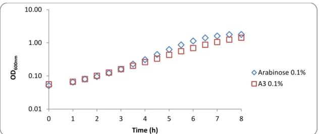
Figure 1.1 - MsmX-dependent ABC importers in B. subtilis……….5 Figure 1.2 - Four distinct folds of ABC transporters………..7 Figure 1.3 - Conformations of the MalEFGK2 transporter (class I importer)……….9 Figure 1.4 - The transport mechanism of Type I importers (exemplified by MalEFGK2)……….…..10 Figure 1.5 - Rearrangements in SBP MalE upon the substrate binding……….11 Figure 1.6 - Transfer of the maltose from MBP to the TM binding site………13 Figure 1.7 - The TMD–MalK interface………..13 Figure 1.8 - The structure of the NBDs, as exemplified by the MalK dimer of the maltose transporter MalEFGK2………....15 Figure 1.9 - Schematic presentation of the msm and mal loci encoding relevant carbohydrate ABC transporters in selected streptococci……….19 Figure 1.10 - Schematic summary of carbohydrates utilization by ABC transporters in S. mutans (A), S. pneumoniae (B) and S. suis (C)……….19 Figure 3.1 - Schematic illustration of the in vivo system fine-tuned………..37 Figure 3.2 - Schematic representation of the amyE locus and the yxkf-msmX operon in the chromosome of several B. subtilis strains used in the work……….38 Figure 3.3 - Growth of B. subtilis ISN1 (ΔamyE::Pspank(hy)-spec) in CSK medium using arabinose and arabionotriose as the sole carbon and energy source……….42 Figure 3.4 - Growth of B. subtilis IQB672 (ΔmsmX::cat ΔamyE::Pspank(hy)) in CSK medium using arabinose and arabionotriose as the sole carbon and energy source………42 Figure 3.5 - Growth of B. subtilis ISN673 (ΔmsmX::cat ΔamyE::Pspank(hy)-msmX) in CSK medium using arabinose and arabionotriose as the sole carbon and energy source………..42 Figure 3.6 - Growth of B. subtilis ISN8 (ΔmsmX::cat ΔamyE::Pspank(hy)-msmK) in CSK medium using arabinose and arabionotriose as the sole carbon and energy source………..44 Figure 3.7 - Growth of B. subtilis ISN9 (ΔmsmX::cat ΔamyE::Pspank(hy)-malK) in CSK medium using arabinose and arabionotriose as the sole carbon and energy source………..45 Figure 3.8 - Growth of B. subtilis ISN16 (ΔmsmX::cat ΔamyE::Pspank(hy)-ycjV) in CSK medium using arabinose and arabionotriose as the sole carbon and energy source………..46 Figure 3.9 - Growth of B. subtilis IQB676 (ΔmsmX::cat ΔamyE::Pspank(hy)-msmX(Glu3Ser, Ile364Ser)) in CSK medium using arabinose and arabionotriose as the sole carbon and energy source……….48 Figure 3.10 - Growth of B. subtilis ISN2 (ΔmsmX::cat ΔamyE::Pspank(hy)-msmX-His6) in CSK medium using arabinose and arabionotriose as the sole carbon and energy source……….48
Figure 3.11 - Growth of B. subtilis ISN3 (ΔmsmX::cat ΔamyE::Pspank(hy)-msmK-His6) in CSK
medium using arabinose and arabionotriose as the sole carbon and energy source………..50
Figure 3.12 - Growth of B. subtilis ISN4 (ΔmsmX::cat ΔamyE::Pspank(hy)- HD73_RS21400-His6) in CSK medium using arabinose and arabionotriose as the sole carbon and energy source……..50
Figure 3.13 - Growth of B. subtilis ISN5 (ΔmsmX::cat ΔamyE::Pspank(hy)-ugpC-His6) in CSK medium using arabinose and arabionotriose as the sole carbon and energy source……….50
Figure 3.14 - Growth of B. subtilis ISN6 (ΔmsmX::cat ΔamyE::Pspank(hy)-malK-His6) in CSK medium using arabinose and arabionotriose as the sole carbon and energy source……….51
Figure 3.15 - Growth of B. subtilis ISN7 (ΔmsmX::cat ΔamyE::Pspank(hy)-yurJ-His6) in CSK medium using arabinose and arabionotriose as the sole carbon and energy source……….51
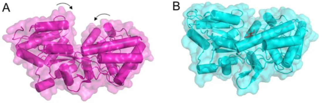
Figure 3.16 - Representation of MsmX-His6 and MsmX-LEHis6 3-D structure model……….53
Figure 3.17 - Ribbon representation of the MalK dimer………..53
Figure 3.18 - Representation of UgpC-His6 and YurJ-His6 3-D structure model………54
Figure 3.19 - Schematic representation of the modifications made in the genetic system………..55
Figure 3.20 - Schematic representation of the amyE locus and the yxkf-msmX operon in the chromosome of several B. subtilis strains, constructed with the redesigned genetic system, used in the work……….….56
Figure 3.21 - Growth of B. subtilis ISN10 (ΔmsmX::cat ΔamyE::Pspank(hy)-msmX-LEHis6) in CSK medium using arabinose and arabionotriose as the sole carbon and energy source………58
Figure 3.22 - Growth of B. subtilis ISN13 (ΔmsmX::cat ΔamyE::Pspank(hy)-ugpC-LEHis6) in CSK medium using arabinose and arabionotriose as the sole carbon and energy source………58
Figure 3.23 - Growth of B. subtilis ISN15 (ΔmsmX::cat ΔamyE::Pspank(hy)-yurJ-LEHis6) in CSK medium using arabinose and arabionotriose as the sole carbon and energy source………...59
Figure 3.24 - Growth of B. subtilis ISN11 (ΔmsmX::cat ΔamyE::Pspank(hy)-HD73_RS21400-LEHis6) in CSK medium using arabinose and arabionotriose as the sole carbon and energy source………....60
Figure 3.25 - Growth of B. subtilis ISN12 (ΔmsmX::cat ΔamyE::Pspank(hy)-msmK-LEHis6) in CSK medium using arabinose and arabionotriose as the sole carbon and energy source………61
Figure 3.26 - Growth of B. subtilis ISN14 (ΔmsmX::cat ΔamyE::Pspank(hy)-malK-LEHis6) in CSK medium using arabinose and arabionotriose as the sole carbon and energy source………61
Figure 3.27 - Growth of B. subtilis ISN17 (ΔmsmX::cat ΔamyE::Pspank(hy)-ycjV-LEHis6) in CSK medium using arabinose and arabionotriose as the sole carbon and energy source………61
Figure 3.28 - Representation of YurJ-H6 and YurJ-LEH6 3-D structure model……….………...62
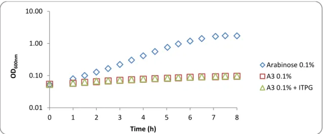
Figure 3.29 - Representation of UgpC-H6 and UgpC-LEH6 3-D structure model……….63 Figure 3.30 - Western-Blot analysis of B. subtilis cell extracts, obtained from CSK medium with
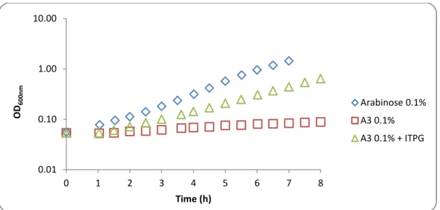
Figure 3.31 - Western-Blot analysis of B. subtilis cell extracts, obtained from CSK medium with arabinose 0.1% in non-inducing conditions………..65 Figure 3.32 - Western-Blot analysis of B. subtilis cell extracts, obtained from CSK medium with arabinotriose 0.1% in inducing conditions……….66 Figure 3.33 - Western-Blot analysis of B. subtilis cell extracts, obtained from CSK medium with arabinotriose 0.1% in non-inducing conditions………..67
Tables Index
Table 2.1 - List of plasmids used in this work………..……..….27
Table 2.2 - List of oligonucleotides used in this work………....28
Table 2.3 - List of B. subtilis strains used or constructed during this work…….………...29
Table 3.1 - List of MsmX homologs selected for this work…….………...35
Table 3.2 - Growth of different B. subtilis strains ISN1, IQB672 and IQB673 in the presence of distinct saccharides as sole carbon and energy source………..………….41
Table 3.3 - Growth of different B. subtilis strains IQB642, IQB677 and IQB678 in the presence of distinct saccharides as sole carbon and energy source………..……….43
Table 3.4 - Growth of different B. subtilis strains ISN8, ISN9 and ISN16 in the presence of distinct saccharides as sole carbon and energy source……….….44
Table 3.5 - Growth of different B. subtilis strains IQB673, IQB676 and ISN2 in the presence of distinct saccharides as sole carbon and energy source……….47
Table 3.6 - Growth of different B. subtilis strains ISN3, ISN4, ISN5, ISN6 and ISN7 in the presence of distinct saccharides as sole carbon and energy source……….……….49
Table 3.7 - Growth of different B. subtilis strains IQB676, ISN10, ISN13 and ISN15 in the presence of distinct saccharides as sole carbon and energy source………57
Table 3.8 - Growth of different B. subtilis strains ISN11, ISN12, ISN14 and ISN17 in the presence of distinct saccharides as sole carbon and energy source………..59
Abbreviations, Symbols and Notations
a. a. – Amino acidABC - ATP-binding cassette ADP – Adenosine diphosphate ATP – Adenosine triphosphate A3 – 1,5-α-L-Arabinotriose
bla - Beta-lactamase gene
BLAST – Basic Local Alignment Search Tool bp – Base pairs
cat – chloramphenicol acetyltransferase
gene
CRD – C-terminal regulatory domain CSK – C medium supplemented with potassium succinate
CUT1 – Carbohydrate Uptake Transporter-1 DTT - Dithiothreitol
dNTP – Deoxyribonucleic acid ECF – Energy coupling factor
EDTA - Ethylenediaminetetraacetic acid HRP - Horseradish peroxidase
IPTG – Isopropyl- β-D-galactopyranoside I-TASSER - Iterative Threading ASSEmbly Refinement
kDa - KiloDalton
LB – Luria-Bertani medium
LMW - Low Molecular Weight mRNA – Messenger ribonucleic acid MW – Molecular weight
NBD – Nucleotide-binding domain OD – Optical Density
ORF – Open Reading Frame PBS – Phosphate buffer saline PCR – Polymerase Chain Reaction Pi - Inorganic phosphate
PMSF – Phenylmethylsulfonyl fluoride PTS – Phosphotransferase system RBS – Ribosome binding site RNA – Ribonucleic acid rpm – Revolutions per minute SBP – Solute-binding domain
SDS-PAGE - Sodium dodecyl sulphate polyacrylamide gel electrophoresis
SpecR – Spectinomycin resistance gene
TIR - Translation initiation rate TAE - Tris-acetate-EDTA
TMD – Transmembrane domain
Tris - Tris(hydroxymethyl)aminomethane UV – Ultraviolet light
Amino acids – three and one letter code
Amino acid Three letter code One letter code
alanine ala A
arginine arg R
asparagine asn N
aspartic acid asp D
cysteine cys C
glutamic acid glu E
glutamine gln Q glycine gly G histidine his H isoleucine ile I leucine leu L lysine lys K methionine met M phenylalanine phe F proline pro P serine ser S threonine thr T tryptophan trp W tyrosine tyr Y valine val V
Bases – one letter code Base Letter Adenine A Citosyne C Guanine G Thymine T
Chapter 1
General Introduction
1. General Introduction
1.1. Bacillus subtilis – An overview
Bacillus subtilis is a Gram-positive and rod-shape bacteria that like other members of the
genus Bacillus, is able to form an endospore in order to survive in extreme environmental conditions. This bacterium is found in the soil, water sources or in association with plants and animal gastrointestinal tract (Priest, 1993; Casula and Cutting, 2002). In these environments the major source of carbohydrates for microorganisms is plant biomass, which is constituted by cellulose and hemicellulose polymers. Therefore B. subtilis, and the other microorganisms, from these habitats possess a wide variety of extracellular polysaccharide degrading enzymes, cellulases and hemicellulases. The resulting degradation products (mono-, di- and oligosaccharides) are imported to the cells through different transport systems, including specific ABC transporters.
Kunst et al in 1997 sequenced the entire genome of B. subtilis, opening the doors for a full use of this bacterium as a model of Gram-positive bacteria in fundamental and applied research. For example in industrial microbiology, specifically in “white biotechnology”, which relies on microorganisms and enzymes to synthesize products that are easily degradable, and during their production require less energy and create less waste (Chauhan et al, 2012). Carbohydrate-, lipid- and protein-degrading enzymes, antibiotics, fine biochemicals (vitamins) and insecticides (Harwood, 1992) are examples of the use of Bacillus spp in the “white biotechnology” associated with the vast diversity of the metabolism of this organism.
Hemicellulases and cellulases are the major industrially important enzymes right after the proteases (Polizeli et al, 2005; Dhawan and Kaur, 2007), due to the wide abundance of hemicellulose and cellulose in nature. Microbial mannanases, which degradate hemicellulose, are mainly produced by Gram-positive Bacillus species (Mabrouk and Ahwany, 2008; Meenakshi
et al, 2010) in extracellular medium. These enzymes can act in wide range of pH and
temperature, being used in multiple applications in pulp and paper, pharmaceutical, food, feed, oil and textile industries (Chauhan et al, 2012).
1.1.1. AraNPQ ABC importer and the Multitask MsmX ATPase
Quentin et al, 1999 performed in silico an inventory and assembly of the ABC transporter systems in the complete genome of Bacillus subtilis with an in silico analysis, estimating the existence of at least 78 ABC transporters based on the identification of 86 NBDs in 78 proteins, 103 MSD proteins and 37 BPD proteins representing 5% of the protein-coding genes of this organism. The uptake of sugars is undertaken by at least 10 ABC systems, being one of this importers the AraNPQ. The AraNPQ system is encoded by the araABDLMNPQ-abfA operon (Sá Nogueira et al, 1997), where AraN is the high-affinity substrate-binding protein (BPD) and AraP and AraQ are the two transmembrane domains (TMDs). In this operon are present genes encoding for enzymes that play a role in arabinose catabolism and degradation of arabinooligosaccharides (Inácio et al, 2008). This system is regulated at the transcriptional level by induction in the presence of arabinose and repression by glucose (Sá-Nogueira et al, 1997; Sá-Nogueira and Mota, 1997). AraNPQ is the sole transporter for arabinotriose, and α-1,5-arabinotetraose is only partially accountable for the uptake of α-1,5-arabinobiose (Ferreira and Sá-Nogueira, 2010)
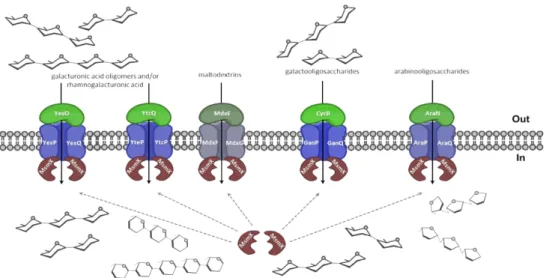
The AraNPQ transporter in the context of the arabinose operon lacks a gene encoding for the nucleotide-binding domains (NBDs) or ATPase, which is necessary to provide energy to the system. Ferreira and Sá-Nogueira (2010) identified the ATPase MsmX, encoded in a monocistronic gene in another locus of the chromosome, as the NBD partner of the AraNPQ transporter. Later, the MsmX was also identified as the NBD partner of the CycB-GanPQ transporter responsible for the uptake of galactooligosaccharides, and YesOPQ and YtcQP-YteQ transporters that are involved in the transport of galacturonic acid oligomers and/or rhamnose-galacturonic acid disaccharides (Ferreira and Sá-Nogueira, unpublished data). This ATPase is also related with another importer in B. subtilis, the maltose and maltodextrins transporter MdxEFG, in a previous study conducted by Schönert et al (2006). Clearly the data show that MsmX is a multitask ATPase shared by multiple sugar ABC transporters (Ferreira and Sá-Nogueira, 2010 and unpublished data; Schönert et al, 2006) (Figure 1.1).
Figure 1.1 – MsmX-dependent ABC importers in B. subtilis. The ABC-type importer AraNPQ is involved in the uptake of α-1,5-arabinooligosaccaharides (arabinotriose, arabinotetraose and some arabinobiose). The ABC-type importer MdxEFG is involved in the uptake of maltodextrins. The CycB-GanPQ ABC-type importer is responsible for the uptake of galactooligosaccharides. The YesOPQ and YtcQP-YteQ transport systems are involved in the uptake of galacturonic acid oligomers and/or rhamnogalaturonic acid (adapted from Ferreira and Sá-Nogueira, unpublished data).
1.2.
Membrane Transports
Selective permeability to nutrients and metabolites is an essential feature for cell survival, requiring transporters in the membrane with different characteristics in the function and structure, each one adapted to the type of solute translocated. Membrane transporters are distinguished based on the energy source used and therefore are classified in four major groups: protein channels, primary active transporters, secondary transporters and group translocators.
Channels allow the passage of solutes through facilitated diffusion in a process that is energy-independent. In Gram-negative bacteria, porin proteins form a TM-spanning aqueous pore constituting a channel in the outer-membrane that allow diffusion of several substrates. Another example are the cytoplasmic membrane channels which are gated and controlled by voltage (Ren et al, 2001) or membrane tensions, for instance the MscS channel in E. coli that function as a protector of the cells from hypo-osmotic shock (Levina et al, 1999; Bass et al, 2002; Davidson et al, 2008).
Primary active transporters constitute a large and diverse protein family that transport substrates across the membrane against a concentration gradient, which depends upon the energy withdrawn from chemical, electrical or solar energy sources. The largest and most widespread family of this class is the ATP-binding cassete (ABC) transporters that obtain energy through the hydrolysis of ATP molecules for the translocation of the substrate.
Secondary active transporters use energy provided by ion gradients to drive transport, being examples uniporters, antiporters and symporters (Davidson et al, 2008). Group translocation causes chemical changes in the substrates, being one example of this family the phosphotransferase system (PTS), which is exclusive to prokaryotes and plays an important role in the uptake of sugars. In this system the transported carbohydrate is phosphorylate once it reaches the cytoplasmic side of the membrane by phosphoenolpyruvate (PEP) that act as both the phosphate donor for sugar phosphorylation and the energy source for sugar accumulation (Postma et al, 1993; Robillard and Broos, 1999; Tchieu et al, 2001; Saier et al, 2002; Jaehme et
al, 2015).
1.3. ABC Transporters
1.3.1.
An overview
ABC-type transporters constitute a large and diverse superfamily of ATP-dependent protein complexes, which play an important role in organisms from the three domains of life Bacteria, Archaea and Eukarya (Eitinger et al, 2011). They are characterised by a highly conserved ATP-binding cassette that hydrolyses ATP molecules and provide free energy that is converted into trans-bilayer movement of substrates, by the transmembrane domains, as import to the cytoplasm or export from the cytoplasm (Locher, 2009). The type of substrates transported by this systems varies in a wide range from small inorganic and organic molecules, such as amino acids, sugars, nucleosides, vitamins and metal clusters to larger organic compounds, as peptides, lipid molecules, oligonucleotides and polysaccharides (Wilkens, 2015). The importance of the ABC transporters goes beyond the uptake of nutrients or export of toxic waste, for example ABC importers have important roles in the maintenance of cell integrity, responses to environmental stresses, cell-to-cell communication and cell differentiation, and in pathogenicity (Eitinger et al, 2011) and ABC exporters are involved in the drug resistance of
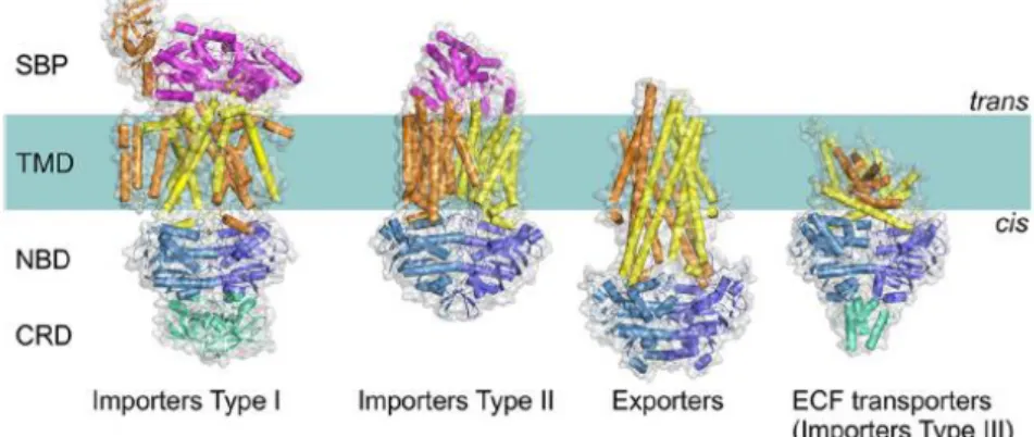
All ABC transporters have a core with the same modular architecture: two transmembrane domains (TMDs) or subunits and two nucleotide-binding domains (NBDs) or subunits. So far four types of ABC transporters have been identified based on the TMDs folds as determined by the crystal structures: importers type I and type II, ECF transporters (importer type III) and exporters. The available structures of ABC transporters provided fundamental knowledge about the transport mechanism and the great structural diversity of this systems (ter Beek et al, 2014).
1.3.2. Types of ABC Transporters
Canonical (protein-dependent) ABC transporters can be distinguish in two major groups: importers and exporters. Within the importers group there are two classes (I and II) and the energy coupling factor (ECF) transporters considered the third class of importers, although it is structurally and functionally more distinct (Eitinger et al, 2011 and Erkens et al, 2011). Until a few years ago it was believed that ABC importers only existed in bacteria and archaea, however recent studies showed evidence of the presence of class I and II Importers in plants, namely
Arabidopsis thaliana and Oryza sativa (Eitinger et al, 2011). ABC exporters exists in bacteria,
archaea and eukaryotes, being the only type of ABC transporters present in higher eukaryotes, and are involved in the transport of hydrophobic compounds such as lipids, fatty acids, cholesterol, drugs and large molecules as proteins (ter Beek et al, 2014). This type of transporters plays an important role in mammals since its defects are associated with several diseases such as, immune deficiency and cancer, cystic fibrosis, genetic conditions including Tangier and Stargardt disease (Wilkens, 2015). The following image (Figure 1.2) illustrates the four types of ABC transporters.
Figure 1.2 - Four distinct folds of ABC transporters. The components of the general architecture are
the two NBDs (blue and sky blue) that are attached to two TMDs (orange and yellow). Additional domains (green) that often have a regulatory function (C-terminal regulatory domain [CRD]) are present in some transporters. In Type I and II importers, the SBPs (or SBDs; magenta). ECF, energy coupling factor (Adapted from ter Beek et al, 2014).
ABC Importers were originally divided in two categories, type I and type II, due to differences in the size and overall architecture of the core of the transporters (Locher, 2009; Oldham et al, 2008), which is associated with the type of substrates transported. In general, importers type I contain less transmembrane helices than the type II, are smaller and use a single type of mechanism of transport the “alternating access model” (Jardetzky, 1966; Rice et al, 2014). This is associated with the transport of substances in bulk (although not exclusively) as amino acids and sugars (ter Beek et al, 2014), being examples the methionine transporter MetNI, the maltose transporter MalFGK2 and molybdate transporters ModBC (from Archaeoglobus fulgidus and Methanosarcina acetivorans) (Rice et al, 2014).
Importers of type II are more specialized in the transport of compounds in small quantities such as, metal chelates and vitamins (Davidson et al, 2008; Eitinger et al, 2011). Complexes that fall in this category are the vitamin B12 transporter BtuCD, the heme transporter HmuUV and the molybdate transporter MolBC (HI1470/1) from Haemophilus influenzae (Rice et
al, 2014), which possess differences in the transport mechanism that are also associated with
the substrate size ranging from vitamin B12 to molybdate (Rice et al, 2013 and 2014).
Energy coupling factor (ECF) transporters were recently considered as a third class of ABC importers (Rodionov et al, 2009), due to the energy withdrawn from ATP hydrolyse, although displaying significant difference with the other classes for instance in lacking a substrate-binding protein (SBP) presenting instead a EcfS or S component (ter Beek et al, 2014). These complexes have a critical role in micronutrient uptake in bacteria and archaea, being examples the folate and the hydroxymethyl pyrimidine transporters from Lactobacillus brevis (Wang et al, 2013; Xu et al, 2013; Rice et al, 2014).
Briefly there are ABC transporters that evolved to perform different functions than membrane transport, the so-called non-canonical ABC transporters, being examples the chloride channel CFTR, and the sulfonylurea receptor SUR (Wilkens, 2015).
The AraNPQ transport system, the system evaluated in this study, is an ABC importer type I so our focus is in this class of transporters.
1.3.3. Transport Mechanism
Type I importers mechanism of transport was first proposed by Jardetzky in 1966 (and further elaborated by Tanford, 1982) as the “alternating access” model and ever since other mechanisms with a few shared features were proposed, namely “switch” model (Higgins et al, 2004), and “constant contact” model (Sauna et al, 2007; Siarheyeva et al, 2010).
The maltose importer (MalEFGK2) from E. coli is one of the best-characterized Type I
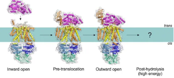
systems, and may be used to explain the transport mechanism employed by this type of transporters. Chen (2013) proposed a mechanism for the maltose transporter based on the “alternating access” model, deduced from the interpretation of X-Ray crystallography experiments that captured the complex in several conformations: inward-facing, pre-translocation, and outward-facing conformations (Figure 1.3). Biochemical experiments and structural studies using spectroscopic techniques such as, electron paramagnetic resonance, (Davidson et al, 1992; Chen et al, 2001; Lu et al, 2005; Grote et al, 2008, 2009; Orelle et al, 2008, 2010; Bordignon et al, 2010; Jacso et al, 2012; Böhm et al, 2013; Chen, 2013), support this proposed mechanism (ter Beek et al, 2014).
Figure 1.3 – Conformations of the MalEFGK2 transporter (class I importer). Structures have been
determined for the inward-facing, pre-translocation, and outward-facing conformations (Protein Data Bank accession nº: 4JBW, 4KHZ, and 4KI0). Adapted from ter Beek, et al (2014).
The proposed mechanism for type I importers, exemplified by the MalEFGK2 transporter,
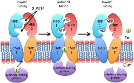
is the following (Figure 1.4): the binding of a substrate-loaded MBP (MalE) to the TMDs causes a conformational change in these domains that is propagated to the NBDs, bringing the two monomers (NBDs) into closer proximity (the pre-translocation state). This proximity allows the ATP to bind to the NDBs causing the dimer to close with two ATP molecules located at the interface, which in turn causes conformational changes in the TMDs allowing the cavity to open toward the outside (outward open/facing) and the substrate to binds to a specific site on the TM (MalF). The NDBs promote the hydrolysis of the ATP molecules and the consequent release of its products (Pi and ADP) triggers the dimer to move apart, that in turn propagate the conformational change to the TMDs allowing them to expose their cavity to the cytoplasm with subsequent release of the substrate (ter Beek, et al 2014).
Figure 1.4 – The transport mechanism of Type I importers (exemplified by MalEFGK2). The
inward-facing type I transporter (e.g., MalFGK2) binds to the substrate through the SPB. NBDs dimerize and result in the outward-facing conformation, allowing the substrate to contact with the TMDs. ATP is hydrolyzed and product release, together with NBD dissociation, resets the transporter to the inward-facing conformation. Adapted from Wilkens (2015).
1.3.4. The solute binding protein (SBP)
In Type I transporters (and also in Type II) the substrate is delivered to the transmembrane domains through a soluble substrate-binding protein (SBP) located in the trans-side of the membrane (Quiocho and Ledvina, 1996; Berntsson et al, 2010). The SBPs present in Gram-negative bacteria constitute soluble proteins with 30-40 kDa that are freely diffused through the periplasm, while in Gram-positive bacteria and archaea they are anchored to the membrane through a lipid or a separate TM helix (Sutcliffe and Russell, 1995; Biemans-Oldehinkel et al, 2006; Eitinger et al, 2011; Wilkens, 2015).
SPBs are able to bind to a wide variety of substrates therefore displaying different affinities that range from the nanomolar to the micromolar. The sequence and size of the SPBs are also determined by the type of binding subtract, however the general architecture is highly conserved with two symmetrical domains or lobs that are connected via a hinge region (Quiocho and Ledvina, 1996; Davidson et al, 2008; ter Beek et al, 2014; Eitinger et al, 2011).
The maltose-binding protein (MalE), a constituent of the maltose importer (MalEFGK2)
from E. coli, share the same mechanism of substrate binding than the others SPBs: the Venus fly trap model (Figure 1.5; Quiocho and Ledvina, 1996; ter Beek et al, 2014). Basically in the absence of a ligand, the two lobes adopt predominantly an open conformation that changes to a close conformation when the subtract binds to the SPBs becoming trapped inside. The substrate is released into the TMDs through the interaction of each lobe of the SPBs with the respective two domains of the TMDs.
Figure 1.5 - Rearrangements in SBP MalE upon the substrate binding. (A) In the substrate-free form (Protein Data Bank accession no. 1ANF), the cavity between two protein lobes connected by the hinge is accessible. (B) Upon the binding of substrate maltose (dark sticks; Protein Data Bank accession no. 1EZ9), the cavity becomes occluded. Adapted from ter Beek et al, 2014.
1.3.5. The transmembrane domains (TMDs)
The two transmembrane domains form a translocation pore in the membrane that allows the passage of the substrate from the cis-side to the trans-side. This domains are mainly constituted by 4 to 10 membrane-spanning α-helices (Eitinger et al, 2011) and in type I importers the two domains can be either identical (homodimers) or structurally similar (heterodimers). In the maltose transporter MalFGK2 the TMDs, MalF and MalG, displaying 8 and 6 helices,
respectively, form a heterodimer, which are structurally related but not sequence related since they only share 13% of amino acid sequence identity (ter Beek et al, 2014; Rice et al, 2014). The TMDs primary sequence is less conserved than the NBDs but they share a similar topology characteristic from each transporter class.
The transmembrane domain MalF plays an important role in the maltose transporter, besides the formation of the pore, due to two unique features: an additional loop (P2) that interacts with the SBP MalE and a substrate-binding site for the maltose (Oldham et al, 2007). The P2 loop acts like a receptor that recognize the SBP inducing an activated conformational change of the MalE, while maintaining the MalE and MalF in close contact throughout the catalytic cycle (Daus et al, 2009; Jacso et al, 2009 and 2012; Rice et al, 2014).
Upon the release of the substrate into the transmembrane domains, one maltose molecule binds to a unique site on the MalF domain composed by 10 residues that interact with the molecule through H-bonds, van der Waals interactions and aromatic ring stacking (Oldham
et al, 2007; Eitinger et al, 2011). These residues were identified from crystallographic studies
and mutagenesis experiments (Chen, 2013; Oldham and Chen, 2011).
MalG, the other transmembrane domain, also displays two important functions alongside with the formation of the translocation pathway. MalG P3 loop, the “scoop”, is inserted in the SPB binding site and promotes the displacement of the sugar, facilitating an efficient transfer into the membrane pore (Figure 1.6; Oldham et al, 2007). Another important interaction of the MalG is the insertion of its C-terminal tail into the MalK dimer interface (the ATPase), more specifically through the interaction with the Q-loop of each monomer, which may represent an important factor for the formation of the catalytic intermediate conformation of the entire transporter (Oldham et al, 2007).
Figure 1.6 - Transfer of the maltose from MBP to the TM binding site.Insertion of the MalG scoop loop into the substrate-binding site of MBP. A maltose molecule is modelled into the binding site on the basis of the crystal structure of open maltose-bound MBP (PDB accession number 1JW5). Adapted from Oldham et al, 2007.
An essential feature of the two TMDs MalF and MalG is the so called “coupling helices” that consists in two short helices per domain with a characteristic “EAA” motif. This feature is an architecturally conserved element that forms the NBD–TMD interface, where it contacts the Q-loop in the NBDs grooves (Figure 1.7; Locher, 2009; Oldham et al, 2007).
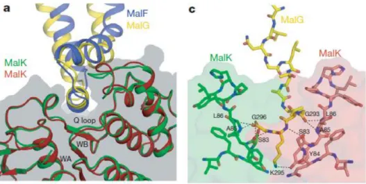
Figure 1.7 - The TMD–MalK interface. a) Docking of the EAA loops into a surface cleft of MalK. The
EAA loops of MalF and MalG are compared by superposition of the two MalK subunits. The MalK dimer is also shown as a transparent surface model. WA, Walker A motif; WB, Walker B motif. c) Insertion of the MalG C-terminal tail into the MalK dimer interface. The two MalK subunits are represented as a transparent surface model except for the interacting Q loops, which are shown in stick model. Hydrogen bonds and salt bridges are indicated by black dashed lines. Adapted from Oldham et al, 2007.
1.3.6. The nucleotide binding domains (NBDs)
The nucleotide binding domains (NBD), also called ATPases, are considered the “motor domains” of the ABC-type transporters, since they provide the necessary energy to induce the conformational changes in the TMDs. The NBDs exhibit a highly conserved structure and sequence with several conserved motifs among all ABC transporters, being considered the hallmark of the family. They function in the dimeric form and depend on magnesium ions for catalysis, each monomer consists of two subdomains: a RecA-like and an α-helical subdomain that are interconnected by two flexible loop regions. The RecA-like subdomain is found in other P-loop ATPases while the α-helical subdomain is unique to the ABC transporters andpresents a more structural diversity (Davidson et al, 2008). In the maltose transporter MalFGK2, the NBDs
are the two MalK units, whose genes are encoded in the transporter operon. MalK ATPase is a model for the NBDs structure in type I importers, as the whole transporter is a model for this type of importers.
NBDs can be identified at the sequence level by a specific set of seven highly conserved motifs (Figure 1.8; ter Beek et al, 2014):
(1) The A-loop helps to position the ATP molecule through stacking with the adenine ring of the conserved aromatic residue (usually a tyrosine);
(2) The P-loop or Walker A motif (GXXGXGK(S/T)) is a phosphate-binding loop that contains the highly conserved lysine residue, which form a network of interactions with two phosphates of the ATP molecule;
(3) The Walker B motif (φφφφDE, where φ is a hydrophobic amino acid) seems to perform two functions: helps to coordinate the magnesium ion via the conserved aspartate residue and polarizes the attacking water through the glutamate residue that functions as a general base (ex: maltose transport; Oldham and Chen, 2011);
(4) The D-loop (motif: SALD) directly follows the Walker B motif, and affect the geometry of the catalytic site helping to form the ATP hydrolysis site through conformational changes;
(5) The H-loop (or switch region) contains a highly conserved histidine residue that forms a hinge between a beta strand and an alfa helix near the C terminus of the NBD, holding together, through interaction the γ-phosphate, the attacking water and the catalytic glutamate for catalysis (Oldham and Chen, 2011);
(6) The Q-loop possess a conserved glutamine residue that binds to the Mg2+ cofactor in
the active site, therefore its mobility is essential to complete the catalytic cycle of the transporter (Daus et al, 2007). It is located at the interface between the RecA-like subdomain and the alfa-helical subdomain, as well as at the interface to the TMDs constituting a major site of interaction with the “coupling helix” of the TMDs (Dawson et al, 2007; Eitinger, et al 2011; ter Beek et al, 2014).
(7) The ‘LSGGQ’ motif (C-loop) is the ABC signature motif, representing a characteristic feature of the ABC superfamily (Schneider and Hunke, 1998). This motif is located in the α-helical subdomain, in the N-terminal end of a long helix that directs the positive charge of the helical dipole toward the γ-phosphate of ATP.
Figure 1.8 - The structure of the NBDs, as exemplified by the MalK dimer of the maltose transporter MalEFGK2 (Protein Data Bank accession no. 3RLF). (A) View along an axis perpendicular to
the membrane plane from the trans-side onto the NBDs (The TMDs and SBP have been removed for clarity). Domains and highly conserved sequence motifs are color-coded: green, α-helical domain; light blue, RecA-like domain; faded gray, regulatory C-terminal domain; red, A-loop; magenta, Walker A; orange, Walker B; blue, D-loop; green, H-loop; cyan, ABC motif; yellow, Q-loop. The ATP analogue AMP-PNP is shown in sticks. (B) The relative positions of sequence motifs in NBDs. Adapted from ter Beek et al, 2014.
B)
A)
In the outward-facing conformation of the maltose transporter the NBDs form a “sandwich dimer” with two molecules of ATP, where one molecule is bound to NBD1 coordinated by P-loop residues from NBD1 and from residues of the signature sequence of NBD2 and vice versa for the second ATP. The number of ATP molecules that need to be hydrolysed to accomplish a complete transport cycle is not universal for all ATP transporters (Davidson and Sharma, 1997).
The interconnection between the NBDs with the TMDs depend on two key elements: the Q-loop from the NBDs and the “coupling helix” from the TMDs (Figure 1.7; Wilkens, 2015). This two elements allow the transference of motion from the NBDs to the TMDs, that come from the rotational movement of the RecA-like domain with respect to the NBD helical domain during the catalysis, which was shown through crystallographic and EPR spectroscopy experiments with the maltose ATP-binding cassette transporter (Khare et al, 2009; Orelle et al 2010; Wilkens, 2015).
Maltose transporter is regulated by IIAglc, an enzyme from PEP-dependent sugar PTS
system, that inhibits transport activity or through the binding of MalT, a transcription factor (activator) that belongs to the maltose transporter (Boos and Shuman, 1998). The inhibition of the transport is accomplish by the binding of two IIAglc with the MalK dimer (Chen et al, 2013),
which prevents the closure of the MalK dimer and maintain MalFGK2 in the inward-facing resting
state (Rice et al, 2014).
1.3.7. Multitask ATPases
ATPases or the NBDs are essential components to the ABC-type transporters, however a closer look to the gene clusters of some species, that encode sugar ABC transporters, demonstrated that the nucleotide-binding domains sequences are occasionally absent from the cluster. Earlyevidence that an ATPase was capable of energizing more than one ABC transporter system was observed with the MsiK ATPase, whose gene was not encoded in an ABC transporter operon. This ATPase is shared by the cellobiose and the maltose ABC transport systems in
Streptomyces reticuli and S. lividans (Schlösser et al, 1997).
In B. subtilis, the MsmX ATPase is the multitask ATPase that provides energy to multiple ABC transporters (Ferreira and Sá-Nogueira, 2010 and unpublished data), while the msmX gene
is not encoded in the transporters operon but in a monocistronic message (see above, section 1.1.1).
Several reports have demonstrated that some sugar ABC transporters systems share an ATPase with greater incidence in Gram-positive bacteria (Schlösser et al, 1997; Ferreira and Sá-Nogueira, 2010; Marion et al, 2011b; Tyx et al, 2011; Tan et al, 2015;) however, the existence of this phenomenon in a Gram-negative bacteria is also observed (Silva et al, 2005; Chevance et al, 2006). So far the only examples of a multitask ATPase in a pathogenic bacteria is the MsmK ATPase in different species of the genus Streptococcus namely, S. pneumoniae (Marion et al, 2011b; Tyx et al, 2011), S. suis (Tan et al, 2015) and S. mutans (Webb et al, 2008).
1.3.7.1. MsmK, a multitask ATPase in Streptococcus species
S. pneumoniae or pneumococcus (a Gram-positive bacteria), is an opportunistic
respiratory human pathogen that can cause diseases as otitis media, meningitis and pneumonia, being the last two diseases a major cause of death. This pathogen possesses a vast ability in the utilization of carbohydrates which may provide a competitive advantage in the bacterial population of the nasopharynx. Since this organism uses as a carbon source only carbohydrates their import is ensured by 30% of the transport mechanisms of the organism encoded by the genome, being ABC transporters particularly important (Tyx et al, 2011, Buckwalter et al, 2012). There are six or seven (depending on the strain) predicted carbohydrate uptake transporter family 1 (CUT1) ABC importers within the pneumococcal genome, which lack in each locus a gene coding for an ATPase required to energize the transporter (Buckwalter et al, 2012). In S.
pneumoniae TIGR4 there are three ABC importers from CUT1 family, the RafEFG, SatABC and
MalXCD transporters, for which MsmK ATPase is the component that provides energy to the systems (Marion et al, 2011b). In S. pneumoniae, RafEFG is responsible for the uptake of raffinose (Rosenow et al, 1999), while SatABC transports sialic acid (Marion et al, 2011) and MalXCD provides the uptake of maltooligosaccharides (Puyet et al, 1993; Abbott et al, 2010). The MsmK ATPase also contributes to pneumococcal colonization of the S. pneumoniae, which suggests that transport of at least one of the carbohydrate substrates is important during colonization (Marion et al, 2011b). An example is the transport of sialic acid that was associated with cell signalling during the chain of events of the biofilm formation, colonization and host invasion of the organism (Trappetti et al, 2009; Marion et al, 2011).
In S. suis, an emerging important pathogen that is causing deadly infections in pigs and in humans, MsmK was also identified as a multitask ATPase that provides energy to MsmEFG and MalXCD transporters. Likewise, the ATPase gene is not encoded in the operons of these ABC transporters, and is responsible for the uptake of raffinose and melibiose through MsmEFG transporter and maltotriose, maltotetraose and maltodextrins (product of glycogen degradation) by MalXCD transporter, thereby contributing to the colonization and the in vivo survival of S. suis (Tan et al, 2015).
In Streptococcus species, namely S. pneumonia, S. pyrogens and S. suis, the CUT1 family of ABC importers are usually encoded in operons lacking the gene that codes for an ATPase. However, in S. mutans the MsmEFGK and MalXFGK carbohydrates transporters of the CUT1 family both possess a gene encoding for the respective ATPase (MsmK and MalK) together with the genes that encode for the two transmembrane proteins and the solute binding domain in the same operon (Webb et al, 2008). The multiple sugar metabolism system (MsmEFGK) is responsible for the uptake of melibiose, raffinose, isomaltotriose, stachyose and isomaltose (Russel et al, 1992; Tao et al, 1993), and the MalXFGK is involved in the uptake of maltotriose, maltotetraose and other maltodextrins which are zymolytic products of pullulan and glycogen (Webb et al, 2008). The special feature about these transporters is that in spite of having their own ATPase, when suppression of one of these proteins occurs the other protein is capable of providing energy to the alternative system (Webb et al, 2008). A schematic presentation of the
msm and mal loci encoding ABC transporters and a schematic of the carbohydrates utilized in S. mutans, S. pneumoniae and S. suis, are represented in Figure 1.9 and 1.10, respectively.
1.3.7.2.
Multitask ATPases from Streptomyces species and Thermus
thermophilus.
Streptomyces species hydrolyse chitin to oligosaccharides being (GlcNAc)2 and
chitosan the main products. In S. coelicolor A3(2) the (GlcNAc)2 uptake occurs via the ABC
transporter DasABC (Saito et al, 2007) and the chitosan-derived oligosaccharides uptake is accomplished by the ABC transporter CsnEFG (Viens et al, 2015). The respective operons of both transporters lack a gene encoding an ATPase, however it was demonstrated that the MsiK protein is the ATPase responsible for providing energy to the system (Saito et al, 2008; Viens et
al, 2015). In S. lividans and S. reticuli the MsiK ATPase is shared by two distinct ABC transporters
responsible for the uptake of the disaccharides cellobiose and maltose (Schlösser et al, 1997). Additionally in S. reticuli the MsiK ATPase was also associated with the ABC transporter responsible for the uptake of trehalose (Schlösser et al, 2000).
Figure 1.9 - Schematic presentation of the msm and mal loci encoding relevant carbohydrate ABC transporters in selected streptococci. The genes within the loci encoding components of the S. suis MsmEFG (A), S.
mutans MsmEFGK (B), S. pneumoniae MsmEFG (C), S. suis
MalXCD (D), S. mutans MalXFGK (E) and S. pneumoniae MalXCD (F) are represented. Arrows indicate the direction of transcription. Gray arrows, genes encoding the ATPase of ABC transporters; spotted arrows, genes encoding solute binding proteins; black arrows, genes encoding permeases of ABC transporters; blank arrows, other genes adjacent. Adapted from Tan et al, 2015
Figure 1.10 - Schematic summary of carbohydrates utilization by ABC transporters in S. mutans (A), S. pneumoniae (B) and S. suis (C). Above
each ABC complexes is a list of known or putative carbohydrates transported by each ABC transporter. Bidirectional arrow means the MsmK and MalK ATPases can energize permeases interactively. Unidirectional arrows mean the degradation of pullulan and glycogen by pullulanases SpuA or ApuA. Adapted from Tan et al, 2015
To date the phenomenon of a multitask ATPase is mainly associated with Gram-positive bacteria however, in spite of being less frequent, it also occurs in Gram-negative bacteria, as reported in Thermus thermophilus. The MalK1 ATPase, which is homologous to MalK from E. coli, and is encoded in a monocistronic gene is responsible for energizing two distinct ABC importers: the trehalose/maltose/sucrose/palatinose (TMSP) ABC transporter (Silva et al, 2005) and the glucose/mannose ABC transport system (Chevance et al, 2006).
1.4.
Scope of the Thesis
In this study we will characterize multipurpose ATPases from both Gram-positive and Gram-negative bacteria and assess their intra- and interspecies interchangeability in the host B.
subtilis. A genetic system is fine-tuned to test the ability of ATPases from other species to
complement MsmX function and establish B. subtilis as model for the study of bacterial multitask ATPases.
Chapter 2
Materials and Methods
2. Materials and Methods
2.1. Substrates
1,5-α-L-Arabinotriose (sugar beet, purity 95%) was purchased from Megazyme International Ireland Ltd., and arabinose from Sigma-Aldrich Co.
2.2. Bioinformatic Analysis
For the identification of MsmX homologs in other Gram-positive and Gram-negative bacteria, bioinformatics tools were used. BLASTp algorithm was the tool chosen to compare MsmX amino acid sequence with the sequence database from the National Center for Biotechnology Information at the National Institutes of Health, Bethesda, Maryland (http://www.ncbi.nlm.nih.gov). Proteins with an identity superior to 40% were considered as targets. An amino acid sequences alignment was made using the multiple sequence alignment program Clustal Omega (EMBL-EBI). Protein 3-D structures were predicted using the online program I-TASSER created by Zang Lab, University of Michigan (Zhang, 2008; Roy et al, 2010; Yang et al, 2015).
2.3. Isolation of chromosomal DNA
Chromosomal DNA was extracted from Escherichia coli K-12 strain based on the method described by Ferrari et al (1982). The strain was grown overnight (37 ˚C, 180 rpm), in liquid Luria-Bertani (LB) medium (Miller, 1972). All subsequent centrifugations were performed at 16060 g. The cells were harvested in two tubes with 2 mL of culture each by centrifugation for 2 minutes, washed once with 50 mM Tris and 5 mM EDTA, and resuspended in 175 µL of 50 mM Tris, 5 mM EDTA, lysozyme 1 mg/mL and RNase 20 µg/mL with incubation at 37 °C for 30 minutes. After, the solutions were vigorously agitated for 5 minutes and further incubated at the same temperature for at least 15 minutes, followed by a centrifugation step of 10 minutes. 100 µL of phenol (saturated with Tris-HCl, pH=8) was added to each tube, followed by centrifugation for 5 minutes. Each aqueous phase was recovered and mixed with 100 µL of chloroform:isoamyl alcohol (24:1), followed by centrifugation for 3 minutes. The upper phase from the two tubes, was mixed and two volumes of absolute ethanol were added. The precipitated DNA was
collected by centrifugation for 20 minutes at 4 °C, dried and further resuspended in 1X TAE buffer.
2.4. DNA manipulation and sequencing
Routine DNA manipulations were performed as described by Sambrook et al (1989). All restriction enzymes were purchased from Thermo Fisher Scientific Inc. and used according to the manufacturer’srecommendations. PCR amplifications were carried out using Phusion® high-fidelity DNA polymerase (Thermo Fisher Scientific Inc.). Oligonucleotides designed in this work or in previous experiments performed in our laboratory, were purchased from Metabion International AG or StabVida, Lda (Table 2.2). DNA from agarose gels and PCR products were purified with the illustra™ GFX™ PCR DNA and Gel Band Purification kit (GE Healthcare). All DNA ligations were performed using T4 DNA ligase (Thermo Fisher Scientific Inc.). Plasmids were purified using the NZYMiniprep kit (NZYTech, Lda). DNA was sequenced using the method of Sanger performed at StabVida, Lda.
2.5. Site-directed mutagenesis by Overlapping PCR
Chromosomal DNA of strain Escherichia coli K-12 was used as template for site-directed mutagenesis by primer extension, using mutagenic oligonucleotides ARA862 and ARA863 and flanking oligonucleotides. This pair of mutagenic primers allowed the insertion of a nucleotide C in codon 320, thereby restoring the wild-type phenotype of the protein YcjV with a total length of 360 amino acids. Primers ARA860 and ARA863 created fragment AB and primers ARA862 and ARA861 generated fragment CD, which through overlapping PCR resulted in a fragment of ycjV gene with HindIII and SphI restriction sites, provided by the flanking primers ARA860 and ARA861. Another fragment containing ycjV gene and NheI and BglII restriction sites, harbored in the flanking primers ARA845 and ARA846, was generated through the junction of fragment AB, created by primers ARA845 and ARA863, and fragment CD created by primers ARA862 and ARA846. For both experiments, two polymerase chain reactions were carried on using 1x Phusion®HF Buffer (Thermo Fisher Scientific Inc.), 0.5 µM primers, 200 µM dNTPs, 1.2 ng/µl of template genomic DNA (in the first reaction) and 0.2 ng/µl of Template DNA (in the second reaction: fragments AB, CD) and 0.05 U/µl of Phusion®High-Fidelity DNA Polymerase (Thermo
Fisher Scientific Inc.) in a total volume of 50 µl. The insertion of the nucleotide was confirmed by DNA sequencing.
2.6. Construction of plasmids and strains
Plasmid pPS1 was obtained by amplification of the HD73_RS21400 gene from chromosomal DNA of strain Bacillus thuringiensis serovar kurstaki str. HD73 (Bacillus Genetic Stock Center, BGSC, Ohio State University), with the oligonucleotides ARA851 and ARA852, bearing unique restriction sites NheI and BglII, and subsequent cloning of this fragment (1082 bp) into pSN74 digested with NheI and BglII. pPS2 was obtained by amplification of the ugpC gene, from chromosomal DNA of the pathogenic strain Staphylococcus aureus subsp. aureus ST398 (a gift from Hermínia de Lencastre, ITQB, Universidade Nova de Lisboa), using oligonucleotides ARA843 and ARA844, which harbor unique restriction sites NheI and BglII, and then the resulting fragment (1079 bp) was inserted between the NheI and BglII sites of pSN74. The amplification of the malK gene, from chromosomal DNA of strain Escherichia coli K-12, with oligonucleotides ARA847 and ARA848, which contain unique restriction sites NheI and BglII, and subsequent cloning of this fragment (1097 bp) into pSN74 NheI- BglII, yielded plasmid pPS3. Another amplification of the malK gene was made using oligonucleotides ARA858 and ARA859, which contain unique restriction sites HindIII and SphI, and cloning this fragment (1220 bp) into pDR111 (a gift from David Rudner, Harvard University) digested with HindIII and SphI, yielded plasmid pPS6. Plasmid pPS4 was obtained by amplification of the yurJ gene, from chromosomal DNA of the wild-type strain Bacillus subtilis 168T+, with oligonucleotides ARA837 and ARA838,
bearing unique restriction sites NheI and BglII, and subsequent cloning of this fragment (1085 bp) into pSN74 digested with NheI and BglII. Plasmid pPS5 was obtained by amplification of
msmK gene using pAM7 as template with oligonucleotides ARA855 and ARA749, which contain
unique restriction sites SalI and SphI, and subsequent cloning of this fragment (1241 bp) into pDR111 digested with SalI and SphI.
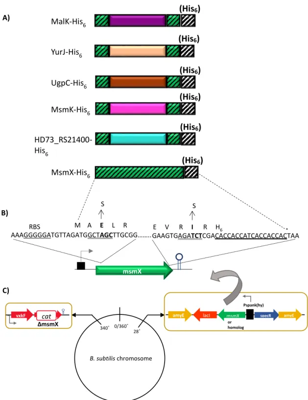
Plasmid pPS7 was obtained by amplification of a DNA fragment from pSN74 containing the lacI gene and the terminal end of the msmX gene with oligonucleotides ARA854 (mutagenic primer contain a unique restriction site BglII and two novel codons) and ARA632. This procedure introduced two novel amino acids (Leu, Glu) into the msmX coding region preceding the C-terminal His-tag were introduced into the msmX coding region of plasmid pSN74. The resulting amplification product was digested with BglII and BamHI and the product (1452 bp) sub cloned
into pSN74 BglII-BamHI, yielding pPS7. msmK gene was amplified from pAM7 with the oligonucleotides ARA773 and ARA774 (Sá-Nogueira, unpublished), which harbour unique restriction sites NheI and BglII, respectively, and digested with NheI and BglII resulting in a fragment with 1112bp. All genes of msmX homologs amplified as described above and digested with NheI and BglII were further cloned into pPS7 NheI-BglII, yielding the following plasmids: pPS8 (HD73_RS21400), pPS9 (msmK), pPS10 (ugpC), pPS11 (malK), and, pPS12 (yurJ). The amplification of ycjV gene from chromosomal DNA of strain E. coli K-12, obtained by site-directed mutagenesis (described in the previous topic), yielded a fragment with unique restriction sites HindIII and SphI and another fragment with unique restriction sites NheI and
BglII. Plasmid pPS13 was obtained by cloning a fragment (1191bp) containing ycjV gene, digested
with HindIII and SphI, into pDR111 HindIII-SphI. ycjV gene digested with NheI and BglII resulted in a fragment with 1064bp, being subsequently cloned into pPS7 NheI-BglII which yielded plasmid pPS14.
Plasmids and oligonucleotides used in this work are listed in Table 2.1 and 2.2, respectively. Oligonucleotides ARA411, ARA430, ARA442, ARA662, ARA741 and ARA841 were used for DNA sequencing (see Appendices 6.2 to 6.17)