MARIA DE FÁTIMA
CAMÕES SOBRAL DE
BASTOS
A NEUROTOXICIDADE DO ALUMÍNIO E SISTEMAS
NEURONAIS DE FOSFORILAÇÃO
ALUMINIUM NEUROTOXICITY AND NEURONAL
PHOSPHORYLATION SYSTEMS
MARIA DE FÁTIMA
CAMÕES SOBRAL DE
BASTOS
A NEUROTOXICIDADE DO ALUMÍNIO E SISTEMAS
NEURONAIS DE FOSFORILAÇÃO
ALUMINIUM NEUROTOXICITY AND NEURONAL
PHOSPHORYLATION SYSTEMS
tese apresentada à Universidade de Aveiro para cumprimento dos requisitos necessários à obtenção do grau de Doutor em Biologia, realizada sob a orientação científica do Prof. Doutor Edgar Figueiredo da Cruz e Silva, Professor Associado do Departamento de Biologia da Universidade de Aveiro
Apoio financeiro do POCTI no âmbito
do III Quadro Comunitário de Apoio. Apoio financeiro da FCT e do FSE no âmbito do III Quadro Comunitário de
o júri
presidente Prof. Dr. José Joaquim de Almeida Grácio
Professor Catedrático do Departamento de Engenharia Mecânica da Universidade de Aveiro
Prof. Dr. James Patrick O’Callaghan
Professor Associado, Center for Neuroscience, School of Medicine, West Virginia University, Morgantown, West Virginia, United States of America
Prof. Dr. Edgar Figueiredo da Cruz e Silva
Professor Associado do Departamento de Biologia da Universidade de Aveiro
Prof. Dra. Etelvina Maria de Almeida Paula Figueira
Professora Auxiliar do Departamento de Biologia da Universidade de Aveiro
Prof. Dra. Odete Abreu Beirão da Cruz e Silva
Professora Auxiliar da Secção Autónoma das Ciências da Saúde da Universidade de Aveiro
Prof. Dra. Diane Bemis Miller
Investigadora Principal, Chronic Stress and Neurotoxicity Laboratory, Health Effects Laboratory Division, National Institute of Occupational Safety and Health, Centers for Disease Control and Prevention, Morgantown, West Virginia, United States of America
agradecimentos Quero expressar um especial reconhecimento ao meu orientador Professor
Doutor Edgar F. da Cruz e Silva pelo imprescindível apoio científico, incentivo e acompanhamento permanentes que tornaram possível a realização do trabalho científico apresentado nesta dissertação.
Gostaria também de manifestar a minha gratidão à Professora Doutora Odete A. B. da Cruz e Silva pela colaboração na orientação deste trabalho científico e pela oportunidade que me deu em participar noutros projectos que
contribuíram para o meu enriquecimento científico.
Aos meus colegas de laboratório quero agradecer a ajuda que de uma forma ou de outra todos prestaram, assim como o companheirismo e a boa
disposição que manifestaram, e em particular a alguns com quem estabeleci verdadeiras relações de amizade.
Agradeço ao Centro de Biologia Celular e ao Departamento de Biologia da Universidade de Aveiro o bom acolhimento concedido. Agradeço ainda a todos os docentes e não docentes que de algum modo contribuíram para a
realização deste trabalho.
O desenvolvimento do trabalho experimental foi possível graças ao apoio financeiro das seguintes instituições:
- FCT – Programa PRAXIS XXI (Bolsa BD/11334/97)
- FLAD – Bolsa para acção de formação no estrangeiro (2004) - FCG – Bolsa de curta duração (2004)
Um agradecimento e reconhecimento muito especial á minha família pelo incentivo constante e apoio incondicional dado ao longo destes anos. A sua compreensão, carinho e ajuda permitiram-me realizar este sonho.
Este foi um período em que vivi dos momentos mais felizes da minha vida e passei pelos mais difíceis; agradeço a todos que me ajudaram a ultrapassar os obstáculos que foram surgindo e a levar esta etapa a bom termo.
palavras-chave Alumínio, neurotoxicidade, expressão de proteínas, proteína fosfatase 1,
neurofilamentos, cultura de células.
resumo O alumínio é o terceiro elemento mais abundante na Terra. Uma vez que se
encontra distribuído ubiquamente pelo meio ambiente e é utilizado em vários produtos e processos, a população humana está inevitavelmente exposta diariamente a este metal. De facto, o alumínio tem sido relacionado com diversas doenças neurodegenerativas como: a esclerose lateral amiotrófica, a demência de Parkinson (DP), a doença de Alzheimer (DA) e a encefalopatia relacionada com a diálise.
A fosforilação de proteínas é um dos principais mecanismos reguladores intracelulares da maior parte das vias de sinalização nas células eucarióticas. Este processo dinâmico regula o estado de fosforilação e/ou a actividade das proteínas através de um balanço entre as proteínas cinases, que fosforilam, e as proteínas fosfatases (PP) que desfosforilam as proteínas.
A proteína fosfatase 1 (PP1) é uma fosfatase específica para serina/treonina que está envolvida em importantes mecanismos celulares tais como o ciclo celular, contracção muscular e apoptose, entre outros. A PP1 tem três isoformas conhecidas, denominadas PP1α, PP1β e PP1γ,. O gene que codifica para a isoforma gama pode sofrer splicing alternativo originando a
isoforma ubíqua PP1γ1 e a isoforma enriquecida no testículo PP1γ2.
A fosforilação anormal de proteínas tem sido associada a várias patologias, incluindo cancro, diabetes e várias doenças neurodegenerativas (DP, doença de Huntington e DA). Uma das proteínas que se encontram anormalmente fosforiladas na DA são, por exemplo, os neurofilamentos (NF).
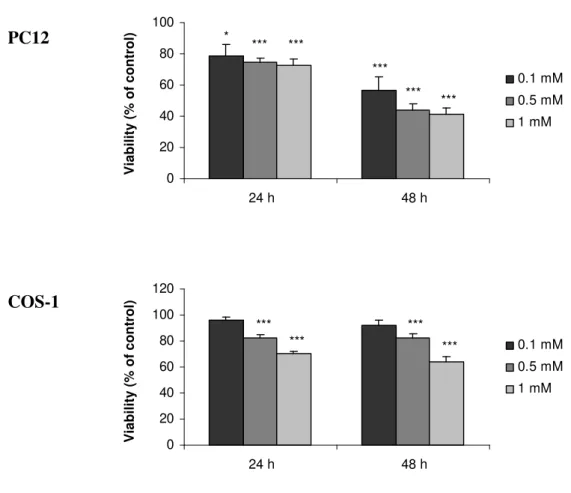
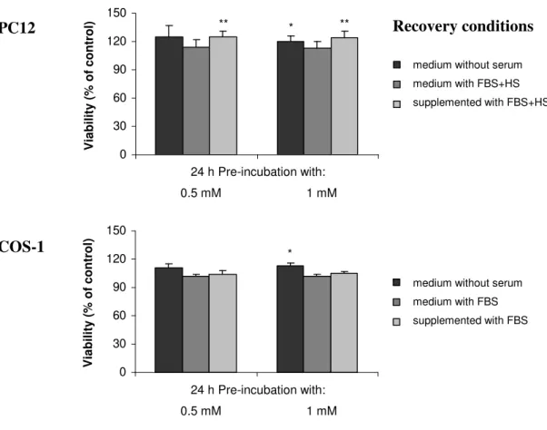
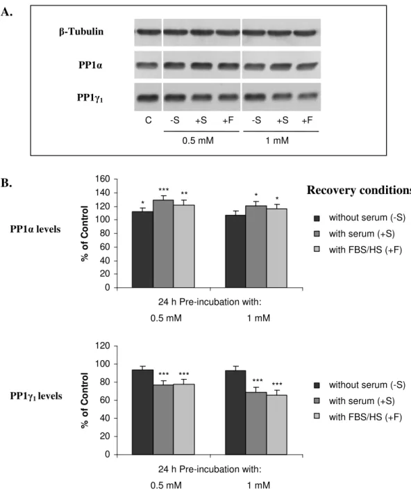
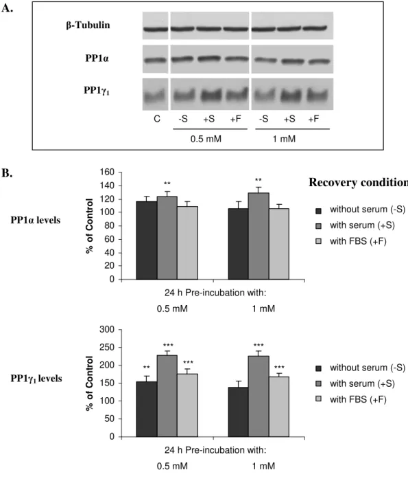
Neste contexto, avaliou-se o efeito do alumínio na expressão de proteínas (PP1, NF) e realizou-se um estudo comparativo entre duas linhas celulares com características diferentes, as células PC12 e COS-1. Observou-se que o alumínio induziu um decréscimo na viabilidade celular, assim como na
expressão e actividade de ambas as isoformas da PP1 em ambas as linhas celulares. Verificou-se que este efeito podia ser revertido retirando-se o alumínio. Um estudo semelhante foi ainda realizado num sistema neuronal utilizando culturas primárias de neurónios corticais. A expressão de ambas as isoformas da PP1 permaneceu inalterada após a exposição ao alumínio. No entanto, o alumínio induziu um decréscimo da expressão dos NF e da sinaptofisina, uma proteína que marca os terminais sinápticos. Por fim,
estudou-se o efeito do alumínio na expressão de outras proteínas em sistemas
in vitro e in vivo utilizando a tecnologia SELDI-TOF MS. Esta tecnologia
permitiu detectar várias proteínas cuja expressão foi alterada devido ao alumínio. Com este trabalho pretendeu-se contribuir para um melhor conhecimento da neurotoxicidade do alumínio.
keywords Aluminium, neurotoxicity, protein expression, protein phosphatase 1,
neurofilaments, cell culture.
abstract Aluminium is the third most abundant element on Earth. Aluminium is
ubiquitous in the environment and is used in a variety of products and processes, thus, daily exposure of the general population to this metal is unavoidable. Indeed, aluminium has been implicated with various neurodegenerative disorders like: amyotrophic lateral sclerosis
(ALS)/Parkinson’s dementia (PD) complex of Guam, Alzheimer’s disease (AD) and dialysis encephalopathy. Aluminium is known to interfere with several mechanisms of the nervous system, including alteration of cytoskeletal proteins, behavioural abnormalities, neurotransmission systems, oxidative damage, energy metabolism, second messengers, and also to induce neuronal apoptosis.
Protein phosphorylation is a major intracellular regulatory mechanism of all signalling pathways in the eukaryotic cell. This dynamic process regulates the net phosphorylation state and the activity of proteins by a balance between protein kinases, which phosphorylate, and protein phosphatases (PP), which dephosphorylate proteins. Protein phosphatase 1 (PP1) is a serine/threonine specific phosphatase which is involved in the control of important cellular mechanisms such as the cell cycle, muscle contraction and apoptosis, among others. PP1 has three known isoforms termed PP1α, PP1β and PP1γ. The gene for PP1γ produces by alternative splicing a ubiquitously expressed PP1γ1 and a testis-specific PP1γ2 isoform.
Abnormal protein phosphorylation has been associated with various disorders, including cancer, diabetes, and several neurodegenerative disorders (PD, Huntington’s disease and AD). Besides tau other proteins that are abnormally phosphorylated in AD are the neurofilaments (NF).
In this context, the aluminium effect on the expression of proteins (PP1, NF) was evaluated. A comparative study was performed using two cell lines with different characteristics, PC12 and COS-1 cells. It was observed that aluminium induced a decrease in the cellular viability, as well as in the
expression and activity of both PP1 isoforms in both cell lines. This effect was reverted following aluminium withdrawal. A similar study was also performed in a neuronal system, primary cortical neuron culture. The expression of both PP1 isoforms remained unchanged after aluminium exposure. However, aluminium induced a decrease in the expression of NF and of synaptophysin, a protein marker for synaptic terminals.
Finally, the effect of aluminium on the expression of other proteins in in vitro and in vivo systems was evaluated using SELDI-TOF MS technology. In this study, several proteins with altered expression due to aluminium were detected. This work aimed to contribute to the better understanding of aluminium neurotoxicity.
INDEX
PUBLICATIONS 11
ABBREVIATIONS 13
I. GENERAL INTRODUCTION 17
I.1.ALUMINIUMNEUROTOXICITY 17
I.1.1.ALUMINIUM BIOAVAILABILITY, HUMAN INTAKE AND BIOPROCESSING 17
I.1.2.ALUMINIUM SPECIATION CHEMISTRY 19
I.1.3.ALUMINIUM AND NEURODEGENERATIVE DISEASES 21
I.1.3.1.DIALYSIS ENCEPHALOPATHY 22
I.1.3.2.AMYOTROPHIC LATERAL SCLEROSIS AND PARKINSONISM-DEMENTIA COMPLEX OF GUAM 23
I.1.3.3.ALZHEIMER’S DISEASE 24
I.1.4.EFFECTS OF ALUMINIUM ON NERVOUS SYSTEM 28
I.1.4.1.CYTOSKELETAL PROTEIN AGGREGATES 28
I.1.4.2.BEHAVIOURAL ABNORMALITIES 29
I.1.4.3.CHOLINERGIC AND OTHER NEUROTRANSMISSION SYSTEMS 30
I.1.4.4.OXIDATIVE DAMAGE 31
I.1.4.5.ENERGY METABOLISM 32
I.1.4.6.SIGNAL TRANSDUCTION PATHWAYS (SECOND MESSENGERS) 32
I.1.5.ALUMINIUM-INDUCED NEURONAL APOPTOSIS 34
I.2.PROTEINPHOSPHORYLATION 36
I.2.1.PROTEIN PHOSPHORYLATION AS A DYNAMIC PROCESS 36
I.2.2.SERINE/THREONINE PROTEIN PHOSPHATASES 37
I.2.2.1.PROTEIN PHOSPHATASE 1 39
I.2.2.2.PROTEIN PHOSPHATASE 2A 42
I.2.2.3.PROTEIN PHOSPHATASE 2B 44
I.2.2.4.PROTEIN PHOSPHATASE 2C 45
I.2.2.5.NEW PROTEIN PHOSPHATASES:PP4,PP5,PP6 AND PP7 46
I.2.3.PROTEIN PHOSPHATASE INHIBITORS 46
I.2.4.ABNORMAL PHOSPHORYLATION OF PROTEINS 48
8
I.2.4.2.TAU PROTEIN 53
I.3.ALUMINIUMCONTRIBUTIONTOABNORMALPROTEINPHOSPHORYLATION
57
I.3.1.ALUMINIUM AND TAU 57
I.3.2.ALUMINIUM AND ABETA 58
I.3.3.ALUMINIUM AND NEUROFILAMENTS 60
I.4.AIMS 63
II. EFFECT OF ALUMINIUM ON PP1 EXPRESSION AND ACTIVITY IN PC12 AND
COS-1 CELL LINES 67
II.1.INTRODUCTION 67
II.2.MATERIALSANDMETHODS 68
II.2.1.CELL CULTURE 68
II.2.2.EXPERIMENTAL CELL TREATMENTS 68
II.2.3.CELLULAR VIABILITY 69
II.2.4.SDS-PAGE AND IMMUNOBLOTTING 70
II.2.5.PROTEIN PHOSPHATASE ACTIVITY ASSAYS 72
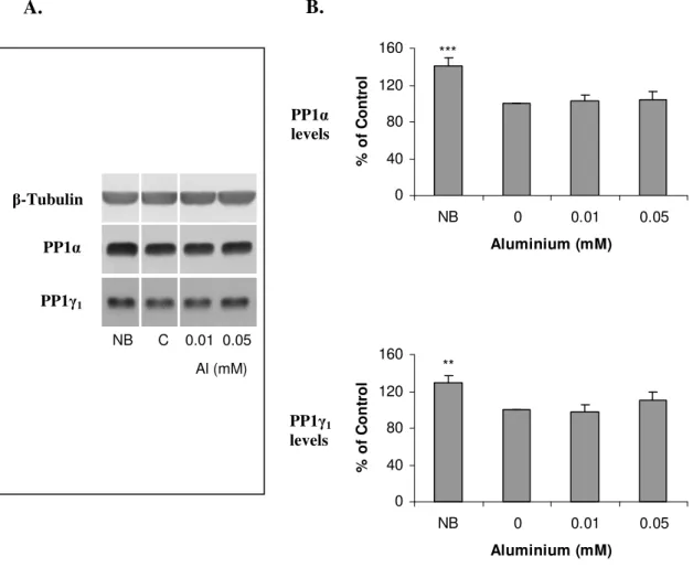
II.3.RESULTS 73
II.3.1.CELLULAR VIABILITY 73
II.3.2.PROTEIN PHOSPHATASE EXPRESSION 78
II.3.3.PROTEIN PHOSPHATASE ACTIVITY 88
II.4.SUMARYOFRESULTS 92
II.5.DISCUSSION 92
III. EFFECT OF ALUMINIUM ON PRIMARY CORTICAL NEURONAL CULTURES 99
III.1.INTRODUCTION 99
III.2.MATERIALSANDMETHODS 100
III.2.1.RAT CORTICAL PRIMARY CULTURES 100
III.2.2.TIME COURSE OF PROTEIN EXPRESSION 100
III.2.3.EXPOSURE OF CORTICAL NEURONS TO ALUMINIUM 102
III.3.RESULTS 102
III.3.1.TIME COURSE OF PROTEIN EXPRESSION 102
III.3.3.ALUMINIUM EFFECT ON PROTEIN EXPRESSION 107
III.4.SUMARYOFRESULTS 111
III.5.DISCUSSION 111
IV. SELDI-TOF MS ANALYSIS OF ALTERED ALUMINIUM-INDUCED PROTEOMIC
PROFILING 117
IV.1.INTRODUCTION 117
IV.2.MATERIALSANDMETHODS 117
IV.2.1.IN VITRO AND IN VIVO SAMPLE PROCESSING 118
IV.2.2.PROTEINCHIP ARRAY ANALYSIS 119
IV.2.3.SELDI-TOFMS ANALYSIS 120
IV.3.RESULTS 121
IV.3.1.ALUMINIUM-INDUCED ALTERED EXPRESSION OF PROTEINS IN VITRO 122
IV.3.2.ALUMINIUM-INDUCED ALTERED EXPRESSION OF PROTEINS IN VIVO 134
IV.4.SUMARYOFRESULTS 142
IV.5.DISCUSSION 143
V. CONCLUDING REMARKS 149
REFERENCES 153
APPENDIX 195
APPENDIXI-SOLUTIONS 195
APPENDIXII–KITS AND METHODS 206
PUBLICATIONS
Nesta dissertação foram utilizados resultados do trabalho publicado abaixo indicado. A autora declara que interveio na concepção e execução do trabalho experimental, na interpretação dos resultados e na sua redacção para publicação.
This thesis contains experimental results included in the publication indicated below. The author of this thesis declares that she participated in the planning and execution of the experimental work, as well as in data interpretation and in the preparation of work for publication.
Amador, Fátima Camões, Henriques, Ana Gabriela, da Cruz e Silva, Odete A. B., da
Cruz e Silva, Edgar F., Monitoring protein phosphatase 1 isoform levels as a marker for cellular stress. Neurotoxicol. and Teratol. (2004) 26, 387-395.
ABBREVIATIONS
Abeta ACN AD ALS ANOVA AP APS ATP BCA BCIP BSA Da DIV DMEM DTT E18 EAM ECL EDTA EGTA FBS (x) g HEPES HPLC HRP HS IC50 IgG Amyloid beta-peptide Acetonitrile Alzheimer’s diseaseAmyotrophic lateral sclerosis Analysis of variance Alkaline phosphatase Ammonium persulfate Adenosine triphosphate Bicinchoninic acid 5-Bromo-4-chloro-3-indolyl phosphate Bovine serum albumin
Dalton Days in vitro
Dulbecco’s modified Eagle’s medium Dithiothreitol
Embryo with 18 days (pre-natal) Energy absorbing molecule Enhanced chemiluminescence Ethylene diamine tetra acetic acid
Ethylene glycol-bis (β-aminoethylether)-N,N,N’,N’-tetra acetic acid Fetal bovine serum
Gravitational acceleration (when referring to centrifugation) N-2-hydroxyethylpiperazine-N’-2-ethanesulphonate
High pressure liquid chromatography Horseradish peroxidase
Horse serum
50% Inhibition concentration Immunoglobulin G
14 MAP MTT NBT NF-nonP NF-H NF-L NF-M NF-P NFT OD OGP P4 P7 PAGE PBS PD PHF PMSF PP RT SDS SEM SPA Tau TBS TBS-T TCA TEMED TFA Tris WR Microtubule-associated protein 3-(4,5-Dimethylthiazol-2-yl)-2,5-diphenyltetrazolium bromide Nitro blue tetrazolium
Neurofilament nonphosphorylated Neurofilament heavy chain
Neurofilament light chain Neurofilament medium chain Neurofilament phosphorylated Neurofibrillary tangles
Optical density
N-Octyl glucopyranoside Rat with 4 days (post-natal) Rat with 7 days (post-natal)
Polyacrylamide gel electrophoresis Phosphate buffer saline
Parkinson’s disease Paired helical filaments Phenyl methylsulfoxide Protein phosphatase Room temperature Sodium dodecyl sulphate Standard error of the mean Sinapinic acid
Microtubule-associated protein τ Tris buffered saline solution Tris buffered saline-Tween 20 Trichloroacetic acid
N,N,N’,N’-tetramethylethylenediamine Trifluoroacetic acid
Tris(hydroxymethyl) aminomethane Working reagent
GENERAL INTRODUCTION
I. GENERAL INTRODUCTION
I.1. ALUMINIUM NEUROTOXICITY
I.1.1. Aluminium bioavailability, human intake and bioprocessing
Aluminium is a ubiquitous element used extensively in contemporary life. In spite of its abundance, aluminium is not an essential element and there is no known biological reaction that requires aluminium. However, the neurotoxicity of aluminium has been recognized for many years. Aluminium is highly abundant and the third most abundant element ubiquitously found in the natural environment, its bioavailability is high and is readily accessible. Humans are exposed daily to aluminium from different sources. Food is the main source of aluminium intake, while drinking water contributes only about 3% of total daily intake. Aluminium in drinking water has two main sources: dissolved aluminium is present naturally as a result of leaching from minerals in the soil and bedrock in the catchment of the water source, this leaching can be greatly enhanced as a result of acid precipitation; and, aluminium is widely used in water treatment as a coagulant, to reduce the number of small particles and to improve the colour of the water. It has been proposed that the safe amount of aluminium in drinking water should be lower than 0.1 mg/l (Martyn et al., 1989). Aluminium is found in beverages such as tea infusions which contain rather large amounts of aluminium, typically 2-6 mg/l (Flaten and Odegard, 1988), and is also found in processed food which contains aluminium-containing food additives. Food can also be contaminated with aluminium by contact through cooking devices, or when wrapped with aluminium foil, especially with the addition of acids such as lemon juice or vinegar. Aluminium is also present in canned drinks and canned food coming from the package (Jagannatha and Valeswara, 1995; Jagannatha, 1994). Humans are also in contact with aluminium through hygienic products like toothpaste and specially anti-perspirants, which have considerable amounts of aluminium that can be absorbed through the skin (Exley, 2004). In medicine, aluminium is used in several medications like
18
buffered aspirin, but especially in antacids used for the treatment of gastric ulcers, with typical consumption in the order of 1 g per day (Lione, 1985; Reinke et al., 2003). Aluminium is also used in dental prothesis and as adjuvant in several vaccines (Brewer, 2006). Another potentially important source of exposure to aluminium is occupational exposure (McLachlan, 1995).
Exposure to aluminium is unavoidable during the entire life span. Humans consume an average of 7.6 mg/day of aluminium from drinking water and food (Yokel and McNamara, 2001). However, the absorption rate of aluminium is relative low, only 0.06-0.1% of ingested aluminium is absorbed across the gastrointestinal tract (Moore et al., 2000). The gut is therefore the first barrier for aluminium uptake. Aluminium absorption is limited by the presence of certain other dietary components such as citrate, which can form a complex with the metal increasing significantly its absorption (Whitehead et al., 1997). Age contributes to gut barrier impairment and so to the aluminium absorption. Indeed, it was reported that young individuals absorb much less aluminium from an aluminium citrate drink than older people (Taylor et al., 1992). In the bloodstream, aluminium binds to the plasma proteins, transferrin (the main protein carrier) and albumin, the remainder complexes with citrate (the main small molecule carrier) (Martin et al., 1987; Fatemi et al., 1991). Aluminium in circulation may distribute through all the body. The main target for accumulation is the skeleton (Kerr et al., 1992), although, liver, kidney, muscle and heart also accumulate aluminium (Walker et al., 1994). It has been proposed that aluminium could enter the brain from systemic circulation by three different routes: blood-brain barrier (BBB), nasal-olfactory pathway (by olfactory nerves) and cerebrospinal fluid (through the choroid plexus) (Perl and Good, 1987; Yokel et al., 1999). The BBB route is generally considered to be the most plausible mode of entry (Yokel, 2002). The potential mechanisms of distribution of substances across the BBB, the second barrier to aluminium, are the same as those across any cell membrane: diffusion, carrier/receptor mediated transport by facilitated diffusion and active transport. One of the most probable mechanisms of aluminium access into the brain is by transferrin-receptor-mediated endocytosis (Roskams and Connor, 1990), the iron transport protein by excellence. Another important carrier for brain aluminium influx may be monocarboxylate transporter, a proton co-transporter located at the BBB (Gerhart et al., 1997; Yokel et al., 2002). However, aluminium alone affects the permeability of the BBB enhancing its
apparent lipophilicity, thus contributing to an increase of its transmembrane diffusion (Meiri et al., 1993), although this depends on the physicochemical properties of the metal coordination sphere (Favarato et al., 1992). In the brain, aluminium competes with other elements, such as Fe, Ca and Mg, present in several proteins and enzymes altering their function. Indeed, some aluminium persists in the brain for a long time. Brain aluminium efflux, presumably Al-citrate, was suggested to be mediated by the monocarboxylate transporter, through the BBB (Yokel et al., 1999). Absorbed aluminium is primarily eliminated via the kidneys and approximately 2% excreted in bile (Alfrey, 1986a; Yokel and McNamara, 2001).
I.1.2. Aluminium speciation chemistry
The chemical speciation of aluminium in aqueous solution is of particular interest, as the form of aluminium regulates its solubility, bioavailability and consequently its toxicity. One factor determining the form of aluminium in solution is pH (Figure I.1). Aluminium is a strong hydrolyzing element and is generally insoluble at neutral pH. Its solubility is enhanced under acidic or alkaline conditions and in the presence of appropriate ligands. Thus, in the range of physiologic pH values (between 6 and 8) aluminium is generally insoluble.
Figure I.1 – Distribution of the hydrolysis products of aluminium as a function of pH (adapted from
Meiri et al., 1993). Pe rc e n t o f to ta l (1 0 0 µ M ) pH 100 80 60 40 20 0 0 2 4 6 8 10 12 Al(OH)4 -Al3+ Al(OH)3 Al(OH)2+ Al(OH)2+
20
In acidic solutions (pH<5), aluminium exists as an octahedral hexahydrate, Al(H2O)63+, usually abbreviated as Al3+ and sometimes referred in the literature as free
aluminium. As pH increases, Al(H2O)63+ undergoes successive deprotonations to yield
different species such as Al(OH)2+ and Al(OH)2+ with decreasing solubility. In neutral pH
solutions, the amorphous Al(OH)3 is produced and precipitates. At alkaline pH this
precipitate redissolves to form tetrahedral aluminate, Al(OH)4-, the primary soluble
aluminium species at a biological pH>6.2. The main species at pH<5 is the octahedral hexahydrate, Al(H2O)63+ and at pH>6.2 the tetrahedral Al(OH)4-. At pH between 5 and 6.2
all species co-exist. Thus, at pH 7.4 the main species is the insoluble Al(OH)3 although,
the prominent soluble species is the Al(OH)4- (Martin, 1986; Meiri et al., 1993; Gupta,
2005).
It should be pointed out that a solution of AlCl3 at physiological pH contains free
aluminium (Al3+) at concentrations much lower than the indicating concentration, because AlCl3 forms insoluble hydroxyl complexes at that pH. Indeed, when aluminium inorganic
salts, such as chloride, sulphate, hydroxide or perchloride, are dissolved in water at a calculated concentration of 10 mM, the aluminium concentration is about 50 µM. The use of Al-lactate or Al-aspartate increases the soluble aluminium concentration to approximately 55-330 µM and the use of Al-maltolate or gluconate increases the soluble aluminium concentration to 4-6 mM. Aluminium complexes of low solubility can be biologically relevant; however, free aluminium is the species which is bioactive, the species that links to proteins or ligands, even at low concentration (Martin, 1986; Meiri et
al., 1993; Gupta, 2005). Thus, the solution concentration is merely informative, as at
physiological pH the bioactive aluminium concentration is much lower.
Aluminium bioavailability, concerning diet and intestinal absorption, depends on which complexes it forms. Several compounds in the diet, including ascorbic acid, citric acid, lactic acid and malic acid, may increase aluminium absorption in the intestine by elevating the pH of aluminium hydroxide precipitation (Partridge et al., 1989). On the other hand, phosphate is also an important dietary factor, forming complexes even at low pH and making aluminium less available for absorption (Driscoll and Schecher, 1988). It has been suggested that the presence of phosphates in the diet is probably the “natural” mechanism whereby aluminium is prevented from entering the circulation (Martin, 1986). Aluminium has also been reported to displace other ions of physiological relevance.
Aluminium is a small ion with an ionic radius of 54 pm and can replace divalent metals such as Ca, Mg and Zn, whose ionic radii are 72, 74 and 100 pm, respectively, and hence is thought to be responsible for executing various toxic effects (Martin, 1996).
Aluminium binds strongly to oxygen-donor ligands, particularly if they are negatively charged. Inorganic or organic phosphates, carboxylate and deprotonated hydroxyl groups are strong Al3+ (aluminium) binders. Thus, aluminium binds to the phosphor groups of DNA or RNA, influences DNA topology and affects gene transcription (Lukiw et al., 1998). Phosphate groups of cell membranes are also targets for aluminium binding (Van Rensburg et al., 1995). As well as phosphorylated proteins, aluminium has been reported to influence various functions of enzymes including protein kinases and phosphatases (Shetty et al., 1992; Amador et al., 2004). Overall, the form and speciation of aluminium may be critical to its biological actions.
I.1.3. Aluminium and neurodegenerative diseases
Acute aluminium exposure is of low toxicity. In humans, oral doses up to 7.2 mg/day are routinely tolerated without any signs of harmful short-term effects. However, intake of large amounts of aluminium can lead to a wide range of toxic effects, including microcytic anaemia (Touam et al., 1983; Jeffery et al., 1996; Garbossa et al., 1998), osteomalacia (Bushinsky et al., 1995; Jablonski et al., 1996; Jeffery et al., 1996), glucose intolerance of uraemia (Banks et al., 1987) and cardiac arrest (Starkey, 1987). Elderly persons with elevated serum aluminium levels exhibit impaired complex visual-motor co-ordination and poor long-term memory (Bowdler et al., 1979).
Evidence suggests that trace metal homeostasis plays a crucial role in the normal functioning of the brain and any disturbance in it can exacerbate events associated with neurodegenerative disorders. In fact, aluminium has been implicated in several neurological and other disorders, namely dialysis encephalopathy (Alfrey et al., 1976; Savory and Wills, 1984; Kerr and Ward, 1988), amyotrophic lateral sclerosis (ALS)/Parkinsonism-dementia (PD) complex of Guam (Perl et al., 1982; Kurland, 1988) or Alzheimer’s disease (AD) (Perl and Brody, 1980; Perl, 1988; Xu et al., 1992a).
22
I.1.3.1. Dialysis encephalopathy
Dialysis encephalopathy is a fatal brain disorder occurring in some patients with chronic renal failure undergoing inadvertent parenteral exposure to aluminium (Alfrey et
al., 1980). The chronic symptoms include speech disorders, neuropsychiatric
abnormalities and multifocal myoclonus (Dewberry et al., 1980). More subtle symptoms of the condition include disturbances of tetra-hydrobiopterin metabolism and abnormalities in a number of psycho-motor functions (e.g., visual spatial recognition memory), all occurring at mildly elevated serum aluminium levels (59 µg/l) and in the absence of chronic dementia (Altmann et al., 1989). Patients with dialysis dementia were shown to have markedly elevated serum aluminium levels with increased concentrations in many tissues namely, kidney, liver, bone, heart and throughout the cerebral cortex (Alfrey et al., 1980; Kerr and Ward, 1988; Meiri et al., 1993). Investigators reported a correlation between the aluminium concentration in water used to prepare the dialysate fluid and the incidence of dialysis dementia (Savory and Wills, 1984). Tissue accumulation of aluminium to levels high enough to cause toxicity is mainly due to a combination of high exposure, partly directly into bloodstream (thus bypassing absorption in the gastrointestinal tract) and these patients’ lack of kidney function, which is the main excretion route for aluminium. The mechanism of neurotoxicity in dialysis encephalopathy has not been established. However, severe acute aluminium intoxication cases have been reported to respond to chelation therapy with desferrioxamine to lower serum aluminium, combined with hemodialysis (Vaan Landeghem et al., 1997; Nakamura et al., 2000). Indeed, desferrioxamine has been reported to accelerate clearance of brain aluminium (Yokel et al., 2001). It was also proposed that ascorbate, desferrioxamine and Ferralex-G in combination as a “molecular shuttle chelation” may provide a useful pharmacotherapy in the potential treatment of aluminium overload disease (Kruck et al., 2004). Dialysis encephalopathy syndrome resulting from acute intoxication of aluminium caused by the use of an aluminium-containing dialysate was common occurrence prior to 1980. However, using modern techniques of water purification, such acute intoxication can now be avoided (Rob et al., 2001). Nevertheless, aluminium toxicity is a known adverse effect in patients with end-stage renal disease due to oral intake of aluminium-containing phosphate binders (Wills and Savory, 1989). Indeed, a fatal case of aluminium
encephalopathy in a patient with severe chronic renal failure not on dialysis but due to intake of large doses of antacids containing aluminium for at least 3 years, was recently reported (Zatta et al., 2004). A similar report of an autopsy of a 59-year-old female aluminium encephalopathy patient who had chronic renal failure and took 3.0 g hydroxy-aluminum gel per day for the control of serum phosphorus level during a 15-year period, was published by Shirabe and colleagues (2002). Furthermore, it was reported recently significant aluminium toxicity in a non-hemodialysis patient who chronically injected intravenously oral methadone solution heated in an aluminium-based cooking utensil (Friesen et al., 2006).
The aluminium contamination of total parenteral nutrition (TPN) solutions is also a matter of great concern. It was reported that the neurological development of premature infants who had received a TPN solution containing a high level of aluminium was impaired compared with infants who had received an aluminium-depleted TPN solution (Bishop et al., 1997). Considering that aluminium in TPN solutions is highly bioavailable and that renal function of infants is impaired, the aluminium contamination of TPN solutions may cause serious brain damage (Kawahara, 2005).
I.1.3.2. Amyotrophic lateral sclerosis and Parkinsonism-dementia complex of
Guam
It has been postulated that aluminium plays a role in the aetiology of two severe neurodegenerative diseases, amyotrophic lateral sclerosis (ALS) and Parkinsonism-dementia (PD) complex of Guam. ALS/PD are characterized by the selective degeneration of motor neurons, the presence of neurofibrillary tangles (NFT) and neuropil threads in the brain, these features result in a clinical syndrome of progressive weakness, culminating in respiratory failure and death (Wakayama et al., 1993a). Both diseases are observed at very high incidence among the Chamorro people of Guam (Garruto et al., 1990; Oyanagi, 2005). A high incidence of ALS is also found in two other areas, western New Guinea and the Kii Peninsula of Japan (Yase et al., 2001). The soils and drinking water of Guam and the two other affected areas are very low in calcium and magnesium but very high in aluminium, iron and silicon (Gajdusek and Salazar, 1982). Aluminium was found to accumulate in the tangle-bearing neurons in post-mortem brains of patients with ALS/PD
24
(Perl et al., 1982). Garruto and Yase (1986) suggested that chronic nutritional deficiencies of calcium and magnesium may lead to increased absorption of aluminium (and other metals), resulting in the deposition of aluminium in neurons. These deposits could interfere with the structure of neurons and eventually result in NFT (Garruto, 1989). The elevated concentrations of aluminium in ALS/PD patients suggested that aluminium and the depletion of calcium and magnesium may play key roles in the pathogenesis of ALS/PD (Yase et al., 2001). The dramatic decrease in the incidence of ALS/PD on Guam with a change in dietary habits and local water supplies has given support to this theory (Garruto et al., 1990). Although, the remarkable clustering of motor neuron diseases (MND) was thought to have disappeared, the southern Kii Peninsula remains a high-risk area for MND, especially if the emigrants who developed MND one to four decades after leaving the focus are included (Yoshida et al., 1998). It has been reported that experimental animals chronically fed a low-calcium and/or magnesium and high-aluminium diet showed neuronal loss in the spinal anterior horn and cerebral cortices (Florence et al., 1994). Further mice with a similar diet (low-Ca/Mg and high-Al) exhibited the deposition of aluminium, the deposition of hyperphosphorylated tau proteins (the basic structure of NFT) and neuronal loss (Kihira et al., 2002). Recently, it was also reported that mice fed a similar diet also exhibited ALS-like skin and CNS changes (Kihira
et al., 2004).
I.1.3.3. Alzheimer’s disease
The association of aluminium with Alzheimer’s disease (AD) has more than 25 years, however it is still a controversial issue and the mechanisms of aluminium toxicity in this disease are not yet established. AD is a neurological disorder affecting elderly people, as first described by Alois Alzheimer in 1906. Patients with AD exhibit progressive mental deterioration manifested by memory loss, inability to calculate, visual-spatial disturbances, confusion and disorientation. The neuropathological characteristics include: cortical and subcortical atrophy; intraneuronal accumulation of neurofibrillary tangles (NFT), which are composed of paired helical filaments (PHF) of hyperphosphorylated tau proteins (Figure I.2); dystrophic neurites surrounding the extracellular deposits of amyloid beta-peptide (Abeta) in plaques (neuritic plaques or senile plaques) (Figure I.2); formation
of neuropil threads; loss of synaptic function; oxidative stress and apoptosis, leading to neuronal loss (Glenner and Wong, 1984; Grundke-Iqbal et al., 1986a; Markesbery, 1997; Christen, 2000; Dickson, 2004; LeBlanc, 2005). These events are observed mostly in the hippocampal and cortical regions of AD brains. The etiological factors of AD are not clearly elucidated, although current hypotheses include genetics, head trauma, oxidative stress, infectious agents and environmental factors, including aluminium toxicity.
Figure I.2 – Neurofibrillary tangles and senile plaques. In both AD hallmarks aluminium was found to
accumulate.
One of the first studies relating aluminium with AD was published in 1973 by Crapper and colleagues (Crapper et al., 1973). In this study they demonstrated that AD brain tissues showed a 2 to 3-fold increase in aluminium concentrations when compared to normal control tissues. Later, it was reported that the aluminium content of human brain is around 6.2-9.8 µg/g (dry mass brain) (Xu et al., 1992a). However, it has been reported that AD patients have elevated concentrations of aluminium (9.0-11.0 µg/g dry weight) in some regions (cortex, mesial temporal and temporal cortex) of their brains compared to controls (Crapper et al., 1973; Solomon et al., 2001; Andrasi et al., 2005; Gupta et al., 2005). Aluminium tends to accumulate more in the cortex and hippocampus, both in normal and AD brains (McDermott et al., 1979). Aluminium deposition is progressive, being higher in severe AD than in moderate AD and relatively low in normal brain (Jagannatha et al., 1999). Even though, the increased content of aluminium in NFT has been established (Perl and Brody, 1980; Lovell et al., 1993; Shin et al., 1994; McLachlan, 1995; Tokutake et al., 1995), the presence of aluminium and silicon in the central region of
26
senile plaque cores in the cortex of AD patients is a controversial issue. Landsberg et al., (1992) stated that they could not find aluminium in senile plaques from autopsy AD brain material, and hypothesized that the occurrence of aluminium found previously in plaques by Candy and colleagues (1986) had been caused by contamination from dyes used to stain the plaques. However, Good and Perl (1993) stated that the Landsberg and co-workers (1992) study did not contradict the aluminium hypothesis, since it was clear from the literature that aluminium was more often associated with the NFT than with the plaques (Zatta, 1993). Indeed, Tokutake and co-workers (1995) found aluminium contained in lipofuscin granules with silicon, probably as aluminosilicate, in senile plaques of brain with AD. Moreover, Exley (2005) reported the detection of aluminium associated with Abeta in AD brains. Overall, whether the presence of aluminium in the brain of AD patients is a cause or a result of the condition remains unknown.
Moore and co-workers (2000) showed that under normal physiological conditions, the ability of the gastrointestinal tract to exclude aluminium is reduced in AD, possibly leading to greater systemic exposure to aluminium. Additionally, a higher frequency of a genetic variant of transferrin, TfC2, has been found in AD patients compared with non-demented controls, suggesting that this factor may be involved in an aberrant transport of aluminium in these patients (Van Rensburg et al., 1995; Zambenedetti et al., 2003). Moreover, ferritin, the iron storage protein, isolated from the brains of AD subjects, has 6-fold higher aluminium content than normal age-matched controls (Fleming and Joshi, 1987). Indeed, aluminium has been shown to accumulate in rat brain ferritin (Sakamoto et
al., 2004), which has been reported to be a component of the senile plaques in AD
(Grundke-Iqbal et al., 1990). From this evidence we may conclude that the aluminium absorption, uptake and accumulation are augmented in patients with AD. On the other hand, the incidence of AD is increased in regions where people are more exposed to aluminium. Although, drinking water contributes only with a minor portion of the total daily oral intake of aluminium, it was found that a significant relationship exists between the number of AD cases and the levels of aluminium present in drinking water (McLachlan
et al., 1996; Gauthier et al., 2000; Flaten, 2001). Additionally, two metal chelation
therapies have been proposed for AD patients, one using Feralex-G, and another using desferrioxamine (Shin et al., 2003; House et al., 2004) A “molecular shuttle chelation” using both desferrioxamine and Feralex-G in combination with ascorbate was also
proposed as a therapy for aluminium overload diseases that may include AD patients (Kruck et al., 2004).
The aluminium hypothesis has also been disputed based on the following features: (i) not all patients with AD have high brain levels of aluminium and the senile plaques that are common in AD are not seen in experimental aluminium toxicity (Wisniewski et al., 1980; Bjertness et al., 1996); (ii) the incidence of cognitive impairment and AD symptoms is not increased, but only dialysis dementia in renal patients is observed with increased aluminium levels (Alfrey, 1986b); (iii) it has been shown that NFT in AD, which are composed primarily of paired helical filaments (PHF) are made up mainly of hyperphosphorylated tau (a microtubule associated protein), in contrast to aluminium-induced neurofibrillary degeneration (NFD), which consists of aggregated hyperphosphorylated neurofilament (an intermediate filament of the neuronal cytoskeleton) (Munoz-Garcia et al., 1986; Erasmus et al., 1993). However, abnormally phosphorylated tau has been found in aluminium-induced neurofilamentous aggregates, together with abnormally phosphorylated neurofilament protein (Singer et al., 1997; Huang et al., 1997). Also, elevated levels of phosphorylated neurofilament proteins were found in cerebrospinal fluid of AD patients (Hu et al., 2002). In addition, immunoreactivity to phosphorylated epitopes of neurofilaments was observed in AD tangles (Sternberger et al., 1985). Nevertheless, the neurotoxic effects of aluminium are beyond any doubt, and aluminium as a factor in AD cannot be discarded, especially concerning the most elderly (over 75), or until the uncertainty about the neuropathological evidence is resolved (Gupta et al., 2005). Consequently, as a result of continued concern about the neurotoxicity of aluminium, the U.S. Environmental Protection Agency has put aluminium on its contaminant candidate list (U.S. Environm. Prot. Agency, 2002), the U.S. Food and Drug Administration implemented labelling requirements for aluminium in large and small volume parenterals (US Food and Drug Adm., 2002) and Canada established operational guidance limits for drinking water aluminium on the basis of the precautionary principle (Health Canada, 2002).
28
I.1.4. Effects of aluminium on nervous system
The mechanisms by which aluminium induces neurotoxicity still remain to be elucidated. However, a wide range of aluminium effects have been reported in relation to the neuronal response to aluminium exposure. Intraneuronal neurofilamentous aggregates formed due to aluminium have been observed, mainly in rabbits. In rodents a number of neurochemical and neurophysiological alterations following in vivo or in vitro exposure to aluminium have been observed. Aluminium has been shown to affect behaviour, cholinergic activity, lipid peroxidation, glucose metabolism and signal transduction.
I.1.4.1. Cytoskeletal protein aggregates
Intracisternal inoculation of aluminium into rabbit brain induces intraneuronal neurofilamentous aggregates (Klatzo et al., 1965; Savory et al., 1999; He and Strong, 2000). Injected rabbits had neurological symptoms with paralysis of their skeletal muscles and died with tetanic spasm just over 10 days after the administration of aluminium (Gotow, 2000). Aluminium-induced neurofilamentous aggregates are characterized by argentophilic masses in neuronal perikarya (area surrounding the nucleus), proximal axonal enlargements and proximal dendrites, which are aggregates of abnormally phosphorylated neurofilament proteins (Troncoso et al., 1986; Gotow et al., 1995; Gotow, 2000). It was suggested that there is a relationship between the phosphorylation state and the structural organization of those neurofilaments (Gotow et al., 1995). Abnormally phosphorylated tau has also been found in aluminium-induced neurofilamentous aggregates in rabbits (Savory et al., 1996; Singer et al., 1997; Huang et al., 1997). Savory and co-workers (1996) studied the time course of cytoskeleton protein phosphorylation in aluminium injected animals and found that the argyrophilic bodies appeared 24 h after aluminium maltolate administration, with a predominance of neurofilament proteins. Non-phosphorylated, phosphorylation independent epitopes appeared first, followed at about 72 h by phosphorylated forms. Tau was also detected at the 72 h mark, although the characteristic epitopes of AD become most distinct at 6-7 days following aluminium injection (Savory et al., 1996). In addition, aluminium-intoxicated rabbits also exhibit dendritic degeneration in motor neurons (Wakayama et al., 1993b).
The direct injection of aluminium compounds into the rabbit central nervous system mimics abnormalities found in human neurodegenerative diseases. The rabbit/aluminium model system provides a means of elucidating mechanisms of neurodegeneration (Bharathi
et al., 2006), particularly those involving apoptosis and abnormal cytoskeletal proteins
(Savory et al., 1999, 2003; He and Strong, 2000; Ghribi et al., 2001a). Rabbits inoculated intracisternally with aluminium exhibited many of the clinical, histological and ultrastructural characteristics of ALS including argentophilic perikaryal inclusions and neurofibrillary tangle-like morphologies (Wakayama et al., 1996). The altered conditions observed in aluminium injected rabbits also mimic a number of neuropathological and biochemical changes present in AD and related human neurodegenerative disorders, like amyloid precursor protein, Abeta, alpha 1-antichymotrypsin and ubiquitin-like immunoreactivities in neurofibrillary degeneration-bearing neurons (Huang et al., 1997). It has been proposed that phosphorylation of cytoskeletal proteins induces the formation of neurofilamentous aggregates, particularly in human neurodegenerative disorders. Given that these aggregates are hyperphosphorylated, phosphorylation alone would make these protein accumulations unstable because of the predominance of negative charges on the phosphate groups. Therefore it can be postulated that a positively-charged species would represent an inherent factor for both the formation and stabilization of the neurofibrillary aggregates, in AD as well as in experimental aluminium-induced neurofibrillary degeneration; aluminium is a strong candidate for this role in the latter (Savory et al., 2001).
I.1.4.2. Behavioural abnormalities
Animals exposed to aluminium exhibit behavioural abnormalities like spatial disorientation, lower activity and higher emotionality (Miu et al., 2003; Roig et al., 2006). Deficits in cognitive and motor function (Oteiza et al., 1993), as well as changes in learning and memory (Julka et al., 1995; Kaneko et al., 2006), have also been noted. In a study using young and old rats exposed to aluminium, while no significant effects of aluminium exposure between groups could be detected on behaviour, the total number of synapses in the CA1 fields of hippocampal decreased with age and aluminium exposure (Colomina et al., 2002). Platt and co-workers (2001), using histochemical and
30
immunocytochemical studies, suggest that the enhancement of inflammation and the interference with cholinergic projections may be the mode of action through which aluminium causes learning and memory deficits. Additionally, an aluminium impairment of hippocampal long-term potentiation, a model for synaptic plasticity underlying some forms of learning and memory, has been reported in rats both in vivo and in vitro (Platt et
al., 1995).
I.1.4.3. Cholinergic and other neurotransmission systems
The cholinergic system is an important component of the neuronal circuitry of learning and memory mechanisms (Alkon et al., 1991; Cain, 1998). Aluminium alters the cholinergic transmission, which is reflected in neurobehavioral deficits (Julka et al., 1995). Significant decrease in choline acetyltransferase (ChAT) activity after chronic aluminium treatment has been observed in the parietal cortex, hippocampus and striatum of rat brain (Gulya et al., 1990). Following aluminium exposure the inhibition of ChAT, the reduction of acetylcholine levels and a significant decrease in high-affinity choline uptake were observed (Julka et al., 1995). It was also reported that under oxidizing conditions aluminium potentiated the inhibition of the high-affinity choline uptake observed following lipid peroxidation (induced by ascorbate/iron) (Amador et al., 2001). The effect of aluminium on the acetylcholinesterase (AChE) activity is a controversial issue. An activation of AChE by aluminium in vivo and to a lesser extent in vitro was reported by Zatta et al. (2002). However, an inactivation of the same enzyme was reported by Julka and co-workers (1995). It was suggested that the difference in activity indicates that aluminium speciation may play a relevant role in producing toxicological effects (Zatta et
al., 2003). Indeed, prolonged treatment with aluminium chloride resulted in inhibition of
AChE in rat brain (Dave et al., 2002). However, Kaizer and co-workers (2005) verified an activation of AChE in different mouse brain regions after exposure to aluminium and citrate. Still, the AChE activity in mice exposed only to aluminium was verified inhibited in the hypothalamus and enhanced in the striatum.
Various authors reported the interference of aluminium with other neurotransmission systems namely: glutamatergic (Platt et al., 1994; Nayak and Chatterjee, 2001; Yang et al., 2003), GABAergic (Trombley, 1998; El-Rahman, 2003), serotonergic
(Kumar, 2002) and dopaminergic systems (Tsunoda and Sharma, 1999; Milanese et al., 2001).
I.1.4.4. Oxidative damage
Aluminium, a metal without redox capacity in biological systems, has been shown to exacerbate oxidative damage initiated by reactive oxygen species (ROS) generating systems. Aluminium has been shown to promote iron-induced lipid peroxidation (Gutteridge et al., 1985) and to potentiate lipid peroxidation induced by ascorbate/iron inducing system (Amador et al., 2001). Aluminium promotion of melanin-initiated oxidative damage was also demonstrated (Meglio and Oteiza, 1999), as well as the lipid peroxidation initiated by xanthine/xanthine oxidase system, another ROS generating system (Golub et al., 2002). Mundy and co-workers (1997) reported that aluminium pre-treatment potentiates the ROS production induced by iron in primary neuronal cultures. Moreover, the concentration of lipid peroxidation products was found to increase in rat brain following aluminium lactate injections (Ogasawara et al., 2003). Rats treated similarly exhibited changes on oxidative stress markers (glutathione transferase, glutathione reductase and peroxidase, reduced and oxidized glutathione, superoxide dismutase, catalase and thiobarbituric acid reactive substances) in different neural areas, indicating that aluminium acts as pro-oxidant (Esparza et al., 2003). Orally administered aluminium-maltolate complex was shown to enhance oxidative stress in the organs of mice (Kaneko et al., 2004). More recently, chronic aluminium exposure in drinking water was observed to specifically enhance oxidative, as well as inflammatory events in the mouse brain (Becaria et al., 2006). Enhanced lipid peroxidation after long-term exposure to low levels of aluminium on different mouse brain regions was also reported (Kaizer et al., 2005). Increased aluminium in plasma and erythrocytes, and increased superoxide dismutase activity in erythrocytes of rat exposed to aluminium was also described (Guo et
al., 2004). The mechanisms proposed for aluminium promotion of lipid peroxidation
involve alterations in lipid substrates that enhance their susceptibility to oxidative damage, and changes in the physical properties of membranes (Oteiza, 1994; Van Rensburg et al., 1995). The mechanism is based on aluminium binding to phospholipid headgroups within the cell membrane and promotion of changes in the arrangement of membrane lipids,
32
including packing of fatty acids which facilitate the propagation of lipid peroxidation (Oteiza, 1994). The aluminium-induced changes in the membrane physical properties include alterations of membrane fluidity and lipid rearrangement through lateral phase separation (Verstraeten et al., 2002). Indeed, lipid peroxidation was shown to facilitate aluminium accumulation in rat nerve terminals (Amador et al., 1999). The alteration of the structure and function of cell membranes induced by aluminium was also reported in human erythrocytes, leading to alterations of its shape (Suwalsky et al., 2004).
I.1.4.5. Energy metabolism
Due to its high reactivity, aluminium is able to interfere with several biological functions, including enzymatic activities in key metabolic pathways. Thus, aluminium may compromise energy production via the Krebs cycle by activating α-ketoglutarate dehydrogenase and succinate dehydrogenase, while inhibiting aconitase (Zatta et al., 2000). Glucose metabolism is also impaired by aluminium which is a strong inhibitor of some enzymes of the glycolysis pathway. Aluminium inhibits the activities of hexokinase and glucose-6-phosphate dehydrogenase (Cho and Joshi, 1989; Exley et al., 1994). Moreover, a reduction in glucose metabolism in rat brain following chronic aluminium exposure was observed (Clauberg et al., 1994). However, recently Kaur and Gill (2006) reported that chronic aluminium exposure enhanced the activity of glucose-6-phosphate dehydrogenase.
I.1.4.6. Signal transduction pathways (second messengers)
Signal transduction pathways, including inositol 1, 4, 5-triphosphate (IP3) and
cAMP-mediated signalling, appear to be targets of aluminium action both in vivo and in
vitro. These signalling pathways regulate important functions such as cell differentiation
and proliferation, neurotransmitter release and synaptic plasticity. Moreover, IP3 is also
involved in long-term potentiation, i.e. a mechanism underlying memory formation (Berridge, 1986). The IP3 signalling system starts with the binding of a neurotransmitter or
phosphatidylinositol-4, 5-diphosphate (PIP2)-specific phospholipase C (PLC), mediated by
a guanine nucleotide-binding protein (G-protein). Activated PIP2-PLC catalyses the
hydrolysis of PIP2 into the second messengers diacylglycerol and IP3. Aluminium was
found to inhibit receptor-stimulated IP3 production in neuroblastoma cells, in a
concentration-dependent manner (Shi and Haug, 1992; Shi et al., 1993). Although, it was suggested that receptor, G-protein, or receptor-G-protein interactions are not affected by aluminium (Shafer et al., 1993), the competitive inhibition of PIP2-PLC by aluminium
(100 µM AlCl3 or aluminium lactate) was verified in different rat brain regions (Nostrandt
et al., 1996). Indeed, aluminium at 500 µM was reported to inhibit the PIP2-PLC activity
by approximately 80% (Shafer et al., 1994). The aluminium inhibition of PLC and consequent decrease on IP3 accumulation is not age-dependent, as verified by the similar
response obtained from cortical homogenates of 7 day old and adult rats (Mundy et al., 1995). Haug and co-workers (1994) proposed that following interiorization of aluminium by the cell, metal interactions decrease the accumulation of inositol phosphates, especially IP3 and derangements of intracellular calcium homeostasis. Moreover, the same authors
referred that if present as a phosphate-like fluoro-aluminate, a stimulatory role of aluminium ions is displayed in G protein-coupled transmembrane signalling. Indeed, direct stimulation of G proteins by aluminium tetrafluoride was reported to induce an increase in inositol phosphates formation and 45Ca2+ efflux (Lo Russo et al., 1997). Furthermore, aluminium at low concentrations (1.25 µM) was found to have a stimulating effect on oligodendrocyte cell cultures by enhancing the production of IP3, stimulating G
protein-linked signal transduction and increasing protein synthesis (Golub et al., 2002). These aluminium activating properties were proposed to be attributable to the aluminium ion acting extracellularly.
Aluminium is also known to interfere with intracellular calcium homeostasis (Shi and Haug, 1992; Haug et al., 1994; Kaur and Gill, 2005). Gandolfi and co-workers (1998) reported that aluminium modifies calcium uptake by the endoplasmic reticulum, accelerates calcium release from mitochondria and strongly inhibits Ca-ATPase activity, with a consequent high-level calcium accumulation inside the cell. In addition, a study on the calcium homeostatic mechanisms in the rat central nervous system revealed that aluminium inhibits Ca-ATPase activity both in vitro and in vivo, inhibits calcium uptake and affects the biological activity of calcium regulatory proteins, calmodulin and protein
34
kinase C (Julka and Gill, 1996). These authors suggested that aluminium disrupts calcium homeostasis by interacting with calcium binding sites. It was reported that aluminium ions bind to calmodulin in the presence of calcium ions, leading to an inactive, reversible conformation, instead of its physiological active form. Structural changes of calmodulin, which occur upon aluminium binding, lead to the impairment of protein flexibility and to the loss of its ability to interact with several other proteins, which may decrease or inhibit the regulatory character of calmodulin (Levi et al., 1998). Disruption of neuronal calcium homeostasis was also found after chronic aluminium toxicity in rats (Kaur and Gill, 2005). These authors reported that chronic aluminium administration caused a significant rise in intrasynaptosomal calcium levels, decreased Ca-ATPase activity and increased calcium uptake via voltage-operated calcium channels. An inhibitory effect on calcium uptake and on Ca-ATPase activity was also described in monkey brain, after chronic aluminium exposure (Sarin et al., 1997). Additionally, in primary neuronal cultures, aluminium was found to potentiate glutamate-induced calcium accumulation (Mundy et al., 1997). The aluminium-induced impairment of calcium homeostasis may conduct the cell towards pathways that are detrimental to its survival, such as apoptosis.
I.1.5. Aluminium-induced neuronal apoptosis
Apoptosis, or programmed cell death, is a normal feature in the development of the nervous system and may also play a role in neurodegenerative diseases and aging (Sastry and Rao, 2000). Apoptosis has been suggested to be responsible for the neuronal cell loss observed in many pathological disorders. Mitochondrial changes following cytotoxic stimuli, including the opening of the mitochondrial permeability transition pore (MTP), represent a primary event in apoptotic cell death. The apoptogenic factor, cytochrome c, is released, probably due to the MTP opening, from mitochondria into the cytoplasm where it binds to another cytoplasmic factor, Apaf-1, and the complex activates the initiator caspase-9 that in turn activates the effector caspase, caspase-3 (Li et al., 1997). Other regulating proteins, such as the anti-apoptotic Bcl-2 and Bcl-XL, and the proapoptotic Bax
are also involved in controlling and initiating apoptosis (Adams and Cory, 2001). Although, mitochondrial dysfunction has been implicated in neuronal cell death, it seems that the endoplasmic reticulum also has an active role in regulating apoptosis (Savory et
al., 2003). In contrast to necrosis, apoptosis is an ordered operation, with characteristic
apoptotic morphological changes that include nuclear condensation and fragmentation, DNA damage, cell shrinkage, membrane blebbing, and the formation of membrane-bound apoptotic bodies (Huppertz et al., 1999).
Apoptosis is an active process controlled by genes which can be activated by a variety of stimuli, including oxidative stress and exposure to hormones, toxins and drugs. Indeed, intracisternal injection of aluminium into rabbit brain leads to biochemical changes suggestive of apoptosis (Savory et al., 1999). Apoptotic neuronal loss was observed after intracisternal administration of aluminium complexes to the rabbit brain (Ghribi et al., 2001a). They also revealed that glial cell-derived neurotrophic factor (GDNF) markedly prevents aluminium-induced apoptosis. Aluminium was found to induce stress in both mitochondrial and endoplasmic reticulum, eventually culminating in the activation of caspases and apoptosis (Savory et al., 2003). Cytochrome c release from mitochondria and binding to Apaf-1 seems to be the trigger that initiates the aluminium-induced apoptosis cascade (Savory et al., 2003). Ghribi and co-workers (2001a) observed cytochrome c release from mitochondria, Bcl-2 down-regulation in both mitochondria and endoplasmic reticulum, Bax translocation into mitochondria, caspase-3 activation and DNA fragmentation, following intracisternal aluminium administration in rabbit brain. In addition, whereas pro-caspase-3 is known to be distributed mainly in the cytoplasm, active caspase-3 was found to be localized mainly in the endoplasmic reticulum following aluminium-induced neurotoxicity in rabbit hippocampus (Ghribi et al., 2002). Aluminium also induced stress in the endoplasmic reticulum in rabbit hippocampus, involving nuclear translocation of gadd 153, a transcription factor important in growth arrest and DNA damage induction, and NF-KB which initiates apoptosis (Ghribi et al., 2001b).
The intracisternal injection of aluminium in rat brain also induced apoptosis as assessed by DNA fragmentation and activation of caspase-3 and caspase-12 (Yang et al., 2004). Aluminium-induced apoptosis was also reported in cultured cortical neurons and SAPK/JNK signalling pathways appear to play an important role in the apoptosis induced by aluminium (Fu et al., 2003). Aluminium maltolate was found to cause death of primary cultured rat hippocampal neurons in a time- and dose-dependent manner, and synapse loss was observed (Kawahara et al., 2003). Aluminium-induced apoptosis was also reported in cultured astrocytes which exhibited altered calcium homeostasis (Guo and Liang, 2001).
36
Moreover, primary cultured astrocytes accumulate aluminium which induces apoptotic features, such as chromatin condensation and fragmentation (Aremu and Meshitsuka, 2005). Apoptosis, including the release of cytochrome c, was also verified in the human cell line NT2 after aluminium maltolate treatment, and it appears that the cytochrome c release results from an opening of the MTP (Griffioen et al., 2004). In addition, aluminium maltolate induced apoptosis in Neuro-2a cells, a neuroblastoma cell line. Besides apoptotic features such as caspase-3 activation, Bcl-2 down-regulation, Bax up-regulation, and nuclear condensation and fragmentation, the detection of aluminium induced p53 up-regulation was indicative of an important role for p53 signalling in apoptosis induced by aluminium (Johnson et al., 2005).
I.2. PROTEIN PHOSPHORYLATION
I.2.1. Protein phosphorylation as a dynamic process
In eukaryotes, protein phosphorylation is probably the most important regulatory event. Structural and regulatory proteins, namely many enzymes and receptors are switched “on” or “off” (activated or not) by phosphorylation and dephosphorylation. This is a dynamic and reversible process controlled by phosphatases and kinases. Protein phosphatases (PP) are enzymes that catalyze the cleavage of phosphate from serine, threonine and tyrosine residues in proteins; they dephosphorylate proteins, changing their shapes and activities. On the other hand, protein kinases phosphorylate proteins by transferring phosphate from ATP to the protein (Figure I.3). Thus, proper regulation of protein phosphorylation requires the coordinated efforts of both protein phosphatases and kinases. The balance between phosphatase and kinase activities regulates different events such as cell proliferation and metabolism, learning and memory, receptor modulation, neurotransmission, muscle contraction or even gene expression (Walaas and Greengard, 1991; Tapia et al., 1999; Genoux et al., 2002).
Figure I.3 - Reversible protein
phosphorylation. The substrate protein
is dephosphorylated by a protein phosphatase and phosphorylated by a protein kinase.
Protein phosphatases and protein kinases are key players in many signal transduction cascades; they are also regulated by a myriad of extracellular and intracellular signals. The human genome encodes a far greater number of serine/threonine protein kinases than of phosphatases. Although, all protein kinases belong to a single family, protein phosphatases are divided into several distinct and unrelated protein/gene families. The serine/threonine-specific protein phosphatases comprise three distinct families. The tyrosine-specific phosphatase family includes the tyrosine-specific phosphatases and the so-called dual specificity phosphatases (capable of dephosphorylating serine, threonine and tyrosine residues). Besides these intracellular phosphatases involved in signal transduction, there are also unrelated non-specific alkaline and acid phosphatases that are usually found either in specialized intracellular compartments or in the extracellular milieu.
I.2.2. Serine/threonine protein phosphatases
Serine/threonine protein phosphatases were initially classified into four classes denominated 1, 2A, 2B and 2C, according to their biochemical characteristics, sensitivity to endogenous inhibitor proteins, dependence upon metal ions and substrate specificity (Ingebritsen and Cohen, 1983). Type-1 phosphatases (PP1) preferentially dephosphorylate the β-subunit of phosphorylase kinase and are inhibited by thermostable protein inhibitor-1 and inhibitor-2 (Cohen, 1989), while type-2 phosphatases (PP2) preferentially dephosphorylate the α-subunit of phosphorylase kinase and are unaffected by inhibitor-1 or inhibitor-2 (Ingebritsen and Cohen, 1983). Type-2 phosphatases are further subdivided into PP2A, PP2B (calcineurin) and PP2C, on the basis of their requirement for divalent
+
Kinases
Phosphatases
PO438
cations. PP2A is active in the absence of divalent cations, while PP2B is dependent upon calcium and stimulated by calmodulin, and PP2C requires magnesium and is okadaic acid-insensitive (Ingebritsen and Cohen, 1983; da Cruz e Silva et al., 1987; Cohen, 1989; Cohen, 1997; Klee et al., 1998). More recently, recombinant DNA and molecular cloning techniques revealed that serine/threonine protein phosphatases are encoded by three gene families, PPP (phosphoprotein phosphatase), PPM (Mg2+-dependent protein phosphatase) and the FCP family; they are defined by distinct amino acid sequences and crystal structures. The PPP family includes the okadaic acid-sensitive phosphatases PP1 and PP2A, and the okadaic acid-insensitive PP2B (Figure I.4), while the PPM family comprises the okadaic acid-insensitive Mg2+-dependent protein phosphatases, including PP2C and pyruvate dehydrogenase phosphatase. The FCP family was more recently recognised through its founding member FCP1 which dephosphorylates the carboxy-terminal domain of RNA polymerase II (Cohen, 2004). This FCP family is also Mg2+ -dependent. Another distinct gene family encodes protein tyrosine phosphatases (PTP), which dephosphorylate phosphotyrosine amino acids.
Figure I.4 – Schematic representation of the PPP gene family. These phosphatases contain a common
catalytic core domain that is conserved among species. PP1 and PP2A are highly homologous enzymes, differing primarily in their N- and C-terminal domains. PP2B differs in that it contains a Ca2+-calmodulin
(CaM) binding site. PP2B contains inserts in the catalytic core that alters the okadaic acid/microcystin toxin binding sites contained in PP1 and PP2A (adapted from da Cruz e Silva, 1998).
Molecular cloning contributed to the protein phosphatases classification in a phylogenetic way, for instance, revealed that PP2A was much more related to PP1 than to
N C
25
67
293 310
348 524
CaM binding domain Insert 7 300 330 PP1 PP2A PP2B Catalytic core