Master of Science
Testing the protective effect of a candidate small
molecule in a Drosophila model of Amyotrophic
Lateral Sclerosis (ALS).
Dissertation submitted in partial fulfillment of the requirements for the degree of
Master of Science in Biomedical Engineering
Adviser: Dr. César Mendes, Principal Investigator, Chronic Diseases Research Center (CEDOC) Co-adviser: Dr. Carla Quintão, Associate Professor,
NOVA School of Science and Technology (FCT NOVA)
Examination Committee Chairperson: Dr. Célia Maria Reis Henriques
Raporteur: Dr. Federico Herrera Garcia
model of Amyotrophic Lateral Sclerosis (ALS).
Copyright © Ana Raquel Carvalho Sousa, Faculty of Sciences and Technology, NOVA University Lisbon.
The Faculty of Sciences and Technology and the NOVA University Lisbon have the right, perpetual and without geographical boundaries, to file and publish this dissertation through printed copies reproduced on paper or on digital form, or by any other means known or that may be invented, and to disseminate through scientific repositories and admit its copying and distribution for non-commercial, educational or research purposes, as long as credit is given to the author and editor.
This document was created using the (pdf)LATEX processor, based in the “novathesis” template[1], developed at the Dep. Informática of FCT-NOVA [2].
First and foremost, I would like to thank my supervisor, César Mendes, for granting me this opportunity and for his constant guidance, encouragement and friendship. I would also like to thank Cláudia Nunes Santos for the unwavering support, motivation and for the time spent discussing perspectives. I am grateful to Carla Quintão for trusting in my effort and accepting to be my cosupervisor. To Regina Menezes and Rita João Ramos for all the help every time I needed.
I felt glad for having so great lab colleagues who provided me with an excellent atmosphere for doing research. I thank Ana Cabrita for the shared "thesis-pressure" moments, English lessons and (of course) napkins. To Anna Hobbiss for all the support and interesting conversations. To Ricardo Custódio for all the great discussions and joyful moments. A special thanks to Alexandra Medeiros for the patience, encouragement, friendship and the great suggestions in every step of this work. She was undoubtedly one of the people who helped me the most in this project.
I would also like to thank my friends, and colleagues at university for their optimism and encouragement which made my stay at studies more enjoyable. Special thanks go to Ana Pinheiro for countless times of understanding, for sharing with me all my successes and failures and for making every silly moment even more fun. Many thanks also to Joana Martins, Beatriz Costa and Joana Inácio for their advice and inspiration. In addition, a special thanks to my roommates, Pedro Carmo, Teresa Antunes and Miguel Duarte, for encouraging me to pursue my objectives, for all the deep conversations during dinner, for always listening and giving me words of confidence and for all the fun moments spent in "4º Esquerdo".
Last but not least, I am extremely grateful to my family, especially my parents, for their love, caring and sacrifices for preparing me for my future. I would not be this stronger without you as my inspiration.
these ‘how’ and ‘why’ questions. Occasionally, I find an answer."
Amyotrophic Lateral Sclerosis is a fatal neurodegenerative disease affecting upper and lower motor neurons. Patients display progressive paralysis with death typically result-ing from diaphragmatic failure. Considerable evidence points out for a significant, but nevertheless complex genetic contribution for both ALS forms, familial and sporadic. Hence, subsequent work has identified a broad set of mutated genes associated with ALS symptoms, including Fused in Sarcoma (FUS).
Not surprisingly, current efforts to develop new treatments for ALS involve the identi-fication of small molecules that counteract the cellular hallmarks of the pathology. Ample evidence suggests that small bioactive molecules such as polyphenols have protective ef-fects in neurodegenerative disorders. In fact, many studies have been supporting the possibility of changing the progression of the neurodegeneration through diet.
Our study had a two-fold objective: to characterize the neuronal and kinematic decay of aDrosophila model of ALS; to test the protective effect of a small molecule previously
identified for its capability to improve cellular growth of a yeast model overexpressing FUS. Since Drosophila has a relatively complex nervous system and a stereotyped set
of motor outputs, it represents a step forward in the validation of this promising small molecule.
Consistent with previous transgenic models, theDrosophila model of ALS
overexpress-ing human FUS alleles (wild type and R521C) exhibited locomotor deficits, impairment of the climbing ability, reduced reproduction and shortened life span, with the degree of severity of mutant FUS phenotypes more aggressive than the wild type form. Fur-thermore, a detailed analysis of the locomotor pattern of the flies modelling ALS, using the custom-made FlyWalker system, revealed that the motor phenotype of these flies is evident after 14 days of FUS wild type expression. In addition, FUS wild type transgenic flies exposed to 10 mM of the small molecule showed a significant increase in the survival rate. Collectively, we conclude that theDrosophila model captures important aspects of
human FUS-based ALS, providing a useful tool to test the efficacy of bioactive molecules. Keywords: Amyotrophic Lateral Sclerosis; Fused in Sarcoma; Neurodegeneration; Polyphe-nols
A Esclerose Lateral Amiotrófica (ELA) é uma doença neurológica degenerativa fatal. Nesta patologia, os neurónios motores que conduzem a informação do cérebro aos múscu-los, passando pela medula espinhal, morrem prematuramente. Deste modo, os pacientes sofrem paralisia gradual e morte precoce devido à perda de capacidades essenciais, como andar, falar, engolir e até mesmo respirar. Com os avanços da genética molecular, alguns genes responsáveis por ambas as formas clínicas da doença (esporádica e familiar) fo-ram caracterizados, incluindo o SOD1 (Superoxide Dismutase 1), o TDP-43 (Transactive Response DNA-binding protein-43) e o FUS (Fused in Sarcoma). Além disso, alguns me-canismos celulares associados à neurodegeneração incluem a agregação de proteínas, o stress do Retículo Endoplasmático, o stress oxidativo e a neuroinflamação.
Atualmente, os esforços para desenvolver novos tratamentos para a ELA involvem a identificação de moléculas que contrariem as características celulares da patologia. Am-plas evidências sugerem que moléculas bioativas, tais como os polifenóis, têm efeitos protetores nas doenças neurodegenerativas. De fato, muitos estudos têm apoiado a possi-bilidade de alterar a progressão da neurodegeneração através da dieta.
Portanto, o nosso estudo teve dois principais objetivos: por um lado, caracterizar o decaimento neuronal e cinemático de um modelo deDrosophila de ELA e por outro, testar
o efeito protetor de uma pequena molécula bioactiva previamente identificada pela sua capacidade de melhorar o crescimento celular de um modelo de levedura que expressa FUS. Uma vez que aDrosophila possui um sistema nervoso relativamente complexo e um
conjunto estereótipado de neurónios motores, representa um excelente modelo animal para a validação desta molécula promissora.
Consistente com os modelos transgénicos anteriores, o modelo deDrosophila de ELA
expressando alelos FUS humanos (tipo selvagem e R521C, uma mutação comum em pacientes com ELA) exibiu neurodegeneração, dificuldades locomotoras, capacidade de subida comprometida e reprodução e vida útil reduzida, sendo os fenótipos dos mutantes mais agressivos do que os da forma selvagem. Além disso, uma análise detalhada do padrão locomotor das moscas que modelam a ELA, usando o sistema FlyWalker, revelou que o fenótipo motor dessas moscas é evidente após 14 dias de expressão do FUS (forma selvagem). Adicionalmente, as moscas transgénicas do tipo FUS selvagem expostas a 10
tantes da doença humana, sendo uma ferramenta importante para testar a eficácia de moléculas bioactivas.
List of Figures xv
List of Tables xvii
Acronyms xix
1 Introduction 1
1.1 Context and Motivation. . . 1
1.2 Amyotrophic Lateral Sclerosis . . . 2
1.2.1 Clinical phenotypes . . . 3
1.2.2 Genetics . . . 5
1.2.3 Pathogenic mechanisms . . . 7
1.2.4 Treatment . . . 11
1.3 Bioactive small molecules . . . 12
1.3.1 Compound C . . . 12
1.4 Drosophila melanogaster as a model system to study FUS-induced ALS dis-ease . . . 13
1.5 Objectives and proposed approaches . . . 14
2 Material and methods 15 2.1 Preparation of the fly food . . . 15
2.2 Fly eye imaging . . . 16
2.3 Life span assay . . . 16
2.4 Climbing assay . . . 16
2.5 Egg laying assay . . . 16
2.6 Quantification of motor neuron projection in the leg NMJ . . . 17
2.7 FlyWalker basis . . . 18 2.7.1 Step parameters . . . 19 2.7.2 Spatial parameters . . . 20 2.7.3 Gait parameters . . . 21 2.8 FlyWalker procedure . . . 22 2.8.1 Fly preparation . . . 22 2.8.2 Data acquisition . . . 22
2.8.3 Movie cropping . . . 23
2.8.4 Fly tracking using the FlyWalker software . . . 23
2.8.5 Data analysis. . . 23
2.9 Overall statistical considerations . . . 25
3 Results and Discussion 27 3.1 Ectopic expression of human FUS alleles leads to neurodegeneration in Drosophila eyes . . . 27
3.1.1 Compound C does not ameliorate the neurodegenerative phenotype in the fly eyes . . . 28
3.2 Conditional expression of human FUS alleles in Drosophila neurons in-creases mortality. . . 30
3.2.1 Exposure to DMSO (corresponding to the 2% used in the 10mM of compound C) is not toxic for the flies . . . 32
3.2.2 The treatment with Compound C extends the life span of FUS[WT] flies . . . 32
3.3 FlyWalker analysis. . . 35
3.3.1 A global statistical analysis suggests that the locomotor defect in-duced by the expression of FUS[WT] is evident at 14 days and the treatment with Compound C partially rescues this motor phenotype 35 3.3.2 Deep statistical analysis . . . 38
3.4 Expression of FUS alleles in the adult flies causes severe motor dysfunction using the climbing assay . . . 48
3.4.1 The treatment with Compound C during seven days does not rescue the climbing ability of flies expressing FUS alleles . . . 48
3.5 The induction of FUS alleles lead to a decrease in laid eggs and emerged larvae . . . 50
3.5.1 The treatment with Compound C does not rescue the egg laying ability and larvae emergence in FUS expressing flies . . . 51
3.6 Flies expressing FUS alleles do not show changes in the motor neuron projections in the leg NMJ . . . 52
4 Conclusions 55 4.1 Summary . . . 55
4.2 Limitations of this work and proposal of possible solutions . . . 56
Bibliography 59 I Fly Basis 71 I.1 Drosophila melanogaster features . . . 71
1.1 The components of the nervous system impacted in ALS pathogenesis.. . . . 3
1.2 ALS clinical phenotypes. . . 4
1.3 Schematic diagram showing functional domains in FUS proteins and FUS mutations identified in ALS. . . 6
1.4 Interaction of inflammatory T cells with microglia and astrocytes in ALS pathology. . . 9
1.5 Glutamatergic neurotransmission and excitotoxicity. . . 11
1.6 Compound C protects the yeast model of ALS from FUS toxicity. . . 13
2.1 Protocol followed to obtain the different concentrations of Compound C tested. . . . 15
2.2 Schematic showing the procedure of the egg laying assay. . . 17
2.3 Schematic of the fTIR optical effect. . . 18
2.4 Image generated by the FlyWalker software. . . 19
2.5 Step pattern of a general fly walking at 40 mm/s (average) obtained with the FlyWalker software. . . 20
2.6 Schematic diagram of some spatial parameters obtained with the FlyWalker software. . . 21
2.7 Schematic diagram of some gait parameters obtained with the FlyWalker soft-ware. . . 22
2.8 Method to obtain residual values. . . 24
3.1 Imaging of day 1 adult fly eye expressing FUS alelles under the control of GMR-gal4 driver. . . 28
3.2 Eye neurodegenerative phenotypes after the treatment with Compound C. . 29
3.3 Lifespan of female flies expressing FUS alleles under the control of ElaV-GS. 31 3.4 Life span of female flies expressing FUS alleles - DMSO toxicity. . . 32
3.5 Lifespan of female flies expressing FUS alleles under treatment with Com-pound C - comparison with vehicle. . . 34
3.6 Selected time points to do the movies using the FlyWalker system. . . 35
3.7 Global PCA: step, spatial and gait motor parameters projection onto one prin-cipal component. . . 36
3.9 Time dependent degeneration induced by FUS wild type - statistically
signifi-cant results. . . 39
3.10 Time dependent degeneration induced by FUS wild type in step parameters. 40 3.11 Time dependent degeneration induced by FUS wild type in spatial parameters. 41 3.12 Time dependent degeneration induced by FUS wild type in gait parameters. 42 3.13 Effect of Compound C on the neurodegeneration of the flies - statistically significant results. . . 44
3.14 Effect of Compound C on the neurodegeneration of the flies in step parameters. 45 3.15 Effect of Compound C on the neurodegeneration of the flies in spatial param-eters. . . 46
3.16 Effect of Compound C on the neurodegeneration of the flies in gait parameters. 47 3.17 Climbing ability of flies expressing FUS alleles under the control of ElaV-GS. 49 3.18 Effect of Compound C on the climbing ability of flies expressing FUS alleles. 50 3.19 Egg laying and larvae emergence of female flies expressing FUS alleles under the control of ElaV-GS. . . 51
3.20 Effect of Compound C on the egg laying ability and larvae emergence of flies expressing FUS alleles. . . 52
3.21 Schematic of the adultDrosophila leg motor system. . . . 53
3.22 Visualization of neuro-muscular junctions (NMJs) in the adult leg. . . 54
I.1 The life cycle ofDrosophila melanogaster. . . . 71
I.2 Examples ofDrosophila phenotypic markers. . . . 73
ALS Amyotrophic Lateral Sclerosis.
CNS Central Nervous System.
ER Endoplasmic Reticulum.
fALS familial Amyotrophic Lateral Sclerosis.
FDA Food and Drug Administration.
FTD Frototemporal Dementia.
fTIR frustrated Total Internal Reflection.
FUS Fused in Sarcoma.
LMN Lower Motor Neurons.
MND Motor Neuron Disease.
NMJ Neuromuscular Junction.
PBP Pseudobulbar Palsy.
PCA Principal Component Analysis.
PLS Primary Lateral Sclerosis.
PMA Progressive Muscular Atrophy.
ROS Reactive Oxygen Species.
sALS sporadic Amyotrophic Lateral Sclerosis.
SOD1 Superoxide Dismutase 1.
TDP-43 Transactive Response DNA-binding protein-43.
C
h
a
p
t
1
I n t r o d u c t i o n
1.1
Context and Motivation
Amyotrophic Lateral Sclerosis is an adult-onset neurodegenerative disorder characterized by the loss of motor neurons in the brain or spinal cord leading to progressive paralysis and consequently death from respiratory failure within 3-5 years after diagnosis on average [2,66].
ALS, like most of the neurodegenerative disorders, starts focally and spreads progres-sively: symptoms begin with weakness or muscle cramping and progress to paralysis of nearly all skeletal muscles. Due to improved care, evidence has shown that survival is increasing [24] but some mechanisms implicated in the death of the motor neurons are still unclear and there is no effective therapy for preventing the neurodegeneration. Thus, ALS disease has a fatal progressive course [66,72,76].
About 10% of cases of ALS are transmitted within families, frequently as dominant traits. Nevertheless, Mendelian genes were also found mutated in individuals with no family history, suggesting a complex genetic contribution to both ALS forms, sporadic and familial. Thus, advances in genetic testing techniques led to the discovery of some ALS causative genes, including Superoxide Dismutase 1 (SOD1), Transactive Response DNA-binding protein-43 (TARDBP or TDP-43) and Fused in Sarcoma (FUS)[66].
Due to the large number of gene mutations associated with this disease, a plethora of toxic mechanisms mediating the degeneration and death of motor neurons have been proposed. Several hypothesis have emerged leading to the identification of genetic and biochemical markers and providing therapeutic targets including protein aggregation, ER stress and impairment of protein degradation, mitochondrial dysfunction and neu-roinflammation, among others [2,66].
In 1995, the Food and Drug Administration (FDA) approved the first drug for ALS treatment known as riluzole, a glutamate antagonist. In May 2017, the FDA approved
edaravone, a potent free radical scavenger. However, these drugs only slow the advance of the disease meaning that the search for a cure continues. It is thus essential to unveil the mechanisms implicated in ALS pathogenesis to find early and specific diagnostic methods and develop effective therapies that allow not only to decrease disease progression but also to deal with the secondary consequences such as respiratory failure. Therefore, phar-maceutical companies and medical centers have been sponsoring clinical trials to test several drugs that are thought to be protective against ALS pathology. Furthermore, re-cent studies have suggested that bioactive compounds such as polyphenols are protective for several neurodegenerative disorders [16,49,72].
One powerful approach for studying the mechanisms underlying neurodegenerative diseases has been the use of animal models. Indeed, invertebrates such as Drosophila melanogaster have been proven extremely valuable as a model organism for human
neu-rodegenerative disorders such as ALS [36]. Hence, genetic studies usingDrosophila have
provided novel insights into disease development. These models are also excellentin vivo
systems for the testing of bioactive molecules [38,42]. Thus, the principal objective of this study was to test the therapeutic benefits of a small bioactive molecule (Compound C) on the progressive neurodegeneration induced by ALS-related FUS alleles, usingDrosophila
as a model organism. Compound C was previously tested for its protective properties in a yeast model overexpressing FUS, one of the genetic hallmarks of the disease.
1.2
Amyotrophic Lateral Sclerosis
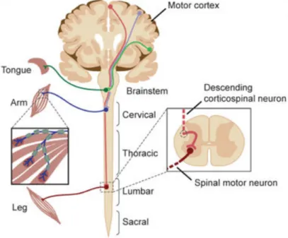
ALS is marked by the degeneration of motor neurons which are a group of efferent neu-rons, within the spinal cord and the brain, that makes synapses with muscle fibers to control muscle activity. There are two main types of motor neurons namely upper mo-tor neurons (UMN) and lower momo-tor neurons (LMN). Upper momo-tor neurons originate in the brain (motor cortex) and project downward to connect with lower motor neurons. The latter localize both in the brainstem and the spinal cord and their axons connect di-rectly with muscles at the Neuromuscular Junction (NMJ). Thus, they connect the Central Nervous System (CNS) with the target muscle to be innervated (Figure1.1) [22,66].
ALS was first described in 1869 by Jean-Martin Charcot. He suggested that “the name reflects both the degeneration of corticospinal motor neurons, whose descending axons in the lateral spinal cord appear scarred (“lateral sclerosis”), and the demise of spinal motor neurons, with secondary denervation and wasting of muscle (“amyotrophy”)” [66]. However, the disease only became well-known when the baseball player Lou Gehrig an-nounced his ALS diagnosis in 1939. Therefore, Amyotrophic Lateral Sclerosis is also known as Charcot disease, Lou Gehrig’s disease or motor neuron disease (MND). There are four other known MNDs: Primary Lateral Sclerosis (PLS), Progressive Muscular Atro-phy (PMA), Progressive Bulbar Palsy (PBP) and Pseudobulbar Palsy [76].
accounting for 90-95% of the cases, which has no genetically inherited component. Ap-proximately 5-10% of the cases are inherited from a family member and are classified as family-type ALS (fALS). The disease is considered familial when there are two or more family members affected. Usually, the onset is earlier in fALS, although the clinical presentation of inherited ALS and sporadic ALS is similar [1,66,69].
Figure 1.1: The components of the nervous system impacted in ALS pathogenesis. De-scending corticospinal motor neurons (upper motor neurons) are the first ones affected by ALS, they project from the motor cortex to synapses in the brainstem and spinal cord. Bulbar or spinal motor neurons (lower motor neurons) projects from the brainstem or spinal cord to skeletal muscles [Adapted from [66]].
ALS is an orphan disease affecting, in general, 1-2 individuals per 100.000 each year in most countries. Since ALS has a rapid lethality, its prevalence is much lower. Although rare, ALS is the most common motor neuron disease affecting people of all races and ethnicities with a higher prevalence among Caucasians [16,34,65].
1.2.1 Clinical phenotypes
Usually, the first symptoms begin between 50 and 65 years old with the median age-onset of 64 years. According to statistics, only 5% of the cases correspond to an age-age-onset younger than 30 years old, while the beginning over 80 years old presents a high incidence (10.2/100.000 in men and 6.1/100.000 in women) which can set aging as a risk factor for the disease [6,76].
ALS has a wide phenotypic variability and often shows clinical overlap with other neurodegenerative disorders being frontotemporal dementia (FTD) the most common.
Thus, identification of specific phenotypes is essential to develop strategies to measure disease progression and improve survival [66].
Different phenotypes can be distinguished based on the body region first affected at disease onset. When ALS symptoms begin in the arms or legs, it is termed "limb onset"ALS. In this case, individuals may have difficulties with simple tasks such as writing or walking. On the other hand, ALS that begins by affecting the muscles of speech, chewing and swallowing is referred as "bulbar1onset"ALS. Limb onset is found in around 80% of the cases but most patients will develop symptoms in both bulbar region and limbs as the disease progresses (Figure1.2) [34,53].
ALS affects both upper and lower motor neurons. However, ALS beginning usually presents symptoms associated with only upper or lower motor neuron involvement lead-ing to a wrong diagnosis. Thus, patients can be diagnosed with primary lateral sclerosis (PLS), presenting only UMN level phenotype, or progressive muscular atrophy, which has only LMN participation (PMA). As the disease progresses, ALS exhibit a detectable involvement of both UMN and LMN, and diagnosis reclassification must be accomplished to discard previous diagnostics of PLS or PMA (Table 1.1) [34,57,66].
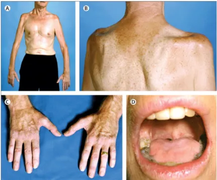
Figure 1.2: ALS clinical phenotypes. (A) Wasting of upper limb leading to an inability to lift arms against gravity. (B) Recessions above and bellow the scapular spine showing wasting of supraspinatus and infraspinatus muscles and partial loss of deltoid muscle. (C) Disproportionate wasting of the thenar muscles and also the first dorsal interossei comparatively with hypothenar muscles in hand – the typical "split-hand."(D) Substantial wasting of the tongue muscles in bulbar onset ALS [Adapted from [34]].
Table 1.1: Clinical presentations in amyotrophic lateral sclerosis. Symptoms are di-vided by affected motor neuron. Both UMN and LMN have to be affected for ALS diagno-sis [34,53].
Upper motor neuron sign in bulbar onset Spasticity;
Spastic dysarthria.
Lower motor neuron sign in bulbar onset
Tongue wasting; Weakness; Fasciculations; Flaccid dysarthria; Later dysphagia. Upper motor neuron sign in limb onset
Spasticity; Weakness;
Brisk deep tendon reflexes. Lower motor neuron sign in limb onset
Fasciculations; Muscle atrophy; Weakness.
Some factors influence prognosis. Low survival is associated with bulbar-onset disease, older ages at the time of disease development and early respiratory muscle dysfunction while high survival is related with the limb-onset disease, younger age at the beginning of the disorder and delayed diagnostic [34,73].
In final disease stages, patients need help with daily life activities and if they develop dyspnea at rest, the death becomes imminent. Usually, patients die from respiratory failure or other pulmonary complications. Sometimes they are kept alive by tracheostomy assisted ventilation developing a profound state motor paralysis known as “totally-locked-in-state” (TLS) in which all voluntary movements are lost [28,48,73].
1.2.2 Genetics
“There is considerable evidence of a genetic contribution to all ALS”[2].
Sporadic ALS refers to the cases that have no familial inheritance. However, techno-logical advances in DNA sequencing revealed the presence of mutations in ALS genes in the sporadic population, confirming the genetic contribution also in this form of the disease. Thus, the cause of sALS probably involves a combination of genetic and environ-mental factors. Familial ALS occurs as a result of mutations in a specific genetic locus and the inheritance is primarily autosomal dominant following a Mendelian pattern [5, 11,66,76].
In the last years, several studies contributed to generate a list of genes whose muta-tions are associated with both types of ALS. Given that one of the principal objectives of this work was to test the effect of a small molecule in a Drosophila model overexpressing FUS, the following topic will discuss the role of this gene [66].
1.2.2.1 Fused in Sarcoma (FUS)
Fused in Sarcoma is a RNA-binding protein, ubiquitously expressed in all cells, that plays an important role in the regulation of RNA transcription, splicing and transport. The N-terminal of FUS protein has properties for transcriptional activation and the C-N-terminal contains domains involved in RNA-protein interactions [58,62].
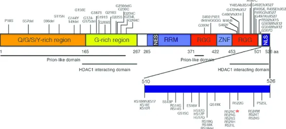
The majority of FUS mutations (accounting for 4% of fALS and less than 1% of sALS) are missense mutations in the C terminus of the protein, where the nuclear localization signal (NLS) is positioned (Figure1.3). The most common mutations in human ALS pa-tients are ’R521H’ and ’R521C’. FUS has nuclear and cytoplasmic expression and shuttles between the cytoplasm and the nucleus. FUS mutations lead to a gain-of-toxicity mech-anism that involves the redistribution of the protein from the nucleus to the cytoplasm [60]. Thus, postmortem analysis of brain and spinal cord tissues from ALS patients carry-ing FUS mutations demonstrated compromised FUS nuclear localization and abnormal cytoplasmic FUS inclusions in neurons and glia. Furthermore, it has been proposed that the overexpression of wild type FUS in vulnerable neurons may be one of the causes of the disease [36,45,56,72].
Figure 1.3: Schematic diagrams showing functional domains in FUS proteins and FUS mutations identified in ALS.The human FUS gene is located on chromosome 16p11.2. The full length human FUS protein contains 526 amino acids that can be further divided into distinct functional domains, such as the “prion-like” or low complexity (LC) domain that contains the Q/G/S/Y-rich region (amino acids 1-165) and the G-rich region (amino acids 165-267), the Arginine-rich motif (RRM, amino acids 285-371), two Arg-Gly-Gly (RGG)-repeat regions (amino acids 371-422 and 453-501), interrupted by a Cys2-Cys2 zinc-finger motif (ZNF)(amino acids 422-453), and a non-conventional nuclear localiza-tion signal (NLS)(amino acids 510-526). Other structural and funclocaliza-tional domains in FUS include the prion-like domains and the HDAC1-interacting domains. R521C mutation is higlighted by the red asterisk [Adapted from [62]].
1.2.3 Pathogenic mechanisms
There are a large number of gene mutations associated with this disease. Thus, several pathogenic mechanisms by which motor neurons degenerate have been proposed and it seems likely that the combination of these mechanisms, instead of a single mechanism, contributes to the neurodegeneration. The identification of toxic mechanisms is essential for understanding disease progression and for the development of effective therapies. The following topics discuss some principal mechanisms [8].
1.2.3.1 Free radical-mediated oxidative stress
In the last years, the interest in reactive oxygen species (ROS) have been increasing. They are natural bioproducts of the normal metabolism of oxygen and can be generated in the human body through several endogenous systems, exposure to various physicochemical conditions or pathophysiological states. However, an imbalance between ROS production and cell’s antioxidant defenses causes ROS accumulation and consequently oxidative damage. In other words, disturbances in the cell’s ability to detoxify reactive species lead to the accumulation of free radicals that can damage cell components including lipids, proteins and DNA, causing mutagenesis. Moreover, due to the accumulation of free radical damage in cells, organisms become aged and more susceptible to several diseases including ALS [19,49,76].
Increased oxidative damage and accumulation of free radicals were found in cere-brospinal fluid (CSF) and urine samples of ALS patients [76].
1.2.3.2 ER stress and unfolded protein response
Endoplasmic reticulum (ER) is the cellular organelle responsible for protein synthesis, posttranslational processing, folding of newly synthesized proteins and delivering the biologically active proteins to their proper target sites. Accumulation of unfolded and misfolded proteins in the lumen of ER (excessive influx) or perturbations in the typical en-vironment necessary for protein folding leads to ER stress. In this regard, a physiological response known as Unfold Protein Response (UPR) is triggered off in the cell to relieve ER stress by transcriptionally regulating ER chaperones and other proteins, attenuating the overall translation rate and increasing the degradation of misfolded proteins. However, if the ER functions are severely affected and it is not possible to restore the cell integrity, the cell undergoes apoptosis [31,59,66].
Previous studies found deposits of granular or amorphous material in the ER lumen of sporadic ALS patients, which can be associated to an accumulation of misfolded proteins, suggesting a relation between ER stress and motor neuron degeneration [31].
1.2.3.3 Protein aggregates
Protein aggregation represents one of the principal pathological hallmarks of ALS. Under normal conditions, cells can handle mutant proteins sufficiently well preventing their toxic gain of function and also their sequestration into inclusions. However, under patho-physiological circumstances, the ubiquitin proteasome system (UPS), that is activated to maintain protein quality control, could become overloaded. This leads to the engulfment of cells, which become defective in the disposal of altered macromolecules [7,46,50].
Recent studies have identified molecular constituents of ALS-linked cellular aggre-gates, including FUS. Despite strong evidence that protein aggregation is a hallmark of ALS, many questions remain about the role, formation and mechanism-of-action of these protein aggregates [7].
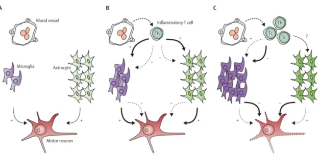
1.2.3.4 Neuroinflammation
The occurrence of a neuroinflammatory reaction is usually found in neurodegenerative disorders (including ALS) and consists of activated glial cells, mainly microglia and astro-cytes, and T cells. Microglia are the resident macrophages2in the nervous system; they constantly monitor the extracellular environment, interacts with astrocytes and neurons and are the first line of defense against infection or injury to the nervous system. Astro-cytes are ectodermal cells that have many complex functions in the nervous system such as regulating extracellular neurotransmitter concentrations, supporting surrounding neu-rons and maintain extracellular ion balance. Finally, T cells are a type of lymphocyte which infiltrates the CNS modulating the neuroinflammatory reaction differently in each stage of disease progression [52].
Several studies have shown that microglial activation is usually triggered by the infil-tration of helper T cells3and cytotoxic T cells4. The presence of T cells is rare in early ALS stages, but readily infiltrate the spinal cord as the disease progresses. At the end stage of disease cytotoxic T cells predominate; thus, some hypothesis suggests that it explains why neuroprotective action ultimately fails (Figure1.4) [52].
In the last years the interest in the contribution of microglia, astrocytes and T cells to motor neurons degeneration has been increasing and has given rise to clinical trials of drugs targeting neuroinflammatory reactions in ALS patients [52,66].
2Macrophages are specialized cells involved in the detection, phagocytosis and destruction of bacteria and other harmful organisms [21].
3Helper T cells are a type of T cell that helps stimulate B cells (type of white blood cell of the lymphocyte subtype) to make antibodies and activates macrophages to kill ingested microorganisms [29].
Figure 1.4: Interaction of inflammatory T cells with microglia and astrocytes in ALS pathology.(A) At asymptomatic stages of the disease, almost no T cells are found in the spinal cord. (B) At early stages of the disease, inflammatory T cells infiltrate the spinal cord from the blood. These T cells are mainly helper T cells which interact with sur-rounding microglia and astrocytes. Here, their neuroprotective action (thick arrow, +) overcome their neurotoxic effect (dashed arrow, -). (C) At later disease stages, cytotoxic T cells predominate in the spinal cord. These cells could trigger the production of a neu-rotoxic environment (thick arrows, -) instead of a neuroprotective environment (dashed arrows, +) surrounding motor neurons leading to their degeneration and consequently death. Th = helper T cell. Tc = cytotoxic T cell [Adapted from[52]].
1.2.3.5 Mitochondrial dysfunction
Mitochondria are rod-shaped organelles considered the “powerhouse” of cells since they convert energy into ATP which is essential for cells metabolism. They also have roles in vital processes including the production of cellular respiration, calcium homeostasis and control of apoptosis. Previous studies reported functional defects and morphological changes of mitochondria in skeletal muscle of ALS patients suggesting that mitochondrial abnormalities could be related to ALS. Moreover, a proposed mechanism suggests that mutant SOD1 is imported into mitochondria damaging this organelle and activating cell death [14,41,76].
1.2.3.6 Impaired axonal structure and disrupted transport
Motor neurons typically have extremely long axons (on the order of meter long) that can be vulnerable to damage. Axons conduct electrical impulses known as action potentials but also transports organelles, RNA, proteins and lipids to and from a neuron’s cell body. Axonal transport is crucial for the survival of motor neurons and can be anterograde or retrograde. It is called anterograde transport when it moves away from the cell body, toward the synaptic structures at the neuromuscular junction, mediated by kinesins. On
the other hand, moving toward the cell body is called retrograde transport and it is mediated by cytoplasmic dynein [17,76].
Dysfunction of both axonal transport, anterograde and retrograde, and axonal cy-toskeletal disorganization, especially of neurofilaments, have been identified as causes of motor neuron degeneration. Defects in axonal transport can lead to an accumulation of neurofilaments, mitochondria and autophagosomes in motor neurons. In this regard, some experiments have shown that in the presence of ALS-linked SOD1 mutants, both anterograde and retrograde transport are slowed, months before degeneration. In addi-tion, several studies have shown that mutation in dynactin, an activator of cytoplasmic dynein, has been associated with a reduction in retrograde transport leading to motor neurons degeneration [17,66].
1.2.3.7 Glutamate-induced excitotoxicity
During glutamatergic neurotransmission, glutamate released from the presynaptic neu-ron activates ionotropic glutamate receptors5in the postsynaptic neuron. The activation of these glutamate receptors leads to the influx of Na+ and Ca2+ into the cell causing depolarization and the generation of an action potential. Afterwards, glutamate is re-moved from the synaptic cleft by excitatory amino acid transporters (EAATS). However, an increased release of glutamate or a failure to rapidly remove synaptic glutamate in-duces excitotoxity6. Nevertheless, it remains unclear if glutamate-induced excitotoxicity is a primary defect responsible for motor neuron degeneration or it is the result of ALS (Figure1.5) [71].
5The ionotropic glutamate receptors are ligand-gated cation channels [71].
6Excitotoxicity describe the neuronal degenerative changes caused by over-stimulation of the glutamate receptors [63,71].
. Figure 1.5: Glutamatergic neurotransmission and excitotoxicity. (A) Under the normal process, glutamate is released from the presynaptic neuron into the synaptic cleft activat-ing the NMDA and AMPA ionotropic receptors in the postsynaptic neuron. This leads to the influx of Na+ and Ca+ ions into the cell causing depolarization and generation of an action potential. (B) Classical excitotoxity is induced by an increase of extracellular gluta-mate concentration. This can be caused by an increased release of glutagluta-mate or failure in reuptake of glutamate into the astrocytes by EAAT2/GLT-1 transporter. Thus, glutamate receptors are excessively stimulated giving rise to an excessive increase of the intracellular concentration of Na+ and Ca+ that can trigger motor neuron death. [Adapted from[71]].
Glutamate excitotoxicity has been associated with ALS due to the detection of high glutamate concentrations in the cerebrospinal fluid (CSF) of several patients with sporadic ALS. Moreover, the main argument for the role of excitotoxity in ALS is that riluzole, a clinical drug that slows the progression of the disease, has anti-excitotoxic properties [15, 71].
1.2.4 Treatment
Currently, there is no available treatment to stop or reverse the progressive degenera-tion of motor neurons in ALS. However, two clinical drugs can slow the progression of symptoms and prevent complications. The first drug approved by the FDA for the treatment of ALS was riluzole, a glutamate antagonist that blocks voltage-gated sodium channels leading to a decrease in the pre-synaptic release of glutamate. In May 2017, the FDA approved edaravone, an intravenous drug that counteracts the excessive oxidative stress in ALS by removing the free radicals in the nervous system. Indeed, these drugs only prolong the survival and delay the use of surrogate approaches such as mechanical ventilation. Hence, more than two decades after the acceptance of riluzole, the search for new therapeutic strategies is essential [20,46,47,51].
1.3
Bioactive small molecules
Many studies have been supporting the possibility of changing the progression or the development of neurodegenerative diseases through diet. Indeed, several findings have been suggesting that bioactive molecules such as polyphenols7, the most abundant in our diet, have protective effects in neurodegenerative disorders [23]. They are thought to be effective in the prevention or reduction of the impact of reactive oxygen species associated with oxidative stress and neurodegeneration because of their strong capability to induce intracellular signaling pathways related to gene expression and cell survival [32]. Hence, actual studies are trying to test their neuroprotective properties and also understand which of the hundreds of natural polyphenols available in our diet provide better effects [39,49,64].
Jimenez-Del-Rioet al. demonstrated that pure polyphenols such as gallic acid, ferulic
acid, caffeic acid, coumaric acid, propyl gallate, epicatechin, epigallocatechin, and epigal-locatechin gallate protect, rescue and, most importantly, restore the impaired movement activity induced by paraquat8inDrosophila melanogaster [32]. Similarly, Maccioniet al.
revealed that standardized phytotherapic extracts, from medicinal plants widely used in Ayurvedic medicine, restored anomalous locomotion (i.e. impaired climbing perfor-mance with unexpected hyperactivity) and electrophysiological responses in aDrosophila
model of ALS [39].
However, it remains unclear what concentrations are necessary to these small molecules reach the brain and what biologically active forms are need to exert beneficial effects. Therefore, more research is necessary to identify the molecular pathways and intracellu-lar targets responsible for the protective effects of polyphenols [3].
1.3.1 Compound C
Edible berries are considered one of nature’s treasure chests not only because they are an enjoyable fruit that provides energy, nutrients and dietary fiber, but also because they con-tain a large number of polyphenols with health-promoting properties. However, berries contain complex polyphenols mixtures making difficult to associate any interesting phar-macological activity to a single small molecule. For this reason, under the European BacHBerry project, more than twenty selected berry extracts, based on their metabolomic profile, were systematically analyzed to identify small molecules that have protective properties against neurodegenerative disorders. These extracts were tested on different
Saccharomyces cerevisiae strains overexpressing proteins related to Alzheimer’s,
Parkin-son’s, Huntington’s or ALS disease that confer toxicity in yeast. After the identification of blackberry (Rubus genevieri) extract as having bioactivity against ALS, it was fractionated
7“Polyphenols are a group of chemical substances present in plants, fruits, and vegetables, characterized by the presence of one or more than one phenol unit per molecule with several hydroxyl groups on aromatic rings” [32].
and the obtained fractions were re-tested in the yeast model of ALS overexpressing FUS protein. All fractions were analyzed for its content by liquid chromatography coupled to mass spectrometry and some isolated small molecules from the bioactive fractions were re-tested. This screening revealed that Compound C displayed significant bioactivity in the yeast ALS model. So, Compound C was identified as a powerful protectant against pathological mechanisms associated with ALS disease in a yeast model overexpressing FUS (Figure1.6) [30].
Figure 1.6: Compound C protects the yeast model of ALS from FUS toxicity. FUS over-expression impairs cellular growth (red) compared with the control (green) and treatment with Compound C rescues cellular growth (blue) [Unpublished results].
1.4
Drosophila melanogaster as a model system to study
FUS-induced ALS disease
As an animal model,Drosophila has several advantages that make it extremely useful and
important for biomedical research. Indeed, the relative simplicity, short lifespan, easy and cheap maintenance, approachable physiology and huge availability of powerful genetic tools make it a very attractive model to the study of several topics such as development, behavior and the basis of human disease [49,67].
In particular,Drosophila models containing ALS-associated transgenes have been
im-portant tools for understanding disease pathogenesis. Previous studies showed that the expression of wild type or mutant human FUS alleles such as FUS[R521C] in the eye, motor neurons or the nervous system of the fly leads to eye degeneration, defects in loco-motion and increase in mortality [36]. Thus, theDrosophila model of ALS overexpressing
human FUS alleles recapitulates the majority of ALS characteristics including progres-sive motor deficits, motor neuron degeneration and early lethality. However, the ALS-like phenotypes in these animal models are highly dependent on transgene expression levels and severity of phenotypes correlate with level of protein overexpression [77].
Interestingly, the homolog of human FUS gene inDrosophila corresponds to cabeza
(caz) gene on X chromosome, that shares 53% amino acid identity to its mammalian counterpart. TheDrosophila Caz protein has 399 amino acids and is expressed in neurons,
glia and muscle cells [62].
1.5
Objectives and proposed approaches
This project had two main goals:
• Characterize the neuronal and kinematic decay of aDrosophila model of ALS;
• Test the effect of candidate small molecule (Compound C) in the motor decay of the
Drosophila model of ALS.
Since FUS is one of the principal candidates in ALS development, our first step was to mimic the human disease by overexpressing human wild type FUS and human FUS with targeted mutagenesis (R521C). Once we profiled the mutant FUS induced degeneration at the kinematic level, we tested the protective effect of a small molecule (Compound C) previously tested for their capability to improve cellular growth of the yeast model overexpressing FUS.
The use of adult Drosophila as a model to test the efficacy of small molecules has
multiple advantages. First, it allows challenging neurons in anin vivo, whole body context.
Second, due to the fly’s relatively short life span, it is possible to follow in a feasible time period the degeneration of motor activity. Finally, we can quantify with high detail a large set of locomotor features of animals undergoing degeneration, allowing detecting any relevant suppressions in the motor decay process.
To achieve the proposed goals, we performed survival, climbing and egg laying assays and we also analyzed the neurodegeneration in the fly eyes. Furthermore, a detailed anal-ysis of the locomotor pattern of the flies modeling ALS disease using the FlyWalker was done. This approach was used to correlate cellular demise with specific kinematic conse-quences to the animals and most importantly, to quantitatively profile neurodegeneration that was critical to detect putative suppression of the neurodegenerative phenotype. Fi-nally, we indirectly quantified the number of motor neurons establishing synapses with leg muscles.
Since the ultimate goal was to ameliorate the locomotor symptoms associated with ALS pathology, we fed the flies with Compound C in these experiments and tested its effect on the motor decay induced by the expression of FUS alleles.
C
h
a
p
t
2
M a t e r i a l a n d m e t h o d s
2.1
Preparation of the fly food
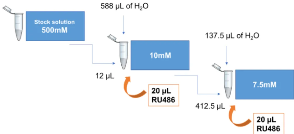
The two concentrations of Compound C tested (7.5mM and 10mM) were obtained through successive dilutions, starting from a stock solution of Compound C dissolved in DMSO (500mM). In the cases that the flies were treated with RU486, 20µL of this compound was added to the 10 mM or 7.5 mM solutions (Figure2.1).
Finally, 150µL of the selected solution were impregnated in the vials containing fly food overnight. With this method, the compound was not mixed in all the food, it just impregnated a few centimeters. However, given that the flies were transferred to new vials twice a week, they just ate the food at the surface.
Figure 2.1: Protocol followed to obtain the different concentrations of Compound C tested.
2.2
Fly eye imaging
Eye phenotypes of one-day-old female flies were evaluated using Leica S6E and the images were acquired on Zeiss Stereo LUMAR stereoscope. The flies were placed at -80ºC for five minutes, before the imaging. For each genotype, 10 to 30 flies were evaluated. Embryos, larvae and pupae were exposed to DMSO or Compound C throughout their development and maintained at 25ºC in incubators without light.
2.3
Life span assay
Flies were crossed in the absence of DMSO, Compound C or RU486 on a standard food medium. Day 1 adult females were transferred on to new experimental vials containing fly food mixed either with DMSO or Compound C dissolved in DMSO, with (+) or without (-) RU486 (1 mM), at a density of 25 flies per vial for each genotype (n=3). Deaths were scored every two days and flies were transferred to fresh food two times a week. All the flies were maintained at 25ºC in incubators under a 12 h light/dark cycle.
2.4
Climbing assay
Motor function was assessed by a negative geotaxis response assay, commonly called climbing assay. Briefly, groups of 10 males of the same age of each genotype were placed into 18-cm-long vials, at room temperature for environmental acclimatization, and 10 min later they were tapped to the bottom of the vial. The climbing time was recorded when 50% of the population (five flies) crossed the 8-cm or the 15-cm finish line. At least 50 flies were tested in five independent groups of males per condition. Results are the average climbing time of these separate trials. The males were exposed to vehicle (DMSO) or Compound C from day one adult-stage and maintained at 25ºC in incubators under a 12 h light/dark cycle.
2.5
Egg laying assay
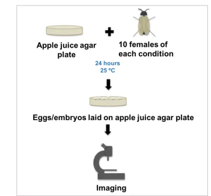
Reproductive outputs of the flies expressing FUS alleles were assessed by quantifying the number of laid eggs and emerged larvae. Briefly, five males and 15 virgin females, for each condition, were used for mating. After seven days of mating, males were discarded. At the selected time-points (seven, 14 and 21 days), ten female flies of each group were randomly selected and placed into apple juice agar plates during 24 hours and then the number of laid eggs was quantified using Leica S6E. Five days later, the number of emerged larvae was also registered (Figure2.2). Adult flies, eggs and larvae were maintained at 25ºC in incubators under a 12 h light/dark cycle. Females were exposed to vehicle (DMSO) or Compound C, both mixed with RU486, from day one adult stage. This experiment was performed two times (n=2).
Figure 2.2: Schematic showing the procedure of the egg laying assay.
2.6
Quantification of motor neuron projection in the leg NMJ
In order to keep leg motor neuron axonal morphology intact for imaging, the legs were dissected and fixated carefully. Five flies per condition were selected and placed into empty tubes. To remove the hydrophobicity of the cuticle, the flies were washed in ethanol (not more than one minute). Afterwards, they were rinsed three times in 0.3% Triton in 1x phosphate buffered saline (PBS) (for one, five and 30 minutes in each wash) to increase penetration of the fixative inside the leg and then, the flies were dissected in this medium. Basically, the head and the abdomen of the flies were removed using forceps and then the coxa-thorax junction was gently but firmly pushed using the tip of fine forceps until the leg detached. Subsequently, the fixation was done in 4% paraformaldehyde (PFA) in PBS overnight at 4ºC (approximately 20 hours total). Later, the legs were washed three times in PBT, for 20 minutes each wash at room temperature. Finally, the legs were mounted into glass slides using 70% glycerol medium. The imaging was done with a Zeiss LSM710 confocal microscope using a 40x water immersion objective.
2.7
FlyWalker basis
It is challenging to quantify walking behavior inDrosophila due to its small size and the
lack of available tools. In order to overcome this limitation, Mendeset al. described an
approach that can be used to examine the walking patterns ofDrosophila melanogaster.
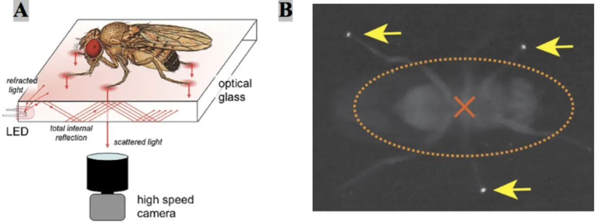
The authors developed an optical touch sensor that is based in frustrated Total Internal Reflection (fTIR) method coupled with high-speed video imaging which detects the fly’s body and leg contacts on the floor during locomotion [43].
Total Internal Reflection occurs when light travels from a medium with a high refrac-tive index, in this case optical glass, to one with a lower refracrefrac-tive index such as air. If the angle of incidence is higher than the critical angle (as compared to the normal of the surface), defined by Snell’s Law, the light is internally reflected rather than refracted. However, when a denser material such as the tarsus of an insect leg touches the surface of the glass, the locally ‘frustrated’ total internal reflection will scatter the light and that can be recorded by a high-speed video camera (Figure2.3) [40,43,44].
Each movie is acquired at 250 frames per second and it can be analyzed frame-by-frame. In each frame, the fTIR effect allows to visualize the fly legs that are in contact with the glass and also the fly body due to the background light (Figure2.3). Afterwards, the tracking of each tarsal contact and the fly body is done with the FlyWalker program (Figure 2.4). This custom-made software evaluates the fTIR signals in terms of pixel intensity in each movie and outputs several graphs and user-defined kinematic param-eters that can be used to describe fly walking behavior with high temporal and spatial resolution. The parameters include step, spatial and gait parameters [43].
Figure 2.3: Schematic of the fTIR optical effect. (A) Detailed description of the fTIR apparatus. Light that comes from LED light sources located at the edges of an optical glass propagates within the glass by internal reflection. Tarsal contacts lead to light scattering which can be recorded by a high-speed video camera. (B) Single frame of a fTIR movie. The fTIR effect can be seen for three legs in stance phase (yellow arrows). The orange dashed ellipse corresponds to the body delimitation; the center of the body is indicated by an orange cross [Adapted from[43]].
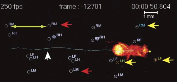
Figure 2.4: Image generated by the FlyWalker software. The fly’s footprints and body center are tracked throughout the video. It is possible to identify the present footprints (yellow arrows), the past footprints (red arrows) and the fly body and trajectory (white arrow). Step length, which is defined as the distance between two consecutive footprints, can also be visualized (green arrows) [Adapted from[43]].
2.7.1 Step parameters
Drosophila has six legs and, in a step cycle, each leg goes through a period of stance phase,
in which the leg is touching the ground, or a swing phase meaning that it is up in the air. Previous findings have shown that, in several insect species, when the speed increases, stance phase duration becomes shorter and swing phase duration remains predominantly constant; at the higher speeds, the duration of both stance and swing phases becomes equal [25,74].
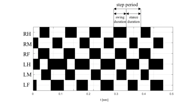
One of the parameters that it is possible to extract from FlyWalker analysis are the step parameters that are related to individual leg movement. These parameters include the duration of stance and swing phases, step frequency (number of steps per second), step period (time taken to complete one leg cycle), step length (distance between two successive footprints of the same leg), average speed and swing speed (Figure2.5) [43].
Figure 2.5: Step pattern of a general fly walking at 40 mm/s (average) obtained with the FlyWalker software.The swing and the stance phases are represented by the black and white regions, respectively. The step period, swing duration and stance duration are indicated by the arrows [Adapted from [43]].
2.7.2 Spatial parameters
The spatial parameters are related to the perception of the surface. They analyze the first and last moments that the leg contacts with the glass surface and how much straight are the stance traces generated between those moments. The onset of these traces, which corresponds to the position where the leg first touches the glass, is termed the Anterior Extreme Position (AEP) while the end of stance traces, before the tarsi enter swing phase, corresponds to the Posterior Extreme Position (PEP). For each stance trace, a smoothed version of the trace is generated and the average of the difference between these two lines allows to calculate the stance linearity index parameter. Thus, faster flies have straighter stance traces (lower stance linearity indexes) when compared with slower flies. Another spatial parameter is the stance straightness index that corresponds to the ratio between the displacement (AEP-PEP vector length) and the path length. Footprint clustering is also a relevant spatial parameter and it is related to the clustering of the AEP’s and PEP’s. This parameter corresponds to the standard deviation from the average position for all AEP’s or PEP’s calculated for each leg. For instance, if the footprint clustering value of the foreleg AEP is small, it means that the AEP coordinates were similar for all the foreleg steps in the video (Figure2.6) [43].
Figure 2.6: Schematic diagram of general spatial parameters obtained with the Fly-Walker software.(A) Representation of the stance strances for each leg of a general fly. AEP and PEP positions are also identified. (B) Method to calculate the stance linearity index. It can be obtained by computing the average difference between an actual stance trace and a smoothed trace. (C) The stance straightness index indicates how much wig-gly is the body center relative to each footprint and corresponds to the ratio between the displacement and the path length. (D) Method to quantify footprint clustering. An average ±STD xy point is generated (blue cross) for each set of AEP/PEP footprints (red circles). The footprint clustering value is calculated as the vector sum of the two STD values (orange arrow) [Adapted from[43]].
2.7.3 Gait parameters
Gait parameters analyze coordination between legs. Insect gaits can be either tripod or tetrapod (Figure2.7), depending on the speed and body load. Flies usually walk using tripod gait which is characterized by three legs in stance phase and three legs in swing phase at any one time. Each group of three legs is constituted, on one side, by the fore and hind legs and, on the contralateral side, by the middle leg. In contrast, a tetrapod gait is characterized by two legs in swing phase and the remaining four legs in stance phase. The two legs in swing phase are localized on contralateral sides and are offset by one segment. It is also observed several non-canonical stance combinations that do not fit in these idealized gaits. In addition, at slow speeds flies occasionally walk with only one leg in swing phase which characterizes the so-called ‘wave gait’ in which individual legs swing in a wave-like pattern from front to back [43].
For each video, a step pattern can be generated (Figure 2.5) and the instantaneous speed and gait characteristics are simultaneously plotted with high temporal resolution (Figure 2.7). The instantaneous speed plot has a wave-like appearance in which the minimum speeds correspond to the transition between phases, when the stance switches
to a different group of legs. For each frame in a video, it is possible to classify if the fly is in a tripod, tetrapod or non-canonical stance (sometimes wave gait is also used). These results are plotted in a gait map that graphically shows the gaits used over time (yellow for tripod, blue for tetrapod, and grey for non-canonical). To quantify the gait maps, the tripod, tetrapod and non-canonical indexes are calculated. These gait indexes correspond to the percentage of frames in a video that displays leg combinations defined by the tripod, tetrapod and non-canonical gait, respectively [43].
Figure 2.7: Schematic diagram of some gait parameters obtained with the FlyWalker software.(A) Representation of the different gaits and leg combinations used by the flies during the walk. Black circles and ‘1’ corresponds to stance phase and white circles and ‘0’ means swing phase. (B) Speed and gait graphs obtained with the FlyWalker software for a general fly walking at 40mm/s on average. The color code corresponds to the gaits used by the fly overtime in all the movie. It is possible to visualize that this female fly used mostly the tripod gait which corresponds to higher values of body velocity [Adapted from [43]].
2.8
FlyWalker procedure
2.8.1 Fly preparation
The flies used to perform the FlyWalker analysis were raised at the same conditions as outlined in the survival assay. Before the acquisition, each group of flies was anesthetized with ice and placed 30 minutes into empty vials to clean their legs.
2.8.2 Data acquisition
The image acquisition was done with a Photron (1024 x 336) or a Point Grey camera
optical glass where the flies walk with optic cleaning fluid before the acquisitions. Each fly was inserted into the fly chamber using a mouth aspirator and the acquisition of the data started immediately after the insertion; thus, flies did not have time to adapt to the surrounding environment. Moreover, the duration of the acquisition did not last longer than one minute. For each time point, approximately seven to ten flies were evaluated.
2.8.3 Movie cropping
The temporal cropping of the movies acquired with thePoint Grey and the Photron
cam-eras was done with the StreamPix 6.5.0.0 and the Photron FASTCAM Viewer 3.6.9.1
(PFV3) software, respectively. The videos were temporally cropped by selecting the first and the last frames, giving rise to small sequences of .png files. The chosen sequences contained a series of five to six step cycles for each leg, where the fly walks straight and from left to right. Then, the sequences acquired with the Point Grey were load in
Im-age J software to be spatially cropped in order to have a smaller imIm-age containing the sequence of interest. The movies acquired with thePhotron camera were cropped
tempo-rally and spatially using the same software, PFV3. In total, approximately 120 movies were cropped.
2.8.4 Fly tracking using the FlyWalker software
The custom-made FlyWalker software, written in MATLAB® R2016b, was used to per-form the fly tracking process. The first step was to load the sequence to be analyzed through this software. Then, the length of the fly was measured, with the option "ruler"available in the interface, and introduced in the software settings. Moreover, the threshold values for the legs and body were defined in the settings to optimize the auto-tracking pro-cess. After the auto-tracking, the detection of the legs and the center of the body were confirmed. If there were detected any defects, it was done hand correction. If not, the evaluation of the movie proceeded. The evaluation process resulted in a set of graphs (including the stance traces, the step pattern, the gait map,...) and also a Microsoft Excel file with all the kinematic parameters that describe the walking patterns of each fly.
2.8.5 Data analysis
The Excel file includes a summary line containing all the parameters and their values. To compare different conditions, these lines were all grouped in another Excel file. Then, for each motor parameter, a scatter graph containing the raw data of the different groups was generated using the Excel tools. From here, it was possible to observe and select the relevant variables for further analysis. The parameters were considered relevant when it was possible to distinguish the groups of interest in the scatter plots.
Based on this first approach, the next step was to calculate the residual values using a script written in R. Given that most of the kinematic parameters vary with speed, the
R script determined the best fit regression model for the control condition and then it computed the residual values for each experimental condition in relation to this regres-sion model. After, the data was expressed as the difference to the residual-normalized line in PCA, heat map and box plots. To perform statistical tests, it was necessary to verify if the data was normally distributed and homoscedastic (i.e. equal variance for all predictors). The normality assumption was confirmed using the Shapiro-Wilk test and the homoscedasticity assumption was confirmed using the Levene’s Test. Afterwards, if the data was considered normally distributed and homoscedastic, the test used was one-way-ANOVA. The null hypothesis of this test is that the means of all groups considered were the same. If the null hypothesis was rejected (p-value < 0.05), it was performed the Tukey’s post hoc test between each group and the control. Otherwise, if data was not normal and homoscedastic, non-parametric tests were performed. In these cases, for comparisons of more than three groups, it was used Kruskal-Wallis analysis of variance. If the null hypothesis, that the median of all groups was the same, was rejected (p-value < 0.05), the Dunn’s post hoc test was performed to see the significant differences between each group and the control. The graphic representations such as the heat maps and the PCA were made with scripts written in Python 3.4 software. The box plots were done with GraphPad Prism software version 7.
2.8.5.1 Residual values - Detailed explanation
To calculate the residual values, a regression analysis was performed in order to find the best fit (linear or non-linear) for each parameter of the control condition. Afterwards, the residuals were obtained by computing the distance of each value (including control values) to the regression model (Figure2.8). There was one residual for each data point. They were positive if they were above the regression model and negative if they were below the regression line. If the point passed through the regression line, the residual at that point was zero.
Figure 2.8: Method to obtain residual values. The best fit model is indicated by the red arrow. A residual is the vertical distance between a data point and the regression line (blue).
2.8.5.2 Principal Component Analysis (PCA)
Principal Component Analysis is a technique commonly used for dimensionality reduc-tion. Briefly, it projects a complex dataset onto a lower-dimensional space that still con-tains most of the information in the large set. Since PCA “ignores” class labels, it can be described as an “unsupervised” algorithm. Thus, the objective of this method is to find directions (called principal components) that maximize the variance of the data. The first principal component explains the most extensive variance in the original data set. The second principal component is determined in the same way, with the condition that it is perpendicular to the first principal component and that it accounts for the next highest variance. This continues until a total of n principal components have been computed, equal to the original number of variables. For visualization, usually the first one, two or three principal components are used to plot the data in an attempt to reveal any groupings [33,70].
2.9
Overall statistical considerations
Most of statistical analyses were done using GraphPad Prism software version 7. For the survival assays, it was performed a Log-rank (Mantel–Cox) test. For climbing and egg laying assays, it was performed a two-way ANOVA followed by a Tukey post-test. In the FlyWalker analysis, statistical tests were performed as outlined above. It was used "ns"for non-significant data. Statistic significant were considered when ∗pvalue ≤ 0.05, ∗ ∗ pvalue ≤ 0.01, ∗ ∗ ∗pvalue ≤ 0.001.
C
h
a
p
t
3
R e s u lt s a n d D i s c u s s i o n
3.1
Ectopic expression of human FUS alleles leads to
neurodegeneration in Drosophila eyes
In order to observe the effects of the expression of human ALS-related FUS alleles in the fly eyes, transgenic flies overexpressing wild type (WT) or mutant (R521C) human FUS in the eyes were generated using the GAL4/UAS system. Comparing to the control (UAS-GFP), the expression of FUS alleles using GMR-gal4, an eye driver, caused severe neurodegeneration inDrosophila eyes characterized by disorganized ommatidia and loss
of pigmentation and mechanosensory bristles. Moreover, the FUS alleles reflected the ex-pected strength, with the phenotype of mutant human FUS more aggressive than the wild type form. Nonetheless, the misexpression of FUS[WT] also induced neurodegeneration (Figure3.1).
These results demonstrate that the expression of human ALS-related FUS alleles causes neurodegenerative phenotypes which is consistent with the gain-of-toxicity mech-anism that has been proposed for ALS disease. Furthermore, they confirm that the FUS[R521C] allele causes stronger degeneration that the wild type version [36].
In addition, it was analyzed the expression of other FUS alleles such as FUS[R518K] and FUS[R521H] in the fly eyes. However, no differences were observed regarding the control which was probably due to problems in transgene generation (data not shown).
Figure 3.1: Imaging of day 1 adult fly eye expressing FUS alelles (B,C) under the control of GMR-gal4 driver (A).
3.1.1 Compound C does not ameliorate the neurodegenerative phenotype in the fly eyes
To try to suppress the neurodegenerative phenotype, flies with the same genotype as above were fed with Compound C during development. Given that Compound C was dissolved in DMSO, another batch of flies was supplied only with DMSO in order to use it as a control for further comparison. Thus, two concentrations of Compound C were tested: 7.5mM and 10mM (Figure3.2).
After the treatment with Compound C, no visible modifications were found in the fly eyes. In addition, the eye phenotypes of the flies fed with DMSO were similar to the ones found for the standard condition (Figure3.1).
Taken together, the results suggest that, in these concentrations, DMSO is not toxic for the flies and the exposure of the fly to a medium supplemented with Compound C does not ameliorate the neurodegenerative phenotypes caused by the expression of FUS alleles in the eyes.
Figure 3.2: Eye neurodegenerative phenotypes after the treatment with Compound C. These flies were supplemented with (A) 7.5mM and (B) 10 mM of DMSO (first row) and Compound C (second row) during development.
3.2
Conditional expression of human FUS alleles in
Drosophila neurons increases mortality
To mimic the human disease inDrosophila, FUS alleles were overexpressed in the neurons
of the fly using the conditional expression system ElaV-GeneSwitch (ElaV-GS). Briefly, following treatment with RU486, the Gene Switch protein was transcriptionally activated and bound to UAS inducing the expression of the respective FUS protein specifically in the nervous system [36].
Then, the effects of the neuronal FUS expression on the life span of the flies were determined by monitoring their survival rate from day one adult stage on. Given that one of the objectives of this project was to test the protective effect of Compound C, it was considered two groups of females flies: one fed with standard food mixed with the vehicle and another one supplied with Compound C (10mM). In each group, three genotypes were considered: GFP (control), FUS[WT] and ElaV-GS;UAS-FUS[R521C], each one treated with or without RU486 (1mM). For each condition, 75 flies were analyzed.
In the group fed with the vehicle, there was very low mortality in the flies expressing the driver alone treated with (+) or without (-) RU486, since >90% flies were viable after 30 days of DMSO exposure in both cases. However, the induction of FUS[WT] and FUS [R521C] in adult neurons caused a decline in the survival rate when compared with the driver alone, even in RU486 untreated flies (-), which was not expected. Furthermore, the induction of FUS[R521C] drastically shortened the life span when compared with FUS[WT]. These findings revealed a mutation-dependent decline in the life span of the FUS[R521C] expressing animals. However, flies expressing FUS[WT] also exhibited a decline in life span, but the degree of severity was less than mutant FUS animals (Figure 3.3).
![Figure 1.6: Compound C protects the yeast model of ALS from FUS toxicity. FUS over- over-expression impairs cellular growth (red) compared with the control (green) and treatment with Compound C rescues cellular growth (blue) [Unpublished results].](https://thumb-eu.123doks.com/thumbv2/123dok_br/15815703.1081065/33.892.187.718.337.696/compound-protects-toxicity-expression-cellular-treatment-compound-unpublished.webp)