ORIGINAL RESEARCH
Low prevalence of fetal-type posterior cerebral
artery in patients with basilar tip aneurysms
Mariana C Diogo,
1Isabel Fragata,
1Sara P Dias,
2Joana Nunes,
3Jaime Pamplona,
1João Reis
11Neuroradiology Department,
Centro Hospitalar de Lisboa Central, Lisboa, Portugal
2
Neurology Department, Centro Hospitalar de Lisboa Central, Lisboa, Portugal
3Neuroradiology Department,
Centro Hospitalar de Vila Nova de Gaia/Espinho, Gaia, Portugal
Correspondence to Dr Mariana Cardoso Diogo, Neuroradiology Department, Centro Hospitalar de Lisboa Central, Rua José António Serrano, Lisboa 1150–199, Portugal; mariana.cardoso. [email protected] Received 10 May 2016 Revised 11 June 2016 Accepted 14 June 2016 To cite: Diogo MC, Fragata I, Dias SP, et al. J NeuroIntervent Surg Published Online First: [ please include Day Month Year] doi:10.1136/ neurintsurg-2016-012503
ABSTRACT
Background Basilar tip aneurysms (BTA) are multifactorial in origin, with luminal forces playing a major role in their formation. Considering the reduced hemodynamic stress on the basilar apex in the fetal-type posterior cerebral artery (fPCA), we hypothesize that BTA should be less common in patients with this variant. Objective To investigate, in a retrospective case– control study, the frequency of fPCA in patients with and without BTA.
Materials and methods We collected clinical and imaging data from consecutive patients with BTA undergoing catheter angiography between July 2010 and July 2015, and from a randomly selected, age- and sex-matched non-BTA control population from our prospective database. Anatomical variants of the distal basilar artery region were assessed in the two groups and compared using parametric and non-parametric tests.
Results Fifty-nine BTA cases and 337 controls were included. fPCA was present in 3% of patients with BTA and 23% in the control group ( p<0.001; OR=0.11, 95% CI 0.03 to 0.48). Basilar tip disposition was cranial in 49% of BTA and 63% of non-BTA cases ( p=0.04; OR=0.57, 95% CI 0.33 to 0.99); a caudal disposition was found in 24% and 6% of cases, respectively ( p<0.001; OR=4.65, 95% CI 2.21 to 9.80). Conclusions We found a statistically significant association between the absence of fPCA and BTA. Our findings underline the importance of hemodynamic stress in the formation of intracranial aneurysms, and suggest that fPCA is a protective variant for formation of BTA.
INTRODUCTION
Intracranial aneurysms are vascular dilatations believed to arise from a focal weakness of the arter-ial wall. In the intracranarter-ial compartment, their prevalence increases with age and varies in differ-ent studies, ranging from 1% to 10%, with saccular aneurysms accounting for 80–90% of all intracra-nial aneurysms.1Although most aneurysms remain asymptomatic, a minority rupture, with high asso-ciated morbidity and mortality rates.2 3 Posterior circulation aneurysms, such as basilar tip aneurysms (BTA), have a higher risk of rupture and are asso-ciated with a worse prognosis.3
Aneurysm formation is a complex and multifac-torial process and its precise pathophysiology is still not entirely understood.1 2 4Established modifiable risk factors include smoking, alcohol consumption, and hypertension,1–3 but genetic predisposition is also an important factor.2 4 Hemodynamic forces
also play a major role in aneurysm formation, enlargement, and rupture, with wall shear stress and transmural pressure linked to mechanical and molecular changes,4–9triggering focal degenerative mechanisms at the vessel wall.10Accordingly, saccu-lar aneurysms are most commonly located at branching points of the major arteries, where hemodynamic stress is highest.5
The fetal origin of the posterior cerebral artery (fPCA) is a common anatomical variant of the circle of Willis, corresponding to a persistent embryonic derivation of the posterior cerebral artery (PCA) from the internal carotid artery (ICA). Although it strictly represents a failure of the pos-terior circulation to develop a connection between the precommunicating (P1) segment and the pre-existing PCA,11 the term has been widely used to describe circles with hypoplastic P1 segments. These different definitions partially account for the reported wide variation in the prevalence of fPCA.12 13This anatomical configuration, in which the basilar artery (BA) feeds only perforators and the cerebellar arteries, has been proved to signi fi-cantly reduce BAflow rates.14 15
We theorize that the fPCA variant by diminishing hemodynamic stress at the basilar apex wall, should confer relative protection against aneurysmal for-mation at the BA bifurcation.
The main purpose of our study was to compare the prevalence of fPCA in patients with and without BTA. A secondary aim was to compare the relative prevalence of unilateral, bilateral, and com-plete and partial fPCA, as well as the type of BA fusion, in patients with and without BTA.
MATERIALS AND METHODS Materials/subjects
In a retrospective case–control study, we analyzed consecutive patients with saccular BTA who under-went digital subtraction angiography (DSA) between July 2010 and July 2015 at the interventional neu-roradiology unit of a tertiary hospital. Patients with non-saccular aneurysms or BA aneurysms in other segments were excluded. Demographic and clinical data were collected for each patient from medical records.
A group of age- and sex-matched patients without BTA who underwent cerebral DSA between January 2011 and July 2015 were randomly selected from our interventional neuroradiology prospective data-base and served as the control population. Patients aged ≥18 years whose examinations had contrast injection on both ICAs and at least one vertebral
artery were included. Exclusion criteria included suboptimal image quality, previous intracranial surgery, and signs of intracra-nial hypertension, intracraintracra-nial vascular occlusion, or vasospasm.
The relevant institutional review boards and ethics commit-tees approved our research protocol.
Imaging studies
All DSA images were reviewed by two independent observers (MCD and IF), with 4 and 10 years’ experience, respectively. Anatomical characteristics and variants of the distal BA region were assessed. We chose to classify fPCA in two subtypes; PCA fetal disposition was considered complete when there was aplasia of the precommunicating (P1) PCA segment, and partial when the P1 segment was hypoplastic (defined as an internal arterial diameter inferior to that of the ipsilateral PCA).14 BA fusion type was classified as symmetrical or asymmetrical; sym-metrical fusions were further subdivided into cranial if the superior cerebellar arteries (SCA) originated from the BA, or caudal if the SCA originated from the PCA (figure 1).11
Statistical analysis
Descriptive statistics were used to characterize the study popula-tion. Continuous variables are expressed as mean and SD. Categorical variables are expressed as absolute (n) and relative (%) frequencies and were analyzed with theχ2or Fisher exact tests, as appropriate. ORs and 95% CIs were used to quantify the strength of associations. All statistical tests were two-tailed, and analyses were performed using IBM SPSS Statistics software (V.23.0), with significance level set at 0.05.
Sample size was calculated for an unpaired case–control study, with a CI of 95% and 80% power, using the OpenEpi calcula-tor, V.3 (available at http://www.openepi.com/SampleSize/SSCC. htm).
RESULTS
Fifty-nine patients with a total of 59 BTA fulfilled the inclusion criteria. There was a female predominance, with 73% (n=43) of patients being women. Our control group comprised 337 patients, 212 (63%) female. Mean age at DSA was 56±13 (range 24–79) for the BTA group and 57±14 (range 20–85) for the control group.
Clinical presentation of BTA was subarachnoid hemorrhage in the majority of patients (59%, n=35). Unruptured aneurysms were diagnosed on CT/MRI performed for other reasons in the remaining cases (table 1).
In the BTA group, no complete fPCA were identified (table 2). Partial fPCA were identified in two patients: one right unilateral fPCA (figure 2) and one bilaterally (figure 3).
A fPCA was identified in 23% (n=79) of patients in the
non-BTA group; this finding was bilateral in 13% of them
(n=10) and complete in 49% (n=39). Basilar tip dispositions were also assessed as cranial, caudal, or asymmetrical. Findings are summarized intable 2.
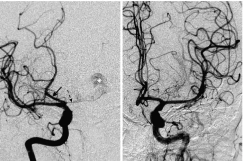
Figure 1 Types of basilar fusion. Digital subtraction angiography— posteroanterior incidences with contrast injection in the vertebral artery (A, right; B, C, left). (A) Symmetrical cranial fusion with both anterior superior cerebellar arteries (ASCAs) originating from the basilar trunk (black arrows). (B) Symmetrical caudal fusion, with both ASCAs originating from the posterior cerebral artery P1 segments (black arrows). (C) Symmetrical fusion with the right ASCA originating from the P1 on the right (white arrow) and from the basilar trunk on the left. Note the previously coiled basilar tip aneurysms (asterisk).
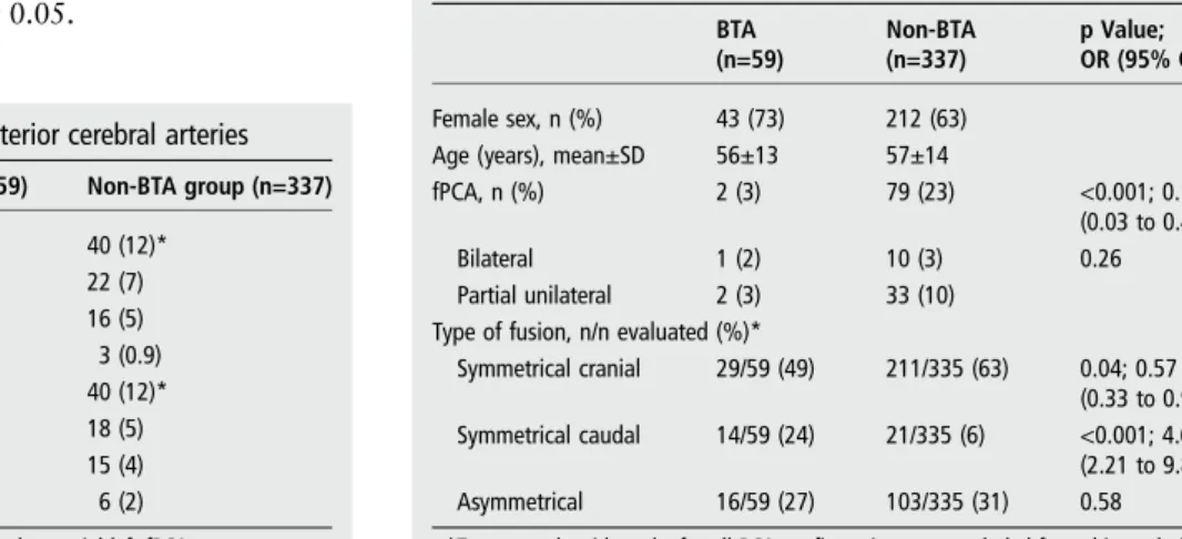
Table 1 Distribution of fetal-type posterior cerebral arteries
BTA group (n=59) Non-BTA group (n=337) Complete fPCA, n (%) 0 40 (12)* Unilateral right, n (%) – 22 (7) Unilateral left, n (%) – 16 (5) Bilateral, n (%) – 3 (0.9) Partial fPCA, n (%) 2 (3) 40 (12)* Unilateral right, n (%) 1 (2) 18 (5) Unilateral left, n (%) 0 15 (4) Bilateral, n (%) 1 (2) 6 (2) *One non-BTA case had a complete right fPCA and a partial left fPCA. BTA, basilar tip aneurysm; fPCA, fetal-type posterior cerebral artery.
Table 2 BTA and non-BTA groups: demographic characteristics and basilar tip variations
BTA (n=59) Non-BTA (n=337) p Value; OR (95% CI) Female sex, n (%) 43 (73) 212 (63)
Age (years), mean±SD 56±13 57±14
fPCA, n (%) 2 (3) 79 (23) <0.001; 0.11 (0.03 to 0.48) Bilateral 1 (2) 10 (3) 0.26 Partial unilateral 2 (3) 33 (10)
Type of fusion, n/n evaluated (%)*
Symmetrical cranial 29/59 (49) 211/335 (63) 0.04; 0.57 (0.33 to 0.99) Symmetrical caudal 14/59 (24) 21/335 (6) <0.001; 4.65
(2.21 to 9.80) Asymmetrical 16/59 (27) 103/335 (31) 0.58 *Two controls with an‘unfused’ PCA configuration were excluded from this analysis. BTA, Basilar tip aneurysm; fPCA, fetal-type posterior cerebral artery.
2 Diogo MC, et al. J NeuroIntervent Surg 2016;0:1–4. doi:10.1136/neurintsurg-2016-012503
DISCUSSION
In our study, BTA were significantly associated with the presence of both P1 segments, and the frequency of fPCA was signi fi-cantly lower in the BTA group than in patients without BTA. Thisfinding is in line with the known the influence of anatom-ical factors on arterial hemodynamics14 15 and aneurysm formation.
Anatomical variants of the circle of Willis are common, and some have long been associated with increased incidence of intracranial aneurysms—namely, of the anterior circulation, sup-porting the idea that vascular anatomy influences aneurysm formation.16 17
There are fewer studies concerning posterior circulation var-iants, with varying results. Kayembeet al,16found no increased incidence of fPCA in the aneurysm group compared with con-trols, but only four of the 44 aneurysms studied involved the basilar tip. More recently, Canet al,10found that a smaller BA diameter and a larger P1–P1 angle were associated with a higher
incidence of BTA, but anatomical variants of the circle of Willis were not assessed. Campos et al,18 identified P1 agenesis in 2.8% of 47 patients with BTA, but hypoplastic P1 segments were not assessed and no data on the prevalence of those var-iants in individuals without BTA were given. In another study investigating the effect of anatomical variations on the incidence of aneurysm recurrence after treatment, Songsaeng et al,19 found a positive association between fPCA and recurrence of posterior communicating artery aneurysms. To our knowledge, this is thefirst study to examine the specific association of per-sistent fPCA and aneurysms of the basilar apex.
It has been established in animal and computational models6 9that hemodynamic forces, especially wall shear stress, can lead to the formation of saccular aneurysms, both by mech-anically affecting the vessel wall and inducing molecular changes that modify it.6 8 9Hemodynamic stress is highest at the bifurca-tion apex of a vessel, such as the tip of the BA.4 8 It is also known that in subjects with unilateral or bilateral fPCA, the Figure 2 Digital subtraction
angiography—right unilateral partial fetal-type posterior cerebral artery (fPCA) in patient with basilar tip aneurysms (BTA). (A) Posteroanterior view after right internal carotid artery (ICA) injection shows opacification of the ipsilateral PCA (black arrow, fPCA); there is also partialfilling of the BTA (asterisk) through a hypoplastic right P1 segment (dashed arrow). (B) Posteroanterior view after left ICA injection with opacification of both the anterior cerebral artery and ipsilateral middle cerebral artery. There is no filling of the posterior circulation.
Figure 3 Bilateral partial fetal-type posterior cerebral artery (fPCA). Anteroposterior projections after contrast injection on the right (A) and left (B) internal carotid artery depict opacification of the anterior circulation and PCA bilaterally, via posterior communicating arteries. (C) Posteroanterior projection after left vertebral artery contrast injection shows presence of hypoplastic P1 segments bilaterally (black arrows).
flow rate in the BA is decreased, in comparison with corre-sponding flow values for a circle of Willis with anterograde feeding of the PCAs from the BA.14 15It would hence be logical to assume that the presence of fPCA, by both reducing the blood flow in the BA, and rendering the high-flow bifurcation into a lower pressure bifurcation, would be associated with a lower incidence of BTA.
In a total of 59 patients with BTA assessed in our study, no complete fPCA were found, with all patients presenting a P1 segment. Two patients (3.4%) presented partial fPCA, one bilat-eral and one unilatbilat-eral right, which was significantly lower than the frequency in the control group ( p<0.001). We did not study the relation between P1 segment diameter and aneurysm size.
Although there is a strong statistical significance, it is not pos-sible to extrapolate from ourfindings that fPCA and aneurysms of the BA apex are mutually exclusive. However, it might be argued that this anatomical variant bestows a relative protection against developing saccular BTA, either owing to wall stress reduction or in combination with other factors already discussed in the literature, such as the P1–P1 angle. We believe that fPCA as an anatomical parameter, with its inherent associated hemo-dynamic consequences, may be a useful tool in predicting the risk of BTA formation. As sheer stress also influences growth and rupture,4fPCA might also be a potential protective factor
in patients with existing BTA.
A study of the BA fusion type, showed a higher prevalence of caudal fusion in the BTA group than in controls, which was stat-istically significant. The relatively higher frequency of caudal BA fusion in patients with BTA is in accordance with previously published data18 19and ourfindings reinforce the importance of
BA fusion type in the formation of BTA.
From a treatment perspective, recognition of variants is para-mount for endovascular planning and follow-up. Our studies as well as previous publications demonstrate that in most patients with BTA, P1 segments are patent,18 allowing for stent place-ment if needed during treatplace-ment.
Our study has some limitations, mainly related to its retro-spective design and the small sample size. Nevertheless, given the lower incidence of BTA, it is a relatively large BTA sample in comparison with previously published series.16–19 In addition, our control group did not comprise healthy individuals, but patients who underwent DSA for a number of different and het-erogeneous pathologies. However, we have no reason to believe that the incidence of posterior circulation variants in these indivi-duals would differ from that of the general population, and our results for fPCA are similar to those of previous reports.12 13
In conclusion, we found a statistically significant association between the absence of a fPCA and BTA. The presence of a fPCA could be an additional morphological parameter to con-sider when assessing the probability of aneurysm formation in high-risk patients.
Further studies, including computationalflow studies, would be needed to assess the probable protective effect of fPCA against the formation of saccular aneurysms at the basilar apex.
Contributors MCD: project development, data collection, manuscript writing,final approval of the version to be published. IF: project development, manuscript writing, final approval of the version to be published. SPD: manuscript writing, statistical analysis,final approval of the version to be published. JN: data collection, final approval of the version to be published. JP: data collection, manuscript correction, final approval of the version to be published. JR: project development, data collection,final approval of the version to be published.
Competing interests None declared.
Ethics approval Approved by the ethics committee of Centro Hospitalar de Lisboa Central.
Informed consent All patients gave informed consent before inclusion in this study. Provenance and peer review Not commissioned; externally peer reviewed. Data sharing statement For any questions or further data requests, contactfirst author.
REFERENCES
1 Caranci F, Briganti F, Cirillo L, et al. Epidemiology and genetics of intracranial aneurysms.Eur J Radiol2013;82:1598–605.
2 Brisman JL, Song JK, Newell DW. Cerebral aneurysms.N Engl J Med 2006;355:928–39.
3 Greving JP, Wermer MJ, Brown RD, et al. Development of the PHASES score for prediction of risk of rupture of intracranial aneurysms: a pooled analysis of six prospective cohort studies.Lancet Neurol2014;13:59–66.
4 Francis SE, Tu J, Qian Y, et al. A combination of genetic, molecular and
haemodynamic risk factors contributes to the formation, enlargement and rupture of brain aneurysms.J Clin Neurosci2013;20:912–18.
5 Bacigaluppi S, Piccinelli M, Antiga L, et al. Factors affecting formation and rupture of intracranial saccular aneurysms.Neurosurg Rev2014;37:1–14.
6 Gao L, Hoi Y, Swartz DD, et al. Nascent aneurysm formation at the basilar terminus induced by hemodynamics.Stroke2008;39:2085–90.
7 Feigin V, Parag V, Lawes CM, et al. Smoking and elevated blood pressure are the most important risk factors for subarachnoid hemorrhage in the Asia-Pacific region: an overview of 26 cohorts involving 306,620 participants.Stroke2005;36:1360–5. 8 Penn DL, Komotar RJ, Sander Connolly E. Hemodynamic mechanisms underlying
cerebral aneurysm pathogenesis. J Clin Neurosci 2011;18:1435–8.
9 Foutrakis GN, Yonas H, Sclabassi RJ. Saccular aneurysm formation in curved and bifurcating arteries. AJNR Am J Neuroradiol 1999;20:1309–17.
10 Can A, Mouminah A, Ho AL, et al. Effect of vascular anatomy on the formation of basilar tip aneurysms.Neurosurgery2015;76:62–6.
11 Lasjaunias P, Berenstein A, TerBrugge K. Clinical vascular anatomy and variations. Springer Science & Business Media, 2013.
12 Kovač JD, Stanković A, Stanković D, et al. Intracranial arterial variations: a comprehensive evaluation using CT angiography.Med Sci Monit2014;20:420–7. 13 van Raamt AF, Mali WP, van Laar PJ, et al. The fetal variant of the circle of Willis
and its influence on the cerebral collateral circulation.Cerebrovasc Dis 2006;22:217–24.
14 Tanaka H, Fujita N, Enoki T, et al. Relationship between variations in the circle of Willis andflow rates in internal carotid and basilar arteries determined by means of magnetic resonance imaging with semiautomated lumen segmentation: reference data from 125 healthy volunteers. AJNR Am J Neuroradiol 2006;27:1770–5. 15 Hendrikse J, van Raamt AF, van der Graaf Y, et al. Distribution of cerebral blood
flow in the circle of Willis.Radiology2005;235:184–9.
16 Kayembe KNT, Sasahara M, Hazama F. Cerebral aneurysms and variations in the circle of Willis.Stroke1984;15:846–50.
17 Tarulli E, Fox AJ. Potent risk factor for aneurysm formation: termination aneurysms of the anterior communicating artery and detection of A1 vessel asymmetry byflow dilution.AJNR Am J Neuroradiol2010;31:1186–91.
18 Campos C, Churojana A, Rodesch G, et al. Basilar tip aneurysm and basilar tip anatomy. Interv Neuroradiol 1998;4:121–5.
19 Songsaeng D, Geibprasert S, Willinsky R, et al. Impact of anatomical variations of the circle of Willis on the incidence of aneurysms and their recurrence rate following endovascular treatment.Clin Radiol2010;65:895–901.
4 Diogo MC, et al. J NeuroIntervent Surg 2016;0:1–4. doi:10.1136/neurintsurg-2016-012503