Both the Caspase CSP-1 and a Caspase-Independent
Pathway Promote Programmed Cell Death in Parallel to
the Canonical Pathway for Apoptosis in
Caenorhabditis
elegans
Daniel P. Denning, Victoria Hatch, H. Robert Horvitz*
Howard Hughes Medical Institute and Department of Biology, Massachusetts Institute of Technology, Cambridge, Massachusetts, United States of America
Abstract
Caspases are cysteine proteases that can drive apoptosis in metazoans and have critical functions in the elimination of cells during development, the maintenance of tissue homeostasis, and responses to cellular damage. Although a growing body of research suggests that programmed cell death can occur in the absence of caspases, mammalian studies of caspase-independent apoptosis are confounded by the existence of at least seven caspase homologs that can function redundantly to promote cell death. Caspase-independent programmed cell death is also thought to occur in the invertebrate nematode Caenorhabditis elegans. TheC. elegansgenome contains four caspase genes (ced-3,csp-1,csp-2, andcsp-3), of which only ced-3has been demonstrated to promote apoptosis. Here, we show that CSP-1 is a pro-apoptotic caspase that promotes programmed cell death in a subset of cells fated to die duringC. elegansembryogenesis.csp-1is expressed robustly in late pachytene nuclei of the germline and is required maternally for its role in embryonic programmed cell deaths. Unlike CED-3, CSP-1 is not regulated by the APAF-1 homolog CED-4 or the BCL-2 homolog CED-9, revealing that csp-1 functions independently of the canonical genetic pathway for apoptosis. Previously we demonstrated that embryos lacking all four caspases can eliminate cells through an extrusion mechanism and that these cells are apoptotic. Extruded cells differ from cells that normally undergo programmed cell death not only by being extruded but also by not being engulfed by neighboring cells. In this study, we identify incsp-3; csp-1; csp-2 ced-3quadruple mutants apoptotic cell corpses that fully resemble wild-type cell corpses: these caspase-deficient cell corpses are morphologically apoptotic, are not extruded, and are internalized by engulfing cells. We conclude that both caspase-dependent and caspase-independent pathways promote apoptotic programmed cell death and the phagocytosis of cell corpses in parallel to the canonical apoptosis pathway involving CED-3 activation.
Citation:Denning DP, Hatch V, Horvitz HR (2013) Both the Caspase CSP-1 and a Caspase-Independent Pathway Promote Programmed Cell Death in Parallel to the Canonical Pathway for Apoptosis inCaenorhabditis elegans. PLoS Genet 9(3): e1003341. doi:10.1371/journal.pgen.1003341
Editor:Hermann Steller, The Rockefeller University, United States of America
ReceivedNovember 14, 2012;AcceptedJanuary 9, 2013;PublishedMarch 7, 2013
Copyright:ß2013 Denning et al. This is an open-access article distributed under the terms of the Creative Commons Attribution License, which permits unrestricted use, distribution, and reproduction in any medium, provided the original author and source are credited.
Funding:This work was funded by the Howard Hughes Medical Institute, the Damon Runyon Cancer Research Foundation, and the Charles A. King Trust. The Caenorhabditis Genetics Center is supported by the NIH Office of Research Infrastructure Programs (P40 OD010440). The funders had no role in study design, data collection and analysis, decision to publish, or preparation of the manuscript.
Competing Interests:The authors declared that no competing interests exist.
* E-mail: [email protected]
Introduction
The elimination of unnecessary or dangerous cells is funda-mental to development, tissue homeostasis and disease mitigation in multicellular organisms. The primary mechanism of cell elimination is apoptosis, a form of cell suicide that was originally defined by evolutionarily conserved morphological characteristics that include chromatin condensation, shrinkage of the cytoplasmic volume and membrane blebbing [1] and by biochemical features like phosphatidylserine exposure and DNA fragmentation [2,3]. Apoptosis serves as a highly controlled mechanism for the removal and degradation of damaged or unnecessary cells, and blocking apoptosis can lead to catastrophic forms of cell death, such as necrosis, which can cause dangerous inflammatory responses [4]. The discovery of the CED-3 caspase as a cell-autonomous executioner of programmed cell death in the nematode Caenor-habditis elegans led to the paradigm that the caspase family of
cysteine proteases drives apoptosis through the cleavage of substrate proteins at specific peptide sequences [5,6]. Indeed, caspases have evolutionarily conserved roles in apoptosis through-out metazoa [7].
prokaryotes, that lack clear caspase homologs [11,12]. Thus, it is possible that apoptosis, as defined morphologically and biochem-ically, can occur in the absence of caspases.
A standard approach to assaying the caspase-dependence of apoptotic stimuli in tissue cell culture is through the pharmaco-logical inhibition of caspases. However, it is difficult to prove that caspase activity is completely blocked in such experiments, and it is possible for caspase inhibitors to trigger non-apoptotic forms of cell death [13]. Studies of caspase-independent apoptosis in metazoans are also complicated by the existence of multiple caspases with potentially redundant functions in promoting cell death. The human genome, for example, encodes at least 10 caspase homologs, seven of which (caspases-2, -3, -6, -7, -8, -9 and -10) have demonstrated roles in apoptosis [14]. The genome of Drosophila melanogaster encodes seven caspase homologs (dcp-1, dronc,drice, dredd,decay, dammand strica) [7], several of which are essential for organismal viability. TheC. elegans genome encodes three caspase homologs (csp-1,csp-2andcsp-3) in addition toced-3 [15]. Therefore, the use of mutant animals or cell lines deleted for one or two caspases might not eliminate all caspases expressed within a specific cell. Furthermore, since caspases have different substrate specificities [16], the use of a chemical substrate-competitive caspase inhibitor might not completely block all caspase activity. Ideally, experiments that test whether apoptosis can occur in the absence of caspases should be performed using mutant animals or cells that are genetically deleted of all caspase homologs. In this regard, C. elegans is an excellent animal for studies of caspase-independent programmed cell death, because: (1) there are several examples ofced-3-independent programmed cell death inC. elegans[17–19]; (2) mutants ofced-3,csp-1,csp-2and csp-3 are viable [18–23]; and, (3) it is relatively easy to generate multiply mutantC. elegansstrains.
Theced-3 caspase gene is required for most programmed cell deaths that occur duringC. elegansdevelopment [5,20]. However, a small number of cells die in animals carrying null mutations of ced-3. The male-specific linker cell, which facilitates the connection of the vas deferens to the cloaca and then dies, undergoes a non-apoptotic cell death that bears morphological features (e.g.,
nuclear membrane crenellation) not seen with other C. elegans programmed cell deaths and that occurs inced-3mutants as well as in animals doubly mutant for ced-3 and csp-1, csp-2 or csp-3 [18,20,24]. We recently showed that a subset of cells fated to die in the C. elegans embryo are eliminated from ced-3 mutants via a caspase-independent shedding mechanism [19]. Interestingly, the shed cells appear apoptotic, exhibiting chromatin condensation, TUNEL-reactive DNA degradation and phosphatidylserine expo-sure despite the absence of all four caspases. Unlike other apoptotic programmed cell deaths ofC. elegans, the shed cells do not undergo phagocytosis by engulfing cells; instead, they are extruded from the developing embryo. By contrast, a small number of apoptotic cell corpses are visible in the heads ofced-3 larvae [17]. Like other programmed cell deaths ofC. elegans, these ced-3-independent cell corpses have a refractile appearance when viewed with Nomarski optics and are not extruded from the animal, suggesting that a ced-3-independent cell-killing activity contributes to these typical programmed cell deaths. The other caspase homologs,csp-1,csp-2andcsp-3, are obvious candidates for driving thisced-3-independent cell-killing activity. However, it has recently been reported thatcsp-2andcsp-3inhibit apoptosis in the germline and soma, respectively [22,23].
To date, the C. elegans caspase homolog csp-1 has no known functionin vivo, including in apoptosis. An isoform of CSP-1 can cleave and possibly activate the CED-3 pro-proteinin vitro[15]. We tested whethercsp-1can promote or inhibit programmed cell death and whether it is regulated by the canonical programmed cell death pathway that activatesced-3. We found thatcsp-1encodes a pro-apoptotic caspase activity that promotes programmed cell death independently of the CED-3 caspase, CED-4 (the Apaf-1 homolog that activates CED-3), and CED-9 (a Bcl-2 family protein that negatively regulates CED-3 activation via inhibition of CED-4). Furthermore, we tested whethercsp-1,csp-2andcsp-3contribute to theced-3-independent cell-killing activity that generates cell corpses in the heads ofced-3mutant larvae and found that these apoptotic cell deaths can occur in the complete absence of caspases. Thus, duringC. elegansdevelopment programmed cell death followed by cell-corpse engulfment is achieved through three redundant pathways: (1) a ced-3-dependent pathway; (2) a csp-1-dependent pathway, which is not regulated by the canonical apoptosis pathway that controlsced-3; and, (3) a caspase-independent pathway.
Results
csp-1promotes the deaths of a subset of somatic cells
fated to die
TheC. elegansgenescsp-1,csp-2andcsp-3are paralogs of the pro-apoptotic ced-3 caspase gene [15], which is required for most programmed cell deaths in the worm [5,20]. Given the conserved role of caspases in apoptosis, we testedcsp-1,csp-2 andcsp-3 for roles – both pro- and anti-apoptotic – in programmed cell death. We used mutations ofcsp-1 (n4967and n5133) andcsp-2(n4871) that completely remove the genomic sequences encoding their respective predicted caspase active sites (SACRG in the CSP-1 protein, and VCCRG in the CSP-2 protein) and therefore eliminate any potential caspase activity encoded by these genes (ref. [19]; Figure 1A).csp-3lacks a caspase active site (ref. [15,22]; Figure 1A); we used thecsp-3deletion mutation n4872, which is likely a null allele [19].
Recently, it was reported that mutations incsp-2andcsp-3cause ectopic cell deaths in the germline and soma, respectively, and hence thatcsp-2andcsp-3inhibit apoptosis [22,23]. We therefore tested whethercsp-1mutants have ectopic cell deaths indicative of a loss of anti-apoptotic function. Using Nomarski optics and a Author Summary
Pmec-3::gfptransgene that expresses GFP in the six touch neurons (AVM, two ALM, PVM and two PLM neurons) in addition to the FLP and PVD neurons, we examinedcsp-1 mutants for missing cells that normally survive. We observed thatcsp-1(n4967)mutants contained a full complement of touch neurons and pharyngeal cells (Table S1). We also noted thatcsp-1(n4967) failed to cause ectopic cell deaths in sensitized animals carrying the loss-of-function mutation n2812 in the anti-apoptotic gene ced-9, a homolog of human BCL2 (Table S1; data not shown). These results indicate thatcsp-1does not have an obvious anti-apoptotic function in the soma. Consistent with a previous report thatcsp-2 does not affect somatic cells [23], a mutation incsp-2did not cause ectopic cell deaths in the somatic cells we examined (Table S1). However, we failed to observe the ectopic cell deaths in csp-3 mutants previously reported [22]. Ectopic somatic cell deaths have also been noted in animals with loss-of-function mutations inced-9 [25] or tat-1 [26,27], which encodes an aminophospholipid translocase required for the asymmetric distribution of phospha-tidylserine on the inner leaflet of the plasma membrane. As expected, we found that ced-9 mutant larvae were missing pharyngeal cells and many touch neurons: more than 80% of PLM neurons were not present inced-9(n2812)larvae (Table S1). However, we failed to detect the previously reported ectopic cell-death defect oftat-1mutants (ref. [26,27]; Table S1); we used the same deletion alleles forcsp-3andtat-1and assayed the same cells that had been characterized in the previous studies.
To determine whether theC. eleganscaspase homologscsp-1,csp-2 orcsp-3promote programmed cell death in the soma, we examined animals carryingcspdeletion mutations for extra cells that failed to undergo programmed cell death in the anterior pharynx. As many as 16 extra cells can be counted in the anterior pharynges of mutants
with strong defects in programmed cell death, e.g.,ced-3(n3692)(ref. [28]; Table 1). Single mutations incsp-1,csp-2orcsp-3failed to cause detectable defects in programmed cell death (Table 1; data not shown). However, we observed that mutations incsp-1(but notcsp-2 orcsp-3) caused the survival of pharyngeal cells in sensitized strains carrying a weak mutation in the caspase geneced-3(Table 1). The partial loss-of-functionced-3mutationsn2427andn2436cause slight and intermediate defects in apoptosis, respectively (ref [17]; Table 1; data not shown). Then4967andn5133mutations, both of which delete the putative active site of CSP-1 (Figure 1A), enhanced the cell-death defects ofced-3(n2427)andced-3(n2436)mutants, increasing the number of extra cells in their anterior pharynges on average by 1.4 and 2.4 cells, respectively (Table 1). These results are consistent with our RNAi experiments in which csp-1B dsRNA (which likely inactivated allcsp-1transcripts) was injected into the gonads of rrf-3(pk1426); ced-3(n2436)animals and caused an enhanced cell-death defect in their progeny (Figure 1C); we used therrf-3mutation to increase sensitivity to RNAi [29]. The cell-death defect conferred by thecsp-1(n4967)mutation was rescued by extrachromosomal arrays carrying a 9 kb genomiccsp-1fragment that included the entirecsp-1 coding region, 1.5 kb of genomic sequence 59 of the csp-1A translational start codon and 3.5 kb of genomic sequence 39of the csp-1A/Btranslational stop codon (Figure 1B; Table S2). These results demonstrate thatcsp-1encodes a detectable cell-killing activity that contributes to programmed cell death inC. elegans. Mutation ofcsp-2 and/orcsp-3neither enhanced nor suppressed the cell-death defects of strains mutant for csp-1 and/or ced-3 (Table 1; Table S3), suggesting thatcsp-1andced-3are the onlyC. eleganscaspase genes with functions in somatic programmed cell deaths.
The development of the anterior part of theC. eleganspharynx involves 16 programmed cell deaths, all of which appear to be Figure 1. The B and/or C isoforms ofcsp-1promote programmed cell death.(A) Representations of the intron-exon organization of the three knowncsp-1mRNA isoforms (A,BandC). Red bars indicate thecsp-1deletion alleles used in this study; arrowheads indicate the SACRG sequence that encodes the caspase active-site. The graphic was generated using the Intron-Exon Graphic Maker (N. Bhatla; www.wormweb.org). (B) Extrachromosomal arrays carrying a wild-type genomic fragment of thecsp-1locus or a mutant version that expresses only theBorCisoforms can rescue thecsp-1(n4967)mutant phenotype. Thecsp-1 PD-only transgene contains two nonsense mutations that encode early stop codons affecting theBandCmRNA isoforms; thecsp-1A-only transgene contains a mutation that changes theB/Cstart codon to an alanine codon; and thecsp-1B/C -only transgene contains two nonsense mutations that encode early stop codons affecting theAisoform. Thecsp-1transgenes were injected into csp-1(n4967); ced-3(n2436)animals, and the resulting independent lines were assayed forcsp-1rescuing activity by counting the number of extra undead cells in the anterior pharynx. The transgenes and their constructions are described in detail in Materials and Methods, and the complete data for each transgenic line are provided in Table S2. (C) RNAi knockdown ofcsp-1phenocopies thecsp-1mutations. dsRNAs targeting thecsp-1pro-domain or thecsp-1Bisoform werein vitrotranscribed and injected into the gonads of RNAi-sensitiverrf-3(pk1426); ced-3(n2436)adult hermaphrodites. Progeny of the injected adults were assayed for extra undead cells in the anterior pharynx. PD, prodomain.
doi:10.1371/journal.pgen.1003341.g001
sensitive to ced-3[17,28,30]. To test whether specific pharyngeal programmed cell deaths requiredcsp-1, we used GFP reporters to visualize the survival of cells fated to die, specifically the sister cells of the M4 and NSM neurons.csp-1was partially required in ced-3(n2427)orced-3(n2436)sensitized strains for the death of the M4 sister cell (Table S4); by contrast, mutation ofcsp-1did not affect the cell deaths of the sister cells of the NSM neurons (data not shown). Likewise, csp-1 did not appear to function in the postembryonic programmed cell deaths of the ventral cord or postdeirid sensilla (Table S4). We conclude thatcsp-1promotes cell death in a subset of cells fated to die duringC. elegansdevelopment.
Thecsp-1Band/orCisoforms are required for the
cell-killing activity ofcsp-1
Thecsp-1locus produces three known mRNA isoforms [15], all of which include the sequence that encodes the presumptive caspase active site (Figure 1A). The csp-1A transcript contains a
long prodomain not present in the other transcripts, and it uses an alternative start site that is 3 kb 59to the start site of thecsp-1Band csp-1Cisoforms. To determine which isoforms are required for the cell-killing activity ofcsp-1, we peformed experiments in which the csp-1rescuing transgene was mutated to express: (1) theAisoform only, (2) theBand Cisoforms only, or (3) a truncated version of csp-1A including only the prodomain (PD). Extrachromosomal arrays engineered to express onlycsp-1-PDor thecsp-1Aisoform failed to rescue the cell-death defect of csp-1(n4967) mutants (Figure 1B; Table S2). By contrast, acsp-1transgene lacking the csp-1Atranslation start codon and predicted to express only the csp-1B and csp-1C transcripts rescued the csp-1(n4967) defect in programmed cell death (Figure 1B; Table S2). Consistent with these results, transgenes expressing thecsp-1BcDNA, but not the csp-1AcDNA, under the control of themec-7promoter efficiently killed touch neurons (Figure 2A–2B; Table 2; data not shown); we also expresed thecsp-1C cDNA under the control of the mec-7 promoter and failed to observe killing of the touch neurons (data
Table 1.The caspase homologcsp-1promotes programmed cell death in theC. elegansanterior pharynx.
genotype extra cells per anterior pharynx±SD n pvalue
The deletion ofcsp-1,csp-2orcsp-3alone does not cause a defect in programmed cell death.
wild-type1 0.1
60.3 14
-ced-3(n3692)1 11.361.1 14 ,0.00001
csp-1(n4967) 0.360.4 16 n.s.
csp-1(n5133) 0.160.2 19 n.s.
csp-1(tm917) 0.160.3 16 n.s.
csp-2(n4871)1 0.2
60.4 12 n.s.
csp-3(n4872)1 0.3
60.6 21 n.s.
Deletion ofcsp-1, but notcsp-2orcsp-3, enhances the defects in programmed cell death caused by partial loss-of-function alleles ofced-3andced-4.
ced-3(n2427)1 1.7
61.2 22
-csp-1(n4967); ced-3(n2427)2 3.0
61.3 38 0.0001
csp-1(n5133); ced-3(n2427) 3.261.5 21 0.0008
csp-1(tm917); ced-3(n2427)2 2.6
61.3 46 0.006
csp-2(n4871) ced-3(n2427)1 1.0
60.8 20 n.s.
csp-3(n4872); ced-3(n2427)3 1.5
61.2 16 n.s.
csp-3; csp-2 ced-3(n2427)1 1.9
61.5 18
-csp-3; csp-1(n4967); csp-2 ced-3(n2427) 3.261.2 17 0.008
ced-3(n2436)1 6.2
61.4 37
-csp-1(n4967); ced-3(n2436) 8.661.6 29 ,0.00001
csp-1(n5133); ced-3(n2436) 8.761.5 19 ,0.00001
csp-1(tm917); ced-3(n2436)2 7.4
61.6 52 ,0.00001
csp-2(n4871) ced-3(n2436)1 5.4
61.0 16 n.s.
csp-3(n4872); ced-3(n2436)1 5.661.5 15 n.s.
csp-3; csp-2 ced-3(n2436)1 4.9
61.7 16
-csp-3; csp-1(n4967); csp-2 ced-3(n2436) 8.261.4 16 ,0.00001
ced-4(n3158)1 5.0
62.6 29
-csp-1(n4967); ced-4(n3158)1 6.5
62.7 32 0.03
1Homozygous for the integrated transgenenIs106[P
lin-11::gfp].
2Includes animals that were either homozygous forunc-75(
+)orunc-75(e950).
3Homozygous for
dpy-20(e1282ts).
For the statistical comparisons betweenced-3(n2427)orced-3(n2436)and double mutants with eachcspallele,pvalues were considered significant if less than 0.01 to correct for multiple comparisons.
not shown). Ectopic expression ofcsp-1Bfrom theser-2dandflp-15 promoters killed the OLL and I2 neurons, respectively (ref. [31]; N. Bhatla and H.R. Horvitz, unpublished results). However, we noted thattm917, acsp-1allele that deletes coding regions of only the csp-1A transcript, enhanced significantly (albeit weakly) the cell-death defects ofced-3(n2427)andced-3(n2436)mutants, increasing the number of extra cells in their anterior pharynges by 0.9 and 1.2
cells, respectively (Table 1). dsRNA targetting thecsp-1Aprodomain (csp-1-PD) caused a similar slight enhancement of the cell-death defect of ced-3(n2436) mutants (Figure 1C), suggesting that, in addition to the more robust cell-killing activity of the csp-1B transcript,csp-1Amight have a weak cell-killing function.
csp-1Bencodes a pro-apoptotic caspase
The proteolytic activity of caspases requires an active-site cysteine. Previously, it was shown that the CSP-1B protein can proteolytically process CED-3 in vitro and that this enzymatic activity required the active-site (SACRG) cysteine of CSP-1B, C138 [15]. We tested in vivo whether C138 was necessary by assaying the touch neuron-killing activity of mutant Pmec-7::csp-1B transgenes in which C138 was changed to a serine. We observed that the ectopic cell deaths were entirely dependent on the caspase active site (Table 2). Thus,csp-1Bpromotes cell death via caspase activity. The cell deaths induced by a Pmec-7::csp-1B transgene resulted in cell corpses with apoptotic characteristics (Figure 2C– 2D). When observed with Nomarski optics, thecsp-1B-induced cell deaths exhibited a refractile button-like appearance (Figure 2C) similar to that of developmental programmed cell deaths. Transmission electron micrographs of the cell corpses showed some contraction of the cytoplasmic volume and considerable condensation of the nuclear chromatin (Figure 2D), which are general characteristics of apoptotic cells, including those generated by ced-3 cell-killing transgenes (ref. [32]; data not shown). We conclude thatcsp-1B encodes a functional caspase that promotes programmed cell deaths with apoptotic morphology.
csp-1Bcell-killing activity is independent ofced-9and
ced-4
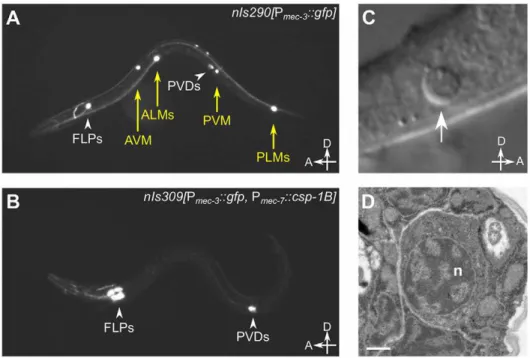
CED-3, like most caspases, is expressed as an inactive zymogen with an inhibitory N-terminal prodomain. Trans-auto-proteolysis Figure 2.csp-1Boverexpression induces ectopic cell deaths.(A) Fluorescence image of a transgenicnIs290[Pmec-3::gfp]larva expressing GFP
from themec-3promoter in the touch neurons (AVM, ALMs, PVM and PLMs, yellow arrows).mec-3is also expressed in the FLP and PVD neurons (white arrowheads). (B) Fluorescence image of a transgenicnIs309[Pmec-7::csp-1B, Pmec-3::gfp]larva expressing CSP-1B from themec-7promoter (which
is expressed in the AVM, ALM, PVM and PLM neurons) and GFP from themec-3promoter. Note the absence of touch neurons. (C) Nomarski differential interference contrast (DIC) image of a refractile PLM cell corpse (arrow) in aced-1(e1735); ced-4(n1162); nIs309L1 larva. (D) Transmission electron microscopic image of the cell corpse in (C). ‘‘n’’, nucleus of the cell corpse; scale bar, 0.5 microns.
doi:10.1371/journal.pgen.1003341.g002
Table 2.Ectopic expression ofcsp-1Bfrom themec-7 promoter can kill touch neurons, and this killing activity requires the conserved cysteine in the putative caspase active site.
% survival
genotype* n AVM ALML/R PVM PLML/R
wild-type[nIs290] 59 100 100 100 100
Pmec-7::csp-1BLine#1[nIs307] 52 71 61 40 13
Pmec-7::csp-1BLine#2[nIs308] 41 49 32 27 16
Pmec-7::csp-1BLine#3[nIs309] 60 2 1 3 0
Pmec-7::csp-1B(C138S)Line#1
[nIs368]
23 100 100 100 98
Pmec-7::csp-1B(C138S)Line#2
[nIs369]
23 100 100 100 87
Pmec-7::csp-1B(C138S)Line#3
[nIs370]
25 100 100 100 100
n, number of animals assayed; for each animal, six touch neurons (AVM, ALML, ALMR, PVM, PLML and PLMR) were scored for survival using the Pmec-3::gfpreporter transgene. *Each strain contained the transgene Pmec-3::gfp, which expressed GFP in the FLP, AVM, ALM, PVM, PVD and PLM neurons.
doi:10.1371/journal.pgen.1003341.t002
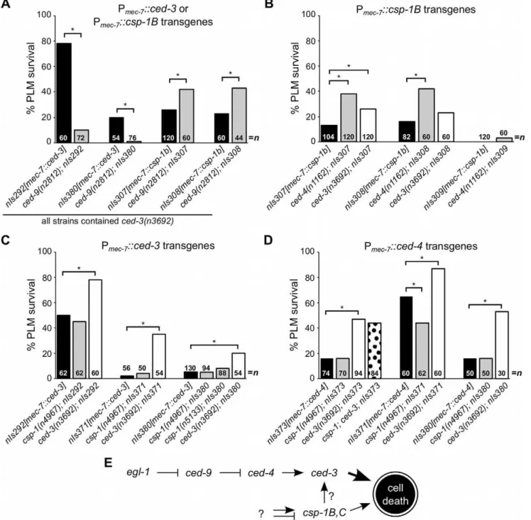
of the CED-3 pro-protein at two aspartate residues removes the pro-domain and yields two subunits that form the active caspase [33]. CED-3 auto-activation is dependent on its prodomain and is facilitated by the association of two CED-3 pro-proteins within an octameric complex formed with the Apaf-1 homolog CED-4 [34– 36]. Under normal cellular conditions, CED-4 is sequestered by CED-9 at mitochondria through a direct protein-protein interac-tion [37–39]. In response to upstream pro-apoptotic signals and the consequent expression of the BH3-domain-only protein EGL-1, which binds to and inhibits CED-9 [40], CED-4 is released from CED-9 and translocates to the nuclear periphery [37,41], where it facilitates CED-3 activation [38]. Thus, the activation of CED-3 is controlled by an apoptosis pathway involving a BH3-domain-only protein, a member of the Bcl-2 family of apoptosis regulators, and a homolog of the apoptosome complex protein Apaf-1. The basic elements of this apoptosis pathway are evolutionarily conserved in mammals and are responsible for the activation of caspases in response to cell-intrinsic apoptotic stimuli [7].
Consistent with the role ofced-9in negatively regulating ced-3 activation, it was previously shown that null mutations of ced-9 enhance the touch neuron-killing activities of Pmec-7::ced-3transgenes [32]. (These experiments were performed using a ced-3(null) background to suppress theced-3-dependent inviability ofced-9(null) animals.) Furthermore, this enhancement is dependent on ced-4 [32], indicating that the absence of CED-9 activates endogenous CED-4 within the touch neurons and that CED-4 activation elevates CED-3 activity. Unlike the CED-3 zymogen, CSP-1B lacks a long prodomain, suggesting that it might be activated via an alternative mechanism (i.e., independently of CED-4 and CED-9). To determine whether these canonical apoptosis regulators control CSP-1B activation, we introduced theced-9(n2812)mutation into ced-3(n3692)strains carrying Pmec-7::csp-1B transgenes and assessed the effect of thisced-9null mutation on PLM survival. In contrast to its effects on Pmec-7::ced-3–mediated PLM killing,ced-9(n2812)failed to enhance PLM killing in Pmec-7::csp-1Bstrains with aced-3(n3692) mutant background (Figure 3A). Instead, ced-9(n2812) partially suppressedcsp-1B-mediated PLM death (Figure 3A). CED-9 has a poorly understood pro-apoptotic activity in addition to its anti-apoptotic role in CED-4 inhibition [42], and it is possible that this ced-9 pro-apoptotic activity contributed to the deaths of cells expressing ectopic CSP-1B. Nevertheless, our results indicate that csp-1B-mediated cell killing, unlikeced-3-mediated cell killing, is not negatively regulated byced-9and suggest that CSP-1B is activated independently of CED-9.
We also observed that the expression of a Pmec-7::csp-1A transgene in ced-3(null) mutant strains failed to cause PLM cell death, even in aced-9(null)background (Figure S1). These results suggest that the CSP-1A isoform (which contains a long prodomain similar to that of CED-3) does not promote programmed cell death, even in the absence of the anti-apoptotic protein CED-9. A role forcsp-1Ain cell death cannot be excluded entirely, as it is possible that endogenous CSP-1A requires a co-factor not present in the touch neurons to mediate cell killing.
Since CSP-1B can proteolytically cleave CED-3in vitro[15], we tested whether the csp-1B cell-killing activing requires the endogenous ced-3 and ced-4 genes. The ced-3(n3692) and ced-4(n1162) mutations weakly suppressed csp-1B-mediated PLM death (Figure 3B), and it is possible that the endogenous csp-1 can in part promote programmed cell death through ced-3. Nonetheless, most csp-1B cell-killing activity was independent of ced-4 and ced-3 (Figure 3B). Loss of endogenous csp-1 failed to suppress PLM death in strains carrying Pmec-7::ced-3or Pmec-7::ced-4 transgenes (Figure 3C–3D). Together, our results are consistent
with a model in whichcsp-1Bpromotes programmed cell death at least mostly independently of and in parallel to the canonical apoptosis pathway (Figure 3E).
csp-1expression in the maternal germline contributes to
embryonic programmed cell death
To determine which C. elegans cells express csp-1, we directly visualized endogenous csp-1 transcripts via fluorescence in situ hybridization (FISH) experiments using Cy5- and ALEXA-labelled probes complementary to the csp-1B transcript (i.e., targeted to allcsp-1transcripts) or to thecsp-1Aprodomain (specific to the csp-1A trancript). To our surprise, csp-1 mRNA was not detectable in the somatic cells of wild-type oregl-1(n1084 n3082) mutant embryos, larvae or adult hermaphrodites (data not shown). By contrast,csp-1transcripts were present in the germlines of L4-stage larval and adult hermaphrodites (Figure 4A–4B). This expression was restricted to the late pachytene stage of meiosis I in both L4 larval gonads (in pachytene nuclei adjacent to differen-tiating sperm) and adult gonads (in pachytene nuclei adjacent to the bend of the gonad arm) (Figure 4A–4B). Bothcsp-1Aand csp-1B/Ctranscripts were expressed in the adult pachytene germ cells, as indicated by the presence of FISH foci recognized by thecsp-1A prodomain probes and foci recognized primarily by the csp-1B probes and only weakly by the csp-1A probes (Figure 4C). Stochastic and ionizing radiation (IR)-induced germline cell deaths occur during the late pachytene stage of oocyte develop-ment in adult gonads [43,44]. However,csp-1(unlikeced-3) was not required for either stochastic or IR-induced germline apoptosis, even inced-3(n2436)strains sensitized for defects in germ-cell death (Figure 4D). In these experiments, apoptotic germ cells were identified using a transgene that expresses a functional GFP::CED-1 fusion protein that envelopes dying cells engulfed by the gonadal sheath [45,46]. We also failed to detect differences in either stochastic or IR-induced germline cell death between csp-1 mutants and wild-type animals in experiments in which apoptotic germ cells were quantified by acridine orange staining or by direct observation of their refractile morphology using Nomarski optics (data not shown). We also noted that the level ofcsp-1 transcript expression in the germline (as determined by FISH) was not affected by either ionizing radiation or by mutation ofegl-1orced-3 (data not shown).
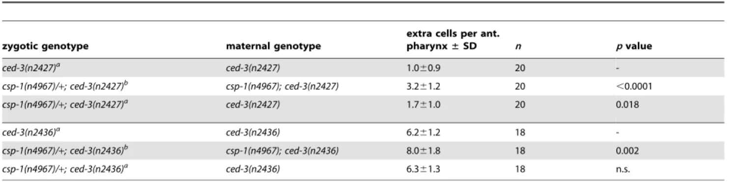
Since we detectedcsp-1expression in the adult germline but not in somatic cells of the embryo, we tested whether maternally suppliedcsp-1transcript was necessary for the zygotic function of csp-1 in programmed cell death. Indeed, in sensitized genetic backgrounds (ced-3(n2427)and ced-3(n2436)),csp-1(+)progeny of csp-1(n4967)hermaphrodites (M2Z+animals) had more undead pharyngeal cells than thecsp-1(+) progeny of csp-1(+) hermaph-rodites (M+Z+ animals) or the csp-1(n4967) progeny ofcsp-1(+) hermaphrodites (M+Z2animals) (Table 3). Thus,csp-1expressed in the maternal germline is necessary for thecsp-1 pro-apoptotic activity in embryonic programmed cell deaths. Given that we could not detectcsp-1expression in either embryos or larvae, it is therefore not surprising that the postembryonic programmed cell deaths of the ventral cord and postdeirid sensilla were unaffected by mutation ofcsp-1(Table S4).
Programmed cell deaths occur in animals completely lacking all caspase genes
mechansm [19]. In that study, we noted that cell shedding from ced-3 mutants occurs independently of csp-1, csp-2 and csp-3: quadruple mutants lacking all four caspases also generate shed cells, indicating that cell elimination by this mechanism is completely caspase-independent [19]. Like most programmed cell deaths, the cells generated by caspase-independent extrusion are apoptotic in appearance. However, unlike caspase-dependent cell corpses, shed cells do not undergo phagocytosis by engulfing cells.
The death of the male linker cell, which also occurs independently ofced-3, is non-apoptotic and requires the heterochronic protein LIN-29, its binding partner MAB-10 [47], and the polyglutamine repeat protein PQN-41 (ref. [18,24]; Table S5). Previously it was shown that this cell death occurs in double-mutant males in which ced-3 and an additional csp gene (csp-1, csp-2 or csp-3) were inactivated [18]. We have now examined males lacking all four caspases and observed that the linker cell died in 100% ofcsp-3; Figure 3.csp-1Bcell-killing activity is not regulated by the canonical programmed cell death pathway.(A–D) The percentages of PLM cells that survive in strains carrying Pmec-7::ced-3, Pmec-7::csp-1Bor Pmec-7::ced-4transgenes. (A)ced-9protects againstced-3- but notcsp-1B-cell-killing
transgenes. (B) The cell-killing activity ofcsp-1Btransgenes is mostly independent ofced-3andced-4. The cell-killing activities of (C)ced-3and (D) ced-4transgenes do not requirecsp-1. PLM survival was scored based on the presence of GFP expressed from themec-3promoter. Asterisks indicate
p,0.05 in a Fisher’s exact test. All strains in (A) containedced-3(n3692). (E) A model depicting the genetic pathways regulating the caspase genes csp-1andced-3(see text).
doi:10.1371/journal.pgen.1003341.g003
csp-1; csp-2 ced-3mutants (Table S5). Thecsp-3; csp-1; csp-2 ced-3 quadruple mutants were viable and fertile. Thus, both zygotic and maternal caspase contributions were eliminated. Our results therefore confirm that this cell death is indeed completely caspase-independent.
In addition, cell corpses are visible in the heads of larvae carrying null alleles ofced-3(ref. [17]; Table 4). All programmed cell deaths in the developing heads of wild-type animals occur embryonically and are engulfed and degraded prior to hatching (ref. [30,48]; Table 4). To detect ced-3-independent programmed cell deaths in larval heads, we used mutations (e.g., ced-1(e1735), ced-6(n2095) or ced-7(n1996)) that cause defects in cell-corpse engulfment and result in the persistence of many embryonic cell corpses into larval stages (ref.
[49,50]; Table 4). Like most wild-type cell corpses, the ced-3-independent cell corpses were refractile in appearance as observed with Nomarski optics and were not extruded from the animal (data not shown). We also observed that larvae mutant forced-4oregl-1 contained similar cell corpses, demonstrating that their generation does not require the canonical pro-apoptotic pathway that mediates most programmed cell deaths (Table 4).
We tested whether the small number of cell corpses visible inced-3 larval heads are generated by the otherC. eleganscaspase genes and found that all double, triple and quadruple caspase mutants that we examined contained a small number of refractile corpses (Table 4). For example, 39% ofcsp-3; csp-1; ced-6; csp-2 ced-3 mutant animals contained at least one refractile cell corpse (Table 4), indicating that Figure 4.csp-1is expressed in late pachytene cells of the L4 and adult hermaphrodite germline.Fluorescencein situhybridization images of gonad arms of (A) an L4 hermaphrodite and (B) an adult hermaphrodite hybridized with Cy5-labelled probes complementary tocsp-1B. The Cy5-labelled probes are visible as green puncta; the gonads are outlined in white. A schematic representation is shown above each micrograph. (C) Fluorescencein situhybridization images of an adult hermaphrodite gonad hybridized with ALEXA594-labelled probes (red puncta) complementary to the region ofcsp-1Athat encodes the prodomain (csp-1A) and Cy5-labelledcsp-1Bprobes (green puncta) that hybridize to allcsp-1transcript isoforms (totalcsp-1). White arrowheads indicatecsp-1A-specific puncta; orange arrows indicatecsp-1B-specific puncta, which are recognized strongly by the totalcsp-1probes but only weakly by thecsp-1A-specific probes. (D) The number of CED-1::GFP-positive apoptotic cells in the gonads of caspase mutants exposed to 0 Gy and 120 Gy of ionizing radiation at the L4 larval stage. The strains were scored at 24 hrs and 48 hrs post L4-stage. Error bars indicate standard deviations.
these programmed cell deaths occur in animals lacking allC. elegans caspases. We observed caspase-independent cell corpses in different regions of the larval head, including positions internal and external to the pharynx, which suggests that multiple cell lineages – at low frequencies – generated caspase-independent cell corpses. Surprising-ly, we discovered that engulfment-competentced-3and3; 1; csp-2 ced-3mutants also contained refractile cell corpses (Table 4). The number of cell corpses perced-3orcsp-1; csp-2 ced-3larva increased until 12 to 24 hours post hatching (see below; data not shown), indicating that at least some of the cell deaths occurred after embryogenesis. Given that all programmed cell deaths in the head normally occur embryonically and that cell corpses are never observed in the heads of wild-type larvae, we concluded that timing of cell
deaths in theseced-3mutants was delayed. Thus, caspase-independent cell corpses can undergo an inefficient programmed cell death with slow kinetics in the absence of CED-3 activity, indicating that these cells likely die via CED-3-mediated apoptosis in wild-type animals.
Caspase-independent cell corpses exhibit apoptotic morphology
Despite the strong causal link between caspase activation and apoptosis, recent studies have demonstrated that many morpho-logical and biochemical changes associated with apoptosis can occur in the absence of caspases [4,19,21]. For example, in C. elegansthe shed cells ofcsp-3; csp-1; csp-2 ced-3quadruple mutants exhibit phosphatidylserine exposure, expression of the
pro-Table 3.csp-1is maternally required for programmed cell deaths that occur embryonically in the presumptive anterior pharynx.
zygotic genotype maternal genotype
extra cells per ant.
pharynx±SD n pvalue
ced-3(n2427)a ced-3(n2427) 1.0
60.9 20
-csp-1(n4967)/+; ced-3(n2427)b csp-1(n4967); ced-3(n2427) 3.2
61.2 20 ,0.0001
csp-1(n4967)/+; ced-3(n2427)a ced-3(n2427) 1.7
61.0 20 0.018
ced-3(n2436)a ced-3(n2436) 6.2
61.2 18
-csp-1(n4967)/+; ced-3(n2436)b csp-1(n4967); ced-3(n2436) 8.0
61.8 18 0.002
csp-1(n4967)/+; ced-3(n2436)a ced-3(n2436) 6.3
61.3 18 n.s.
aHeterozygous forunc-30(e191)/
+.
bHeterozygous forunc-75(e950)/
+. doi:10.1371/journal.pgen.1003341.t003
Table 4.Cell deaths occur in the absence of allC. eleganscaspase genes.
genotype n % with$1 corpse corpses per head±SD
ced-1(e1735) 23 100 21.365.3
ced-1; ced-3(n3692) 49 27 0.360.5
ced-1; ced-4(n1162) 25 36 0.460.6
ced-1; egl-1(n1084 n3082) 26 50 0.660.7
ced-1; csp-1(n4967) 29 100 23.464.7
ced-1; csp-2(n4871) 24 100 20.765.6
ced-1; csp-1(n4967); ced-3(n3692) 30 27 0.360.5
ced-1; csp-1(n4967); csp-2(n4871) ced-3(n3692) 24 21 0.260.4
ced-7(n1996) 21 100 30.164.4
ced-7; ced-3(n3692) 22 23 0.360.6
csp-1(n4967); ced-7 19 100 30.364.3
csp-1(n4967); ced-7; ced-3(n3692) 19 37 0.460.5
csp-3(n4872); csp-1(n4967); ced-7; csp-2(n4871) ced-3(n3692) 32 34 0.460.6
ced-6(n2095) 24 100 19.864.3
csp-3(n4872); csp-1(n4967); ced-6; csp-2(n4871) ced-3(n3692) 36 39 0.460.6
wild-type 28 0 0.060.0
ced-3(n3692) 43 14 0.160.4
ced-3(n2452) 27 41 0.460.5
csp-3(n4872); csp-1(n4967); csp-2(n4871) ced-3(n3692) 25 16 0.260.4
csp-3(n4872); csp-1(n4967); csp-2(n4871) ced-3(n2452) 34 26 0.360.4
The number of refractile cell corpses per head was counted in L1 larvae within one hour of hatching. doi:10.1371/journal.pgen.1003341.t004
apoptotic BH3-only gene egl-1, and chromatin condensation [19]. To determine whether these apoptotic attributes are evident in caspase-independent programmed cell deaths that do not involve extrusion of the dying cell from the embryo, we characterized the cell corpses visible in caspase-deleted larvae (Figure 5 and Figure 6). In most of these experiments, we used strains with the wild-type csp-3 allele, because (1) csp-3lacks a caspase active-site [15]; (2) although previous studies reported that csp-3has an anti-apoptotic function in somatic cells [22], we were unable to replicate those findings (Table S1); and, (3) the presence or absence of acsp-3mutation had no effect on the
frequency or appearance of caspase-independent corpses (Table 4; Figure 6B; data not shown). Like ced-3-mediated programmed cell deaths in wild-type animals, the caspase-independent corpses expressedegl-1, the upstream activator of the canonical apoptosis pathway (Figure 5A). Also, these cell corpses displayed phosphatidylserine on their cell surfaces, as indicated by the phosphatidylserine-binding reporter MFG-e8::Venus (Figure 5B), and exhibited many of the morphological hallmarks of apoptosis, including contraction of cytoplasmic volume and, in some but not all cases, condensation of nuclear chromatin (Figure 5C).
Figure 5. The cell corpses of caspase-deleted mutants are cytologically and morphologically apoptotic. (A) Nomarski DIC and fluorescence images of a cell corpse within the head of aced-1(e1735); csp-1(n4967); csp-2(n4871) ced-3(n3692)L1 larva carrying the integrated transgene nIs342[Pegl-1::gfp], a transcriptional reporter that expresses GFP under the control of the BH3 domain-only encoding gene egl-1. (B)
Nomarski DIC and fluorescence images of a cell corpse within the head of aced-1(e1735); csp-1(n4967); csp-2(n4871) ced-3(n3692)L1 larva carrying the extrachromosomal arraynEx1646[Pdyn-1::mfg-e8::Venus], a fusion protein that binds to cell-surface exposed phosphatidylserine. (C) Representative
transmission electron micrographs of cell corpses fromced-1(e1735); csp-1(n4967); csp-2(n4871) ced-3(n3692)larvae 24 hrs post hatching. ‘‘n’’, nuclei of the cell corpses; scale bars, 0.5 microns. Note the difference in chromatin condensation between the two cell corpses.
doi:10.1371/journal.pgen.1003341.g005
Figure 6. Caspase-independent cell corpses are engulfed and degraded.(A) Nomarski DIC and fluorescence images of a cell corpse from a
ced-1(e1735); csp-1(n4967); csp-2(n4871) ced-3(n3692)L1 larva carrying the integrated transgenenIs400[Pced-1::ced-1DC::gfp], which expresses a
non-rescuing CED-1DC::GFP fusion protein. CED-1 is a transmembrane receptor that is expressed on engulfing cells, binds to apoptotic cell corpses, and is required for phagocytosis [46]. (B) Nomarski DIC and fluorescence images of a cell corpse from acsp-3(n4872); csp-1(n4967); csp-2(n4871) ced-3(n3692)
L1 larva stained with acridine orange (AO), which fluoresces in engulfed cell corpses undergoing degradation in endosomal compartments. (C) The fraction ofcsp-1(n4967); csp-2(n4871) ced-3(n3692)andced-1(e1735); csp-1(n4967); csp-2(n4871) ced-3(n3692)with 0, 1, 2 or.2 cell corpses at different time points post hatching. Asterisks indicate p,0.05 in a Mann-Whitney test comparing the two genotypes at a given time point.
Additionally, we noted that the caspase-independent cell corpses frequently stained with acridine orange (Figure 6A), suggesting that these corpses are engulfed, internalized and degraded via endosomal pathways, as are canonical programmed cell deaths [30,48,49,51]. Indeed, we found that the caspase-independent corpses were recognized by CED-1 (Figure 6B), a receptor expressed on engulfing cells required for the efficient phagocytosis of cell corpses [46,49,50]. The recognition of caspase-independent cell corpses by CED-1 appeared to be functionally important, as ced-1; csp-1; csp-2 ced-3larvae contained more corpses thancsp-1; csp-2 ced-3larvae (Figure 6C). Given thatced-1and other genes that function in cell-corpse engulfment promote programmed cell death [52,53], it is unlikely that the ced-1(e1735)loss-of-function mutation caused additional cell deaths in the caspase-deleted mutants. Instead, the extra cell corpses inced-1mutant larvae likely reflected an engulfment defect, consistent with the comparatively rapid degradation and disappearance of most caspase-independent corpses inced-1(+)larvae within the 36-hour period after hatching (Figure 6C). We conclude that caspases are not required for programmed cell deaths to be recognized by the engulfment machinery, internalized and degraded. In short, many aspects of apoptosis, including phagocytosis – the ultimate fate of apoptotic cells – can occur without caspases. We conclude that a parallel, caspase-independent pathway contributes to programmed cell death inC. elegansand can execute most cellular changes associated with apoptosis.
Discussion
Our experiments revealed unexpected complexities in the execution of apoptosis inC. elegans. While the CED-3 caspase is clearly the primary effector of programmed cell death, we demonstrated the existence of additional caspase-dependent and caspase-independent contributions to developmental apoptosis. Specifically, we observed that maternally-expressed caspase gene csp-1(but notcsp-2orcsp-3) promotes the deaths of a subset of cells programmed to die duringC. elegansembryogenesis (Figure 1 and Figure 4; Table 1 and Table 3). Furthermore, ectopic expression of the csp-1B isoform ofcsp-1 is sufficient to cell-autonomously kill cells that normally survive. These ectopic apoptotic cell deaths require the active site cysteine (C138) of CSP-1B, indicating that a caspase-like proteolytic function is responsible for its cell-killing activity (Table 2). The C. elegans genome therefore expresses at least two pro-apoptotic caspases, CED-3 and CSP-1B, to mediate programmed cell deaths. Nevertheless, the additional caspase activity conferred bycsp-1 cannot account forced-3-independent programmed cell deaths that have been observed inC. elegans. For example, the non-apoptotic death of the male linker cell and the extrusion of shed cells were already known to be independent [18,19]. Here we demonstrate that cells in caspase-deleted animals can undergo an apoptosis-like programmed cell death followed by engulfment, indicating that the complete apoptotic program can occur in the absence of caspases. Thus, in addition to CED-3 and CSP-1B, there are caspase-independent cell-killing activities that contribute to programmed cell deaths.
CSP-1B is regulated by a mechanism distinct from that of CED-3
The caspases CED-3 and CSP-1B appear to be regulated differently. The auto-activation of CED-3 is facilitated by the Apaf-1 homolog CED-4 in a protein-protein interaction that requires the CED-3 prodomain [34–36]. In the absence of a pro-apoptotic signal, CED-9 sequesters CED-4 [37], thereby prevent-ing its association with the inactive CED-3 proprotein. The
CSP-1B proprotein lacks a long prodomain, suggesting that it is not activated through an association with the CED-4 octamer in cells undergoing apoptosis. Consistent with this expectation, we observed that the cell-killing activity ofcsp-1Btransgenes, unlike that of ced-3 transgenes, was not negatively regulated by ced-9 (Figure 3). Furthermore, based on our genetic experiments (Figure 3) and the in vitro studies of Shaham [15], it does not appear that CSP-1B is activated by CED-3. We therefore propose that CSP-1B is regulated by a mechanism different from the canonical programmed cell death pathway that activates CED-3 and that CSP-1B likely promotes cell killing in parallel to CED-3 (Figure 3E).
There are no known or candidate regulators of csp-1. It is possible thatcsp-1is controlled entirely at the transcriptional level and that csp-1 contributes a minor, sub-lethal pro-apoptotic activity to all cells within the C. elegans embryo. Indeed, only using sensitized backgrounds with partial defects in programmed cell death did we detect the pro-apoptotic function of csp-1. Nevertheless, we expect that it will be possible to identify regulators and effectors of csp-1 through genetic screens for mutants that modify the cell-killing activity ofcsp-1Btransgenes.
Do thecspgenes have non-apoptotic functions? Given the minor contribution ofcsp-1to programmed cell death and the lack of a detectable role of csp-2 or csp-3 in apoptosis (Table 1; Table S1; data not shown), it is tempting to speculate that thecspgenes have non-apoptotic functions inC. elegans. InC. elegans, ced-3 functions in axon regeneration following laser axotomy [54]. In mammalian and Drosophila neurons, caspases have functions in dendritic pruning, axon guidance and the synaptic changes underlying long-term depression [14]. Caspase function is also required for the maturation ofDrosophila sperm [55]. Interestingly, we observed robust expression ofcsp-1 in the germlines of L4 and adult hermaphrodites, specifically in the late pachytene nuclei (Figure 4). We also observed temporally and spatially restrictedcsp-2 and csp-3 mRNA expression in the late pachytene nuclei of the L4 larval germline (data not shown), suggesting that the csp genes might have functions in germ cell development. However, mutant hermaphrodites and males carrying all tested combinations ofcsp-1,csp-2andcsp-3, including the triple csp mutant were viable, fertile and failed to exhibit obvious brood-size defects that would suggest abnormalities in sperm or oocyte differentiation (data not shown).
csp-1Bas a tool for the genetic ablation of cells
Genetically encoded cell-killing activities provide an efficient and convenient method for determining cellular function through cell ablation. Killer genes such asced-3have been used under the control of various promoters to ablate specific cells [32,45,56,57]. However, the potent cell-killing activity of ced-3 transgenes can cause organismic inviability, particularly if the promoter expres-sion is not exclusive to a small number of cells (see below).csp-1B overexpression using the mec-7 and flp-15 promoters efficiently killed the touch and I2 neurons, respectively (Figure 2; Table 2; N. Bhatla and H.R. Horvitz, personal communication). The mec-7 and flp-15 promoters are relatively strong, as they also robustly inducedgfpexpression in these cells, such that the neural processes were visible with a dissecting microscope equipped with fluores-cence optics. By contrast, the odr-1 promoter did not produce detectable GFP expression in the neurites of the AWB, AWC and I1 neurons, andcsp-1B under the control of theodr-1promoter failed to kill these cells even when injected at plasmid concentra-tions as high as 100 ng/ml (N. Bhatla and H.R. Horvitz, unpublished results). Thus, high levels ofcsp-1Bexpression might
be required to kill most cells, making the use ofcsp-1B as a cell-ablation tool appropriate in situations in which the promoter sequence strongly drives expression in targeted cells and/or weakly promotes expression in additional cells not intended to be targets. For example, the Pmec-7::csp-1Bconstructs, which were injected at a concentration of 15 ng/ml, producedcsp-1Bexpression outside of the touch neurons that was detectable by fluorescence in situ hybridization. However, this level of csp-1B expression was sub-lethal and did not induce cell death or other cellular defects outside of the touch neurons (data not shown). By contrast, P
mec-7::ced-3 constructs were toxic to the animals when injected at concentrations above 1 ng/ml, suggesting that cells are very sensitive to ectopic ced-3 and that using ced-3 as a cell ablation tool is potentially problematic when promoter expression is not restricted to a small number of targeted cells.
What is the role ofced-3-independent cell-killing activities that have minor contributions to programmed cell death?
Although the csp-1 gene contributes a cell-killing activity to normal programmed cell deaths (Table 1),csp-1and the othercsp genes are not responsible for theced-3-independent programmed cell deaths present in the heads ofced-3 larvae (Table 4). These deaths, like those of the male linker cell (ref. [18]; Table S5) and the embryonic shed cells [19], are caspase-independent – a surprising result in light of our observations that these cell corpses are morphologically apoptotic (Figure 5) and are engulfed (albeit with slower kinetics) like normal programmed cell deaths (Figure 6). Thus, the complete apoptotic program including cell-corpse internalization can occur in the absence of caspases inC. elegans, suggesting that the cellular changes accompanying apoptosis do not require proteolysis by the caspase family of proteases. Moreover, it is clear that apoptotic programmed cell deaths are achieved through the integration of independent cell-killing activities from CED-3, CSP-1B and an unknown caspase-independent source.
Given the minor cell-killing effects of the CSP-1B and the caspase-independent pathways, why might cell-killing activities in addition to that of CED-3 have evolved? It is possible that different cells, even within the set of C. elegans cells fated to die, are differentially sensitive to pro-apoptotic signals and that additional caspase and caspase-independent pathways ensure efficient and complete cell death under diverse environmental and develop-mental conditions. Interestingly, the postembryonic programmed cell deaths of the ventral cord are more sensitive to weak ced-3 mutations than are the embryonic programmed cell deaths in the presumptive anterior pharynx: ced-3 mutations that have weak effects in the anterior pharnyx typically have stronger effects in the ventral cord (ref. [17]; data not shown). We observed a complementary function for csp-1, which promotes apoptosis in the anterior pharynx (Table 1) but not in the ventral cord (Table S4).
In summary, multiple pro-apoptotic caspases function in programmed cell death in C. elegans, Drosophila and vertebrates. Furthermore, as we and others have shown, there are additional caspase-independent contributions to programmed cell deaths in C. elegans. We identifiedC. eleganscaspase-independent cell deaths that are essentially identical to wild-type programmed cell deaths based on their apoptotic appearance and their recognition and internalization by engulfing cells. We expect that caspase-independent pro-apoptotic activities are present in other metazo-ans and that their identification will be of major importance to our understanding of cell death in the contexts of development and disease.
Materials and Methods
Strains
AllC. elegansstrains were cultured as described previously [58] and maintained at 20uC. We used Bristol N2 as the wild-type strain, and the mutations used in our experiments are listed below: LG I.unc-75(e950), ced-1(e1735), csp-3(n4872, tm2260, tm2286), nIs177[Pceh-28::gfp][59]
LG II.csp-1(n4967, n5133, tm917), mab-10(n5117), lin-29(n836) LG III. 4(n1162, n3158), 6(n2095), 7(n1996), ced-9(n1653, n2812), tat-1(tm1034), nIs308[Pmec-7::csp-1B, Pmec-3::gfp], nIs400[Pced-1::ced-1DC::gfp][19]
LG IV.csp-2(n4871), ced-5(n1812), dpy-20(e1282), unc-30(e191), ced-3(n2427, n2436, n2452, n3692), nIs309[Pmec-7::csp-1B,Pmec-3::gfp] LG V. egl-1(n1084 n3082), bcIs39[Plim-7::ced-1::gfp][45], nIs342 [Pegl-1::46NLS::gfp][59],qIs56[Plag-2::gfp]
LG X. ced-8(n1891), bzIs8[Pmec-4::gfp] [22], nIs106[Plin-11::gfp] [52]
Unknown linkage. nIs290[Pmec-3::gfp]; nIs307[Pmec-7::csp-1B, Pmec-3::gfp], nIs368-370[Pmec-7::csp-1B(C138S), Pmec-3::gfp], nIs398 [Pdyn-1::mfg-e8::Venus][19,60]
Extrachromosomal arrays. nEx1646[Pdyn-1::mfg-e8::Venus] [19,60],nEx1465-71[csp-1(+) (pDD027)], nEx1604-9[csp-1B/C only (pDD030)], nEx1614-16[csp-1A only (pDD029)], nEx1617-19[csp-1-PD (pDD028)]
Plasmids
amplicon was digested with BglII and XhoI and then ligated into pL4440. The RNAi plasmid pL4440::csp-1B (pDD061) was con-structed using PCR to amplify thecsp-1BcDNA with the primers 59 -gcgagatctatgccgagaacggacgccaag and 59 -cgcctcgagttacatcgacctt-gaaaagtgcc, which incorporate the restriction sites BglII and XhoI, respectively. The resultingcsp-1Bamplicon was digested with BglII and XhoI and then ligated into pL4440.
RNAi experiments
Thein vitrotranscription, purification, preparation and micro-injection ofcsp-1-PD(pDD060) andcsp-1B(pDD061) dsRNA were performed as described previously [61].
Fluorescencein situhybridization
The fixation of embryos and larval and adult animals, the conjugation of Cy5 or ALEXA594 fluorescent probes toin situoligo probes, and the hybridization of oligos to fixed samples were performed as described previously [62]. All images were acquired using an inverted Nikon TE-2000 compound microscope equipped for fluorescence microscopy (Prior Scientific). Images were acquired with a PIXIS camera (Princeton Instruments) controlled by MetaMorph software (Molecular Devices) and modified for publication with ImageJ software (NIH). The ‘‘total csp-1’’ set of probes included 32 distinct 20-nucleotide sequences complemen-tary tocsp-1B(Biosearch Technologies, Inc). This set of oligos was conjugated to the fluorophore Cy5 (GE Healthcare) and hybridized to all threecsp-1mRNA isoforms (csp-1A,csp-1Band csp-1C). The ‘‘csp-1A’’ set of probes included 32 distinct 20-nucleotide sequences complementary to the region ofcsp-1Athat encodes the prodomain. This set of oligos was conjugated to the fluorophore ALEXA594 (Invitrogen) and hybridized specifically to thecsp-1AmRNA isoform. Probe sequences are listed in Table S6.
Cell-death assays and microscopy
The numbers of undead cells that failed to undergo pro-grammed cell death in the anterior pharynges and postdeirid sensilla of L3 larvae were determined by direct observation using Nomarski optics as described previously [28]. Persistent cell corpses in larval heads also were quantified by direct observation using Nomarski optics; for this assay, larvae were staged by the time of hatching. For other cell-death assays, the ventral cord cells of young adults, the M4 neuron and its undead sister cell of L3 larvae, the touch neurons of L4 larvae, and the germ cell corpses of adult hermaphrodite gonads were identified using previously described GFP reporter transgenes [45,52,59]. For experiments involving ionizing radiation, L4 larvae were exposed to gamma irradiation from a Co-60 source. All strains were analyzed using a Zeiss Axioskop II compound microscope equipped for fluores-cence microscopy. Images were acquired with an ORCA camera (Hammamatsu) controlled by OpenLab software (Perkin Elmer) and modified for publication using ImageJ (NIH).
Transmission electron microscopy
L1-stage larvae were fixed, stained and sectioned for transmis-sion electron microscopy as described previously [43]. Stained sections were imaged with a JEM-1200EX II microscope (JEOL) using an AMT XR41 CCD camera.
Supporting Information
Figure S1 Transgenes that ectopically express csp-1A in the touch neurons lack cell-killing activity in both the presence and absence of the apoptosis regulator CED-9. The percentages of
PLM cells that survive in strains carrying Pmec-7::csp-1Atransgenes. All strains contained theced-3(n3692)mutation, which suppresses ced-9(n2812)inviability. n.s.,p.0.05 in a Fisher’s exact test. (PDF)
Table S1 The deletion ofcsp-1,csp-2orcsp-3does not cause the deaths of cells that normally survive. (A). The touch neurons survive incspmutants. The survival of AVM, ALML/R, PVM and PLML/R was scored using the transcriptional reportersPmec-3::gfp (nIs290)a or Pmec-4::gfp (bzIs8)b. n, animals scored. (B). Mutants carryingcspdeletions have the same number of pharyngeal cells as wild-type animals. The following pharyngeal cells were scored: the neurons I1, I2, I3, MC, MI, M3, M4 and NSM; the epithelial cells e1, e2, and e3; and, the muscle cells m1 and m2. In total, 34 cells were scored per pharynx. n, animals scored; SD, standard deviation.
(DOC)
Table S2 The defect in programmed cell death ofcsp-1(n4967) animals is rescued by transgenes that contain the endogenouscsp-1 promoter and coding regions. Mutations that alter the start of the B and C splicing isoforms ofcsp-1disrupt the rescuing activity of thecsp-1transgene. The transgenes are described in detail in the legend of Figure 1 and in Materials and Methods. A Student’s t-test was used to compare the csp-1(n4967); ced-3(n2436) strains with csp-1 transgenes to the csp-1(n4967); ced-3(n2436) parental strain. p values were considered significant if less than 0.01 to correct for multiple comparisons.
(DOC)
Table S3 The deletion of csp-2 or csp-3 does not modify the defects in programmed cell death ofcsp-1andced-3mutants. The average number of extra, undead cells in the pharynx was determined for each genotype.n, number of animals scored; SD, standard deviation. For the statistical comparisons between ced-3(n2427)orced-3(n2436)and double mutants with eachcspallele,p values were considered significant if less than 0.02 to correct for multiple comparisons.
(DOC)
Table S4 csp-1promotes the programmed cell death of (A) the M4 sister cell but not those of (B) the VC-like cells in the ventral cord or of (C) the V5.praap cell in the postdeirid sensillum. The survival of the M4 sister cell was scored using the integrated transgenenIs177[Pceh-28::gfp]. The number of extra VC-like cells was determined using the integrated transgenenIs106[Plin-11::gfp]. The survival of V5Rpaapp was determined via direct observation using Nomarski optics.
(DOC)
Table S5 The male linker cell dies in animals lacking all four caspases.
(DOC)
Table S6 Sequences of DNA probes used for fluoresencein situ hybridization (FISH) experiments. Thecsp-1Aoligos hybridize to the region ofcsp-1Athat encodes the prodomain and are therefore specific to thecsp-1Aisoform. The ‘‘total’’csp-1oligos hybridize to a region present in all knowncsp-1mRNA isoforms.
(DOCX)
Acknowledgments
We thank Z. Zhou, T. Hirose, and A. Fire for reporter constructs; B. Castor, E. Murphy, and R. Droste for determining DNA sequences; N. An for strain management; and N. Bhatla, A. Corrionero, B. Galvin, T. Hirose, M. Hurwitz, N. Paquin, and H. Schwartz for helpful discussions. Some strains were provided by S. Mitani or by theCaenorhabditisGenetics Center.