Together to Promote Apoptosis in
C. elegans
Takashi Hirose, H. Robert Horvitz*
Howard Hughes Medical Institute, Department of Biology, Massachusetts Institute of Technology, Cambridge, Massachusetts, United States of America
Abstract
The proper regulation of apoptosis requires precise spatial and temporal control of gene expression. While the transcriptional and translational activation of pro-apoptotic genes is known to be crucial to triggering apoptosis, how different mechanisms cooperate to drive apoptosis is largely unexplored. Here we report that pro-apoptotic transcriptional and translational regulators act in distinct pathways to promote programmed cell death. We show that the evolutionarily conservedC. eleganstranslational regulators GCN-1 and ABCF-3 contribute to promoting the deaths of most somatic cells during development. GCN-1 and ABCF-3 are not obviously involved in the physiological germ-cell deaths that occur during oocyte maturation. By striking contrast, these proteins play an essential role in the deaths of germ cells in response to ionizing irradiation. GCN-1 and ABCF-3 are similarly co-expressed in many somatic and germ cells and physically interactin vivo, suggesting that GCN-1 and ABCF-3 function as members of a protein complex. GCN-1 and ABCF-3 are required for the basal level of phosphorylation of eukaryotic initiation factor 2a (eIF2a), an evolutionarily conserved regulator of mRNA translation. The S. cerevisiae homologs of GCN-1 and ABCF-3, which are known to control eIF2a phosphorylation, can substitute for the worm proteins in promoting somatic cell deaths inC. elegans. We conclude that GCN-1 and ABCF-3 likely control translational initiation inC. elegans. GCN-1 and ABCF-3 act independently of the anti-apoptotic BCL-2 homolog CED-9 and of transcriptional regulators that upregulate the pro-apoptotic BH3-only geneegl-1. Our results suggest that GCN-1 and ABCF-3 function in a pathway distinct from the canonical CED-9-regulated cell-death execution pathway. We propose that the translational regulators GCN-1 and ABCF-3 maternally contribute to general apoptosis inC. elegansvia a novel pathway and that the function of GCN-1 and ABCF-3 in apoptosis might be evolutionarily conserved.
Citation:Hirose T, Horvitz HR (2014) The Translational Regulators GCN-1 and ABCF-3 Act Together to Promote Apoptosis inC. elegans. PLoS Genet 10(8): e1004512. doi:10.1371/journal.pgen.1004512
Editor:Andrew D. Chisholm, University of California San Diego, United States of America
ReceivedOctober 11, 2013;AcceptedMay 31, 2014;PublishedAugust 7, 2014
Copyright:ß2014 Hirose, Horvitz. This is an open-access article distributed under the terms of the Creative Commons Attribution License, which permits unrestricted use, distribution, and reproduction in any medium, provided the original author and source are credited.
Funding:This work was supported by the Howard Hughes Medical Institute. The funders had no role in study design, data collection and analysis, decision to publish, or preparation of the manuscript.
Competing Interests:The authors have declared that no competing interests exist.
* Email: [email protected]
Introduction
Apoptosis is a naturally occurring process that eliminates unwanted cells during development and maintains tissue homeo-stasis [1,2]. For example, apoptosis removes most larval tissues of insects during metamorphosis, sculpts the future inner ear in chicks, eliminates the interdigital web in mammals and shapes the endocardial cushion into valves and septa to generate the four-chamber architecture of the mammalian heart [1,2]. Apoptosis also culls nearly 80% of oocytes prior to birth in humans and eliminates cells that receive insufficient cell-survival signals to maintain homeostasis [1]. The improper regulation of an apoptotic program can result in either too much or too little cell death, leading to developmental abnormalities and a wide variety of human disorders, such as cancer, neurodegenerative diseases, autoimmune diseases and developmental disorders [3,4]. It is important to identify mechanisms that regulate apoptosis to understand both animal development and human disorders caused by the dysregulation of apoptosis.
The precise spatial and temporal expression of regulators of apoptosis is known to be crucial for initiating the apoptotic cell-killing program during development and in response to environ-mental stresses, including ionizing radiation, temperature change, nutrient limitation, oxidative stress and viral infection [1,2]. Many
pathway, and the AMPK-related gene pig-1, which mediates a caspase-independent apoptotic pathway [17]. The transcriptional regulation of apoptotic genes clearly plays a crucial role in determining whether specific cells live or die during development. Translational control is also important for the apoptotic process. In mammals, expression of the pro-apoptotic protein APAF-1 and the anti-apoptotic protein X-chromosome-linked inhibitor of apoptosis XIAP are regulated at the translational level by internal ribosome-entry sites (IRES) [18]. Exposure of cultured mamma-lian cells to etoposide or UV light induces APAF-1 expression via IRES-mediated translation, resulting in the activation of the caspase-dependent apoptotic program [19]. The protein level of XIAP is increased via IRES-mediated translation under stress conditions, such as serum starvation [20]. However, the specific translational regulators involved in IRES-mediated translation of APAF-1 and XIAP are unknown. InC. elegans, the RNA-binding protein GLD-1, which is highly expressed in the transition zone and early pachytene regions of the hermaphrodite gonad, inhibits translation of the mRNA of the p53 homologcep-1 by directly binding to thecep-139UTR, thereby preventingcep-1-dependent apoptosis in response to DNA damage [21]. Translational initiation factors have also been reported to be involved in the control of apoptosis inC. elegans. For example, RNAi knockdown of the C. eleganseukaryotic initiation factor-4G IFG-1 induces CED-4 expression in the gonad and increases the frequency of germ-cell death [22,23]. The eukaryotic initiation factor-3 subunit-k eIF-3.K is partially required for the deaths of somatic cells and acts through the caspase CED-3 to promote those cell deaths [24]. Although many studies have shown that both transcriptional and translational regulation of apoptotic genes is crucial for controlling apoptotic programs, how transcriptional and translational mechanisms are coordinated to promote apoptosis remains elusive.
Here we show that the maternally-contributed translational regulators GCN-1 and ABCF-3 act together to promote the cell deaths of possibly all somatic cells and of germ cells in response to ionizing radiation in a pathway distinct from the BCL-2 homolog CED-9-regulated canonical cell-death execution pathway of C. elegans. GCN-1 and ABCF-3 are required to maintain the basal
level of phosphorylation of eukaryotic initiation factor 2 (eIF2a). The functions of GCN-1 and ABCF-3 in the promotion of programmed cell death are evolutionarily conserved betweenC. elegansand Saccharomyces cerevisiae. We show that GCN-1 and ABCF-3 cooperate with the transcriptional regulators CEH-34, EYA-1 and SPTF-3 and the protein kinase PIG-1 to promote the death of a specific somatic cell, the sister cell of the pharyngeal M4 motor neuron. We propose that the evolutionarily-conserved translational regulators GCN-1 and ABCF-3 contribute to apoptosis in general.
Results
The translational regulators GCN-1 and ABCF-3 are required for M4 sister cell death
The C. elegans pharyngeal M4 motor neuron is generated during embryonic development and survives to regulate pharyn-geal muscle contraction in feeding behavior, whereas the M4 sister cell dies by programmed cell death soon after its generation (Figure 1A) [25,26]. We created aPceh-28::gfpreporter transgene
that expresses GFP specifically in the M4 neuron of wild-type animals and in both the M4 neuron and the surviving M4 sister of ced-3 caspase mutants defective in programmed cell death (Figure 1B–1C). This reporter allowed us to easily identify mutants with a defect in M4 sister cell death. [15]. Using this reporter, we performed a genetic screen for mutations that cause a defect in M4 sister cell death. Among our isolates were two non-allelic mutations,n4827and n4927, that caused M4 sister survival in 12% ofn4827mutants and 13% ofn4927mutants (Figure 1D– 1F).
We mapped n4827 to a 175 kb interval of chromosome III containing 18 predicted genes (Figure S1A). We used whole-genome sequencing to identify four strain-specific unique homo-zygous mutations within this interval in n4827 animals (Figure S1A) [27]. Of the four mutations, only one was exonic. This mutation was located in the third exon ofgcn-1, which encodes a homolog of theS. cerevisiaeGcn1p protein. Then4827mutation is predicted to change the tryptophan 164 codon to an opal stop codon, generating a small truncated protein (Figure 1G). A deletion mutation of gcn-1, nc40D, phenocopied the n4827
mutation [28]: 11% ofgcn-1(nc40D)mutants and 12% ofn4827
mutants had a surviving M4 sister, respectively (Figure 1F). The cell-death defect of n4827 mutants was partially rescued by a transgene that expressgcn-1cDNA under the control of thegcn-1 promoter (Figure 1F). These results indicate thatn4827is likely a null allele ofgcn-1and that loss ofgcn-1function causes a defect in M4 sister cell death.
We mapped n4927 to a 5.3 Mb interval of chromosome III (Figure S1B). This interval contains the gene abcf-3, which encodes a homolog of the S. cerevisiae Gcn20p protein [29]. Gcn20p physically interacts with Gcn1p, theS. cerevisiaehomolog of GCN-1 [30]. We determined the sequence ofabcf-3inn4927 animals and identified a mutation that changes the arginine 206 codon to an opal stop codon (Figure 1G). A deletion mutation of abcf-3,ok2237D, that removes most of theabcf-3coding region
phenocopied the n4927 mutation: 13% of abcf-3(ok2237D)
mutants and 13% ofn4927mutants had a surviving M4 sister (Figure 1F). Furthermore, the cell-death defect ofn4927mutants was completely rescued by a transgene carrying only theabcf-3 genomic locus. We concluded thatn4927is likely a null allele of abcf-3and that loss ofabcf-3function causes a defect in M4 sister cell death.
abcf-3encodes an AAA ATPase protein with two AAA domains (Figure 1G). In many proteins AAA domains have ATPase
Author Summary
activity. To determine whether ATPase activity is important for ABCF-3 to promote M4 sister cell death, we generated abcf-3 transgenes carrying mutations that presumably inactivate the ATPase activity of each AAA domain by altering the lysine residues known to be catalytically essential for other AAA ATPases [31]. A wild-type abcf-3 transgene as well as mutant abcf-3 transgenes that changed lysine 217 of the first AAA domain to methionine [abcf-3 (K217M)], lysine 536 of the second AAA domain to methionine [abcf-3 (K536M)] or both lysine residues [abcf-3 (K217M, K536M)] completely rescued the defect in M4 sister cell death of abcf-3(n4927) mutants (Figure 1F). These results support the idea that the ATPase activity of ABCF-3 is dispensable for M4 sister cell death. This result is consistent with studies ofS. cerevisiaeGcn20p, the homolog ofC. elegans ABCF-3. Gcn20p that lacks the ATPase activities of both AAA domains because of mutations in conserved glycine residues (Gly371 and Gly654) or because of the deletion of two AAA domains still retains Gcn20p function comparable to that of wild-type Gcn20p [32].
GCN-1 and ABCF-3 act together to promote M4 sister cell death
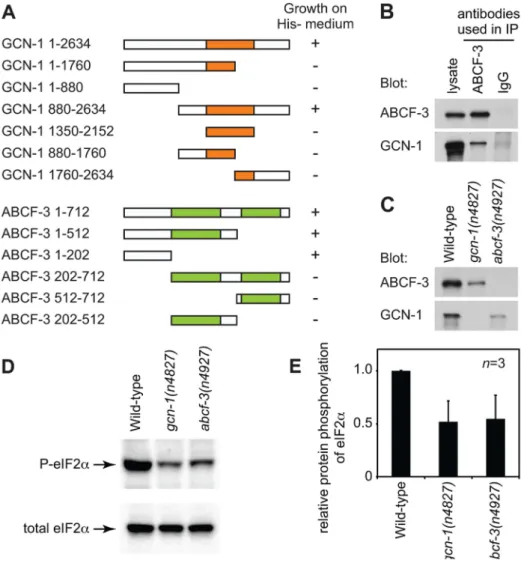
GCN-1 and ABCF-3 are evolutionarily conserved among S. cerevisiae,C. elegansand humans (Figure 1H, Figure S2 and S3). Expression ofS. cerevisiae GCN1, the homolog ofC. elegans gcn-1, and GCN20, the homolog of C. elegans abcf-3, under the control of theabcf-3promoter rescued the defect in M4 sister cell death of C. elegans gcn-1 and abcf-3 mutants, respectively, indicating that S. cerevisiae GCN1 and GCN20 are functional homologs ofC. elegans gcn-1andabcf-3, respectively (Figure 1F). S. cerevisiae Gcn1p has a domain (amino acids 1350–2152) similar to that of translation elongation factor 3 (EF3). The EF3-like domain is highly conserved among species (Figure 1H and Figure S2) and is necessary and sufficient for binding to Gcn20p [32]. We therefore tested whether C. elegans GCN-1 can physically interact with ABCF-3 using the yeast two-hybrid assay (Figure 2A). Full-length GCN-1 (1–2634) interacted with full-length ABCF-3 (1–712). To identify the protein domains important for GCN-1 to bind to ABCF-3, we generated a series
Figure 1.gcn-1(n4827)andabcf-3(n4927)cause a defect in M4 sister cell death.(A) Schematic representation of the M4 cell lineage in the wild type and mutants defective in M4 sister cell death. X, programmed cell death. (B–E) Merged epifluorescence and Nomarski images of the pharynx in wild-type,ced-3(n717),gcn-1(n4827)andabcf-3(n4927)animals expressingPceh-28::gfp. Arrow, M4 neuron. Arrowhead, surviving M4 sister. Scale bar, 20mm. Panels B and C from ref. 15. (F) The percentages of M4 sister survival in animals of the indicated genotypes. (G) Genomic organizations and protein structures ofgcn-1andabcf-3, including the locations and natures of the mutationsn4827andn4927. Orange box, EF3-like domain. Green boxes, AAA domains. Black bars, sequences deleted in allelesgcn-1(nc40D) andabcf-3(ok2237D). (H) Comparison of amino acid sequences of the entire protein or the EF3-like domain of GCN-1 and of the entire protein, the N-terminal domain, the first AAA domain or the second AAA domain of ABCF-3 among yeast,C. elegansand humans. Clustal W was used to align amino-acid sequences and to calculate identity and similarity.
of deletion constructs of GCN-1 and assayed each for ABCF-3-binding activity using the yeast two-hybrid assay. GCN-1 fragments not containing entire the EF3 domain (1–1760, 1– 880, 880–1760 and 1760–2634) or containing only the EF3 domain (1350–2150) failed to bind ABCF-3, whereas GCN-1 fragments containing the EF-3 domain and surrounding regions (880–2634) bound ABCF-3. These results suggest that GCN-1 physically interacts with ABCF-3 but that unlike in yeast the EF3-like domain is not sufficient for GCN-1 to bind to ABCF-3.
We also defined the domains of ABCF-3 important for ABCF-3 to bind to GC1 (Figure 2A). ABCF-3 fragments lacking the N-terminal region (202–712, 512–712 and 202–512) failed to bind GCN-1, whereas ABCF-3 fragments containing the N-terminal region (1–712, 1–512 and 1–202) bound GCN-1, suggesting that the N-terminal portion of ABCF-3 (which does not include the first AAA domain) is necessary and sufficient for binding to GCN-1, just as the N-terminal region ofS. cerevisiaeGcn20p is necessary
and sufficient for binding to Gcn1p, theS. cerevisiaehomolog of GCN-1.
To determine whether GCN-1 and ABCF-3 interactin vivo, we generated antibodies against GCN-1 and ABCF-3 and performed co-immunoprecipitation experiments. We first tested whether these antibodies specifically recognize GCN-1 or ABCF-3 protein using western blot analysis. The antibodies against GCN-1 or ABCF-3 recognized proteins of the sizes predicted for the GCN-1 or ABCF-3 proteins in wild-type animals but not ingcn-1(n4827) orabcf-3(n4927)animals, respectively, confirming the specificity of these antibodies (Figure 2C). Then we tested whether GCN-1 could be co-immunoprecipitated with ABCF-3. Whole-protein extracts from wild-type animals were subjected to immunoprecip-itation using an anti-ABCF-3 antibody (or normal IgG as a control), and then immunocomplexes were analyzed by western blotting using antibodies against 3 or GCN-1. Both ABCF-3 and GCN-1 were recovered in an immunocomplex purified with
Figure 2. GCN-1 and ABCF-3 proteins are evolutionarily conserved functionally.(A) Schematic representations of the GCN-1 and ABCF-3 proteins used in the yeast two-hybrid binding assays. Orange box, EF3-like domain. Green boxes, AAA domains. The growth of yeast on histidine-minus medium is summarized.+, wild-type growth.2, no or little growth. (B) Western blot analysis of immunocomplexes purified from wild-type animals with an anti-ABCF-3 antibody or control IgG. Both ABCF-3 and GCN-1 are present in the immunocomplex purified with an anti-ABCF-3 antibody but not with the control IgG. (C) Western blot analysis showing levels of ABCF-3 and GCN-1 proteins in fourth-larval stage animals of the indicated genotypes. (D) Western blot analysis showing the levels of eIF2awith phosphorylated serine 49 and total eIF2aof fourth-larval stage animals of the indicated genotypes. (E) Relative intensities of phosphorylated eIF2 to total eIF2 of fourth-larval stage animals of the indicated genotypes. Errors, standard deviations.
the anti-ABCF-3 antibody, whereas neither ABCF-3 nor GCN-1 was recovered in an immunocomplex purified with normal IgG (Figure 2B). We conclude that GCN-1 and ABCF-3 are present in the same protein complexin vivo.
Since GCN-1 and ABCF-3 form a complex in vivo, we
suspected that deletion of either protein might affect the stability of the other protein [29,33]. To test this hypothesis, we examined the levels of GCN-1 and ABCF-3 proteins by western blot analyses of whole-protein extracts prepared from wild-type, gcn-1(n4827) and abcf-3(n4927) animals using antibodies against ABCF-3 or GCN-1. The steady-state level of ABCF-3 protein was decreased ingcn-1(n4827)animals by 3.6 fold compared to that of wild-type animals. Similarly, the steady-state level of GCN-1 protein was decreased inabcf-3(n4927)animals by 4.4 fold (Figure 2C). These results suggest that a lack of ABCF-3 or GCN-1 protein affects the stability of the other protein and support our conclusion that GCN-1 and ABCF-3 are in a protein complex togetherin vivo.
If GCN-1 and ABCF-3 physically interactin vivoto promote M4 sister cell death, GCN-1 and ABCF-3 should act together in the same pathway. Since gcn-1(n4827) and abcf-3(n4927) are likely null mutations, the gcn-1(n4827) mutation would not enhance the M4 sister cell-death defect ofabcf-3(n4927)mutants if gcn-1 and abcf-3 function in the same process or pathway. Indeed, we observed no enhancement of the M4 sister cell-death defect ofgcn-1(n4827) abcf-3(n4927)double mutants compared to that of either single mutant: there was 13% M4 sister survival in gcn-1(n4827) abcf-3(n4927) double mutants, 12% M4 sister survival ingcn-1(n4827)mutants and 13% M4 sister survival in abcf-3(n4927)mutants (Figure 1F). We conclude thatgcn-1and abcf-3 function together in the same process of pathway to promote M4 sister cell death, consistent with our finding that GCN-1 and ABCF-3 physically interactin vivo.
In S. cerevisiae, Gcn1p and Gcn20p are required for the efficient phosphorylation of eukaryotic initiation factor 2 (eIF2a) under both normal conditions and conditions of amino-acid starvation [29,30]. Gcn1p and Gcn20p form a protein complex that activates the serine-threonine protein kinase Gcn2p, which then phosphorylates an evolutionarily conserved serine residue of eIF2a. The amino acid sequences surrounding the eIF2a
phosphorylation site are identical inS. cerevisiae,C. elegansand humans, suggesting a conserved regulatory mechanism of eIF2a
[28]. We tested whether GCN-1 and ABCF-3 promote the phosphorylation of eIF2a in C. elegans using an antibody that specifically recognizes eIF2athat is phosphorylated at serine 49 (P-eIF2a). From wild-type animals cultivated under normal physio-logical conditions, a single band of eIF2awas detected in western blotting analyses using either the anti-P-eIF2a antibody or an antibody that recognized total eIF2a (Figure 2D). In gcn-1(n4827)andabcf-3(n4927)mutants, the phosphorylation levels of eIF2a in physiological conditions were 52% and 54% of the levels in wild-type animals, respectively (Figure 2D and 2E). We conclude that gcn-1 and abcf-3 are required to maintain the steady-state level of the phosphorylation of eIF2a.
The regulation of phosphorylation of eIF2a plays an essential role in the initiation of translation. We therefore directly tested whether gcn-1and abcf-3affect gene expression at the transla-tional level. Since gcn-1and abcf-3 are highly expressed in the gonads at the fourth larval stage, maternally contribute to the death of the M4 sister and affect most programmed cell deaths (see below, Figure 3D and 3I, Table 1 and Table S5), we isolated both wild-type animals and gcn-1 and abcf-3 mutants at the fourth larval stage and performed mRNA-seq and ribosome profiling (Ribo-seq) to generate quantitative genome-wide information concerning mRNA abundance and the locations of mRNAs
occupied by ribosomes [34]. Parallel analyses of data from Ribo-seq and mRNA-Ribo-seq studies allowed us to distinguish differences in mRNA abundance from differences in translational control and to generate a quantitative and comprehensive list of genes the expression of which is likely regulated bygcn-1andabcf-3at the translational level. Loss of gcn-1orabcf-3 function affected the expression of a large number of genes at either the transcriptional or translational level or at both (Figure S4A and S4B and Table S1). Since GCN-1 and ABCF-3 very likely function in translational control, their effects on transcript levels are likely indirect. Changes in gene expression compared to wild-type animals were similar between gcn-1 and abcf-3 mutants, supporting our conclusion that gcn-1 and abcf-3act together (Figure S4C and S4D). The expression of 464 genes or 217 genes changed in both gcn-1andabcf-3mutants compared to wild-type animals at least two-fold (p,0.1) in mRNA-seq or Ribo-seq analyses, respectively (Figure S4E and S4F). Of the 217 genes altered in translational expression, 98 genes showed no alterations in mRNA levels using our standards of a two-fold change and p,0.1 (Table S2 and Table S3). These genes are candidates for being directly regulated by bothgcn-1andabcf-3translationally. These results suggest that gcn-1andabcf-3function together in the translational control of many genes.
In mammals, eIF2aphosphorylation is mediated by at least four different protein kinases: PKR-like endoplasmic reticulum kinase (PERK), general control non-derepresessible-2 (GCN2), double-stranded RNA-activated protein kinase (PKR) and heme-regulated
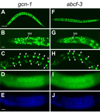
Figure 3. gcn-1 and abcf-3 are ubiquitously expressed. (A–E) Expression pattern ofgcn-1. (A–C) Expression of GFP under the control of thegcn-1promoter in second-larval stage animals and (D and E) FISH ofgcn-1mRNA in third-larval stage animals. Green,gcn-1mRNA. Blue, DAPI staining of nuclei. (F–J) Expression pattern of abcf-3. (F–H) Expression of GFP under the control of theabcf-3 promoter in the second-larval stage animals and (I and J) FISH ofabcf-3mRNA in third-larval stage animals. Green,abcf-1mRNA. Blue, DAPI staining of nuclei. Arrows, M4 neuron. Arrowheads, intestinal cells. Scale bars, 50mm in A and F; 10mm in B, C, G, H; and 5mm in D, E, I and J.
inhibitor kinase (HRI); each of these kinases is activated by a distinct stress signal [18]. These kinases share homology in their kinase catalytic domains, but their effector domains are distinct and are subject to different regulatory mechanisms. Homologs of genes encoding two of these protein kinases exist in theC. elegans genome: the PERK homolog PEK-1 and the GCN2 homolog GCN-2. Y38E10A.8 has a kinase domain similar to that of mammalian eIF2akinases but does not have an obvious homolog. We tested whether these three protein kinases are required for the programmed cell death of the M4 sister. Neither single mutants of each kinase gene nor the triple mutant was defective in M4 sister cell death (Table S4), suggesting that one or more unidentified protein kinase(s) regulated by GCN-1 and ABCF-3 are responsible for phosphorylating eIF2ain the regulation of M4 sister cell death. Alternatively, it is possible GCN-1 and ABCF-3 promote M4 sister cell death through one or more targets other than eIF2a.
gcn-1andabcf-3are expressed ubiquitously
To determine the expression patterns ofgcn-1and abcf-3, we generated transgenes expressing a reporter GFP under the control of the endogenousgcn-1orabcf-3promoter. Bothgcn-1and abcf-3 were expressed in most cells during all stages of development. We observed gcn-1 and abcf-3 expression in head neurons, hypodermal cells, intestinal cells, body wall muscles, and pharyngeal neurons, including the M4 neuron (Figure 3A–3C and 3F–3H). We also used the technique of fluorescencein situ hybridization (FISH) with a level of sensitivity sufficient to detect single mRNA molecules [35] to observe endogenous gcn-1and abcf-3 transcripts. Consistent with the expression of the GFP reporter transgenes,gcn-1and abcf-3mRNAs were observed in most somatic cells. In addition, gcn-1 and abcf-3mRNAs were abundant in the germ cells in the hermaphrodite gonad (Figure 3D, 3E, 3I and 3J). The similar expression patterns of gcn-1andabcf-3are consistent with our observations that GCN-1 and ABCF-3 physically interact and act together to promote the death of the M4 sister.
Sincegcn-1andabcf-3are ubiquitously expressed and required to broadly maintain the basal level of phosphorylation of eIF2a, we tested whethergcn-1 and abcf-3might be involved in other biological processes. We did not observe abnormalities in the morphologies of the hermaphrodite vulva, the male tail or the neurite processes of the M4, I2 and PVQ neurons. However,
the growth rate ofgcn-1andabcf-3mutants from embryogenesis to the fourth larval stage was around 24 hours longer than that of wild-type animals, and the mitotic pachytene region of the hermaphrodite gonad was expanded over the loop regions of the gonads. (data not shown). These observations suggest thatgcn-1 andabcf-3affect biological processes in addition to programmed cell death.
GCN-1 and ABCF-3 promote the deaths of most somatic cells during development and of germ cells in response to ionizing radiation
Given the ubiquitous expression patterns ofgcn-1andabcf-3, we tested whether gcn-1 and abcf-3 promote programmed cell deaths in addition to that of the M4 sister. We examined gcn-1(n4827)andabcf-3(n4927)mutants for defects in the deaths of the NSM sisters, the PVQ sisters, the g1A sisters, the RIM and RIC sisters and multiple cells in the anterior pharynx. gcn-1(n4827) and abcf-3(n4927) single mutants did not exhibit defects in the deaths of these cells (Table 1). However, when either thegcn-1(n4827)or theabcf-3(n4927) mutation was combined with the partial loss-of-function ced-3(n2427) mutation, which sensitizes strains to weak defects in cell death [36], we observed significant cell-death defects for all cell types tested (Table 1). For example, the gcn-1(n4827) and abcf-3(n4927) mutations en-hanced the ced-3(n2427) defect from 16% to 38% and 34%, respectively, for the NSM sister and from 13% to 36% and 45%, respectively, for the g1A sister. We conclude thatgcn-1andabcf-3 promote programmed cell death generally rather than specifically affecting the M4 sister cell death.
We next tested whethergcn-1and abcf-3are involved in the deaths of germ cells in the gonad of the adult hermaphrodite. More than half of germ cells stochastically undergo programmed cell death under normal conditions during oocyte differentiation [37]. We scored the number of apoptotic germ cells using the vital dye acridine orange (AO), which stains nucleic acids within apoptotic cells in living animals [38]. gcn-1(n4827) and abcf-3(n4927) mutants had 9.1 and 9.5 apoptotic germ cells per gonadal arm on average, respectively, similar to wild-type animals, which had 8.6 apoptotic germ cells per gonadal arm (Figure 4I). We also scored the number of apoptotic germ cells by direct observation of the gonads of engulfment-defective ced-1(e1735) mutants, in which cell corpses accumulate because of a defect in
Table 1.gcn-1(n4827)andabcf-3(n4927)enhance the cell-death defects of partial loss-of-functionced-3(n2427)mutants.
Genotype M4 sister NSM sisters PVQ sisters g1A sisters
RIM and RIC sisters
Extra cells±SD (anterior pharynx)
Wild-type 0 0 0 0 0 0.060.0
gcn-1(n4827) 12 0 11 11 01 0.1
60.3
abcf-3(n4927) 13 0 12 12 02 0.160.3
ced-3(n2427) 48 16 4 13 23 1.460.9
gcn-1(n4827); ced-3(n2427) 98 38 161 361 44 3.7
61.13
abcf-3(n4927); ced-3(n2427) 94 34 142 452 46 3.861.33
The percentages of the survival of the M4 sister, NSM sisters, PVQ sisters, g1A sisters and RIM and RIC sisters or numbers of extra cells in the anterior pharynx. More than 100 cells were scored for survival of the M4 sister, NSM sisters, PVQ sisters, g1A sisters and RIM and RIC sisters.
24 animals were scored for extra cells in the anterior pharynx.
All strains were homozygous for the reporter transgene to score survival of the indicated cells except for the extra cells in the anterior pharynx. See Materials and Methods for the reporter transgenes.
1These strains were homozygous forunc-45(r450). 2These strains were homozygous for
dpy-18(e364).
3Student’st-test compared withced-3(n2427),P
cell-corpse engulfment, facilitating a sensitive assay for the deaths of germ cells [37].ced-1(e1735)mutants had an average of 14.4 cell corpses per gonadal arm (Figure S5). ced-1(e1735) double mutants withgcn-1(n4827)orabcf-3(n4927)had nearly identical numbers of cell corpses per gonadal arm, 13.9 and 14.0, respectively (Figure S5). These results indicate that gcn-1 and abcf-3 are dispensable for germ-cell death under physiological conditions.
Since many germ cells undergo apoptosis in response to genotoxic stresses such as ionizing radiation [39], we tested whethergcn-1andabcf-3mediate ionizing radiation damage-induced germ cell death. As assayed with AO, wild-type animals normally contained an average of 8.6 apoptotic germ cells per gonadal arm, while wild-type animals exposed to ionizing radiation contained on average 27.1 apoptotic germ cells (Figure 4A, 4E and 4I). This germ-cell death was completely blocked by a mutation in the caspase gene ced-3 in wild-type, gcn-1(n4837) and abcf-3(n4927) animals (Figure 4B, 4F and 4I). Strikingly, ionizing radiation failed to increase the number of apoptotic germ cells in gcn-1(n4827) and abcf-3(n4927) mutants (10.9 and 10.4 apoptotic germ cells per gonadal arm in gcn-1 and abcf-3 mutants, respec-tively, 24 hours after gamma ray irradiation) (Figure 4C, 4D, 4G, 4H and 4I). These results indicate thatgcn-1 andabcf-3 are required for ionizing radiation-induced germ cell death but not for the stochastic germ cell death that occurs in physiological conditions.
gcn-1andabcf-3gene dosage affects programmed cell death
The gcn-1(n4827) and abcf-3(n4927) mutations partially blocked both the programmed cell deaths of somatic cells (Table 1) and the deaths of germ cells in response to ionizing radiation (Figure 4). Both somatic and ionizing radiation-induced germ cell deaths involve the canonical cell-death execution pathway consisting of the BH3-only gene egl-1, the BCL-2 homologced-9, the pro-apoptotic APAF-1 homolog ced-4, and the caspase gene ced-3 [40]. Interestingly, animals doubly heterozygous forgcn-1andced-3,ced-4oregl-1had a defect in M4 sister cell death (gcn-1/+; ced-3/+18%,gcn-1/+; ced-4/+12% or gcn-1/+; egl-1/+ 17%, respectively) significantly higher than that of singly heterozygous animals (gcn-1/+4%,ced-3/+0%, ced-4/+ 0% or egl-1/+ 1%, respectively) (Table S5). These results indicate that the simultaneous reduction by half of the dosage of gcn-1and of genes in the canonical cell-death execution pathway causes a significant defect in M4 sister cell death. We observed a similar genetic interaction in animals heterozygous forabcf-3and ced-3,ced-4oregl-1(Table S5).
gcn-1andabcf-3maternally contribute to programmed cell death
We observed that maternal gcn-1 and abcf-3 contribute to zygotic programmed cell death. Whilegcn-1(2) animals gener-ated bygcn-1(2)hermaphrodites andgcn-1(2)males exhibited a defect in M4 sister cell death (12% of M4 sister survival),gcn-1(2)
Figure 4.gcn-1(n4827)andabcf-3(n4927)cause a defect in radiation-induced germline cell death.(A–D) Acridine orange (AO) staining of apoptotic germ cells in the posterior gonads of animals of the indicated genotypes. Arrowheads, AO-positive apoptotic germ cells. Scale bar, 10mm. (E–H) Nomarski DIC images corresponding to A–D. Arrowheads, refractile apoptotic cells. (I) Numbers of AO-positive apoptotic germ cells in the posterior gonads of animals of the indicated genotypes. White bars, means and standard errors of the means without ionizing radiation (IR). Black bars, means and standard errors of the means with IR.
animals produced from gcn-1/+ hermaphrodites and gcn-1(2) males did not (0% of M4 sister survival) (Table S5), indicating that maternal gcn-1 is sufficient to promote programmed cell death. gcn-1(2) animals generated by gcn-1(2) hermaphrodites and gcn-1/+males exhibited a defect in M4 sister cell death (13% of M4 sister survival). gcn-1/+ animals generated by gcn-1(2) hermaphrodites andgcn-1(+)males exhibited a very weak defect in M4 sister cell death (4% of M4 sister survival) compared to 12% of M4 sister survival in gcn-1(2) self-progeny of gcn-1(2) hermaphrodites. By contrast, gcn-1/+ animals produced from gcn-1(+)hermaphrodites andgcn-1(2)males exhibited no defect in M4 sister cell death (0% of M4 sister survival) (Table S5). These results indicate that maternalgcn-1is partially required for the M4 sister to undergo programmed cell death. We observed a similar maternal requirement and sufficiency forabcf-3 (Table S5). We conclude that maternal gcn-1 and abcf-3 are sufficient and partially required for the M4 sister to undergo programmed cell death.
GCN-1 and ABCF-3 act independently of the BCL-2 homolog CED-9 to promote programmed cell death and can act in the cell fated to die
To examine interactions between gcn-1 and abcf-3 and the canonical cell-death execution pathway, we performed epistasis analyses between gcn-1 or abcf-3 and ced-9, which functions downstream ofegl-1and upstream ofced-4andced-3in the cell-death execution pathway [40]. Because the ced-9(n2812) null mutation causes ectopic cell deaths and organismic inviability, we used theced-3partial loss-of-function mutationn2446to suppress ced-9(n2812) lethality [41]. We observed that 50% of ced-9(n2812)animals had a surviving M4 sister in theced-3(n2446) mutant background. This increase over the 5% frequency of M4 sister survival in ced-3(n2446) mutants is consistent with the proposal that ced-9 has a cell-killing activity [42]. We observed that gcn-1 ced-9 and abcf-3 ced-9 double mutants were more highly penetrant for M4 sister survival (90% and 89%, respec-tively) than either single mutant in the ced-3(n2446) mutant background: gcn-1 (45%), abcf-3 (40%) and ced-9 (50%), respectively (Table 2). These results indicate that ced-9 is not required forgcn-1andabcf-3to promote programmed cell death. Thus,gcn-1andabcf-3function downstream of or in parallel to ced-9in the regulation of programmed cell death.
We next tested whether the activity ofgcn-1andabcf-3can act cell-autonomously to promote programmed cell death. Previous studies showed that expression of a ced-3, ced-4oregl-1 cDNA under the control of the mec-7 promoter can act cell-autono-mously to cause the deaths of a set of touch neurons, including the
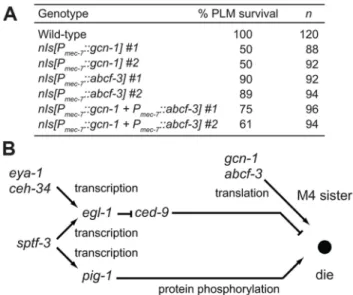
PLML and PLMR cells. We expressedgcn-1andabcf-3cDNAs in the PLM neurons under the control of themec-7promoter. We observed that 100% of the PLM neurons survived in wild-type animals, whereas only 50% or 89% of the PLM neurons survived in animals expressing gcn-1 or abcf-3, respectively, under the control of the mec-7 promoter (Figure 5A). Expression of both gcn-1 and abcf-3 also reduced a survival of the PLM neurons: 61% of the PLM neurons survived. These results indicate that expression ofgcn-1andabcf-3are sufficient to induce cell death and suggest that gcn-1 and abcf-3 acts cell-autonomously to promote programmed cell death.
Our genetic screen for mutants defective in M4 sister cell death identified other genes in addition togcn-1andabcf-3:ceh-34, eya-1, sptf-3and pig-1 [15,17]. We previously showed that the Six family homeodomain protein CEH-34 and the Eyes absent homolog EYA-1 directly drive the transcription of the BH3-only gene egl-1in the M4 sister to promote M4 sister cell-type specific
Table 2.gcn-1(n4827)andabcf-3(n4927)enhance the M4 sister-cell death defect ofced-9(n2812)mutants.
Genotype1 % M4 sister survival
ced-3(n2446) 5
gcn-1(n4827); ced-3(n2446) 45
abcf-3(n4927); ced-3(n2446) 40
ced-9(n2812); ced-3(n2446) 50
gcn-1(n4827) ced-9(n2812); ced-3(n2446) 902
abcf-3(n4927) ced-9(n2812); ced-3(n2446) 892
1All strains were homozygous fornIs177[P
ceh-28::gfp].
2Fisher’s exact test compared withced-9(n2812), P,0.0001.
120 animals were scored for survival of the M4 sister. doi:10.1371/journal.pgen.1004512.t002
Figure 5. GCN-1 and ABCF-3 act cell-autonomously in a pathway distinct from the canonical cell-death execution pathway. (A) The percentages of PLM survival in animals of the indicated genotypes. All strains had the Pmec-4::gfp transgene that expressed GFP in the touch neurons, including the PLM neurons. (B) A model for the pathways that regulate M4 sister cell-specific pro-grammed cell death.gcn-1andabcf-3act in a pathway distinct from the canonicalced-9-dependent cell-death execution pathway to promote M4 sister cell death. See text for details.
death [15] and that the SP1 family transcription factor SPTF-3 directly drives the transcription of both egl-1 and the AMPK-related protein kinase genepig-1, which also promotes M4 sister cell death [17]. We determined how gcn-1 and abcf-3 interact with these genes by examining double mutants. The partial loss-of-function alleles ceh-34(n4796) and sptf-3(n4850) and the null allelepig-1(gm344D)enhanced the M4 sister-cell death defect of gcn-1(n4827) and abcf-3(n4927) null mutants (Table 3). These results indicate thatceh-34,sptf-3andpig-1function in pathways distinct from that ofgcn-1and abcf-3to promote M4 sister cell death.
Discussion
The maternally-contributed translational regulators GCN-1 and ABCF-3 act together to promote the death of the M4 sister in a pathway distinct from the CED-9-mediated cell-death execution pathway
We demonstrated that the translational regulators GCN-1 and ABCF-3 are pro-apoptotic factors that maternally contribute to the programmed cell death of the M4 sister inC. elegans. GCN-1 and ABCF-3 promote the deaths of all somatic cells tested. Essentially all somatic cell deaths are mediated by an evolution-arily conserved cell-death execution pathway consisting of the BH3-only gene egl-1, the BCL-2 homolog ced-9, the APAF-1 homologced-4and the caspase geneced-3 [40]. How dogcn-1 and abcf-3interact with this pathway to regulate apoptosis? We propose thatgcn-1andabcf-3likely act in a novel pathway distinct from the canonical cell-death execution pathway. First,gcn-1and abcf-3 promote apoptosis in the absence of ced-9 activity, indicating thatgcn-1andabcf-3function independently ofced-9 in the regulation of apoptosis and hence do not regulate either ced-9oregl-1. Second sinceced-3andced-4function downstream of ced-9in the cell-death execution pathway,gcn-1andabcf-3could act through these genes to promote cell death. However, our
mRNA-seq and Ribo-seq results indicate thatgcn-1andabcf-3do not have major effects on mRNA abundance or the ribosome footprint density of ced-3 and ced-4 (Figure S6). Our preferred model is that GCN-1 and ABCF-3 function in a pathway that acts in parallel to the canonical cell-death execution pathway, although we cannot preclude the possibility that GCN-1 and ABCF-3 translationally regulate unidentified factors that act throughced-3 or ced-4 without changing the transcriptional and translational levels of the products of these genes. Also, since we used whole animals for our mRNA-seq and Ribo-seq analyses, we would not have detected alterations in CED-3 or CED-4 levels that were specific to a small subset of cells, including the M4 sister.
Our genetic analyses revealed thatgcn-1andabcf-3maternally contribute to the death of the M4 sister, which undergoes programmed cell death during embryogenesis. Maternally-con-tributed factors might act to ensure the rapid deaths of cells during embryogenesis; perhaps zygotic expression of apoptotic genes would be too slow. Also, the maternal effects ofgcn-1andabcf-3 might explain why we discovered new general cell-death genes, despite the fact that many genetic screens have been performed in search ofC. elegansmutants defective in somatic cell deaths. Most such genetic screens have examined F2 animals after mutagenesis, and would have missed maternally-contributed genes that affect general cell death. Perhaps, additional maternal-effect genes with functions in apoptosis exist inC. elegans. Such genes might be efficiently identified by screening in the third generation after mutagenesis.
gcn-1(n4827)andabcf-3(n4927)single mutations appeared to cause a defect in only M4 sister cell death, and these mutations both affected other cell deaths to differing extents in strains sensitized to weak defects in cell death. For example, the death of the M4 sister was most sensitive and the deaths of the PVQ sisters were least sensitive to the gcn-1(n4827) and abcf-3(n4927) mutations among the cells we tested (Table 1). We speculate that sensitivity to perturbation of cell-death genes is different among different cell types. This hypothesis is supported by the observa-tions that penetrance of cell-death defects varies among different cell types in partial loss-of-functionced-3(n2427)mutants and that the extent of the cell-death defect ofced-3(n2427)mutants is well correlated with that ofgcn-1(n4827)andabcf-3(n4927)mutants. Our genetic and biochemical data strongly suggest that GCN-1 and ABCF-3 physically interact in a complexin vivoto promote apoptosis. First, GCN-1 and ABCF-3 interacted in the yeast two-hybrid system. Second, GCN-1 co-immunoprecipitated with ABCF-3 from a total protein extract fromC. elegans. Third, the absence of either the GCN-1 or ABCF-3 protein decreased the steady-state level of the other protein (ABCF-3 or GCN-1, respectively), indicating that an interaction between GCN-1 and ABCF-3 is likely important for the stability of both proteins. Fourth, gcn-1(n4827) abcf-3(n4927) double mutants were not enhanced in the defect in apoptosis compared to each single mutant.
GCN-1 and ABCF-3 play an essential role in germ-cell death in response to ionizing radiation
Although GCN-1 and ABCF-3 promote the deaths of most somatic cells, ingcn-1 or abcf-3mutants only 12% or 13% of animals are defective in M4 sister cell death, respectively, and the cell-death defect of most other cells was observed only in a partial loss-of-functionced-3 mutant background, which is sensitized to weak defects in cell death. Furthermore, loss-of-function ofgcn-1 and abcf-3 did not affect the deaths of germ cells under physiological conditions. By striking contrast, we found that GCN-1 and ABCF-3 play an essential role in germ-cell deaths
Table 3.ceh-34,eya-1,sptf-3andpig-1function independently ofgcn-1andabcf-3.
Genotype
% M4 sister survival
Wild-type 0
gnc-1(n4827) 12
abcf-3(n4927) 13
ceh-34(n4796) 382
sptf-3(n4850) 34
pig-1(gm344D) 20
gnc-1(n4827); ceh-34(n4796)1 523
gnc-1(n4827); sptf-3(n4850) 564
gnc-1(n4827); pig-1(gm344D) 685
abcf-3(n4927); ceh-34(n4796)1 543
abcf-3(n4927); sptf-3(n4850) 584
abcf-3(n4927); pig-1(gm344D) 635
1These strains were homozygous forunc-45(r450). 2Data from ref. 15.
3Fisher’s exact test compared with
ceh-34(n4796),P,0.05.
4Fisher’s exact test compared withsptf-3(n4850),P
,0.001.
induced by ionizing radiation. These results suggest that transla-tional control by GCN-1 and ABCF-3 plays a more important role in germ-cell deaths induced by ionizing radiation than in somatic cell deaths. The hypothesis that translational control is particularly important for cell deaths induced by ionizing radiation is supported by a recent report that a mutation in RNA polymerase I (rpoa-2), which synthesizes ribosomal RNAs, causes a defect in germ-cell deaths induced by ionizing radiation [43].
Ionizing radiation causes DNA double-strand breaks, which lead to the progressive accumulation of mutations and chromo-somal aberrations as damaged cells undergo division, resulting in apoptosis and the demise of genetically damaged cells. In C. elegans, ionizing radiation causes massive deaths of the germ cells during the late pachytene stage of oocyte development in adult gonads, resulting in the elimination of the damaged oocytes [39]. Germ-cell deaths induced by ionizing radiation specifically involve activation of the p53 homolog CEP-1 by the DNA damage response pathway and subsequent CEP-1- dependent transcrip-tional induction of the BH3-only geneegl-1, which activates the cell-death execution pathway regulated by CED-9 [44,45]. How might GCN-1 and ABCF-3 interact with the known DNA-damage response and cell-death execution pathways in the regulation of germline cell deaths induced by ionizing radiation? Our genetic results suggest that GCN-1 and ABCF-3 function independently of CED-9, at least in the regulation of the death of the M4 sister cell. We suggest that as is the case for somatic cell deaths, GCN-1 and ABCF-3 function in a novel pathway independently of CED-9 in regulating the germ-cell deaths induced by ionizing radiation. Alternatively, if GCN-1 and ABCF-3 regulate germ-cell deaths induced by ionizing radiation via a mechanism different from that of somatic cell deaths, it is possible that GCN-1 and ABCF-3 act through egl-1 and its target ced-9, since egl-1 is involved in somatic programmed cell deaths and germ-cell deaths induced by ionizing radiation but not in the stochastic germ-cell deaths that occur under physiological conditions.
The functions of GCN-1 and ABCF-3 in the control of translation are conserved betweenS. cerevisiaeandC. elegans
GCN-1 and ABCF-3 are conserved proteins from yeast to humans. The C. elegans GCN-1 protein has 43% and 53% similarities (23% and 32% identities) to the homologs of S. cerevisiaeand humans, respectively, and the C. elegansABCF-3 proteins has 57% and 69% similarities (40% and 49% identities) to the homologs of S. cerevisiae and humans, respectively (Fig-ure 1H). The yeast GCN-1 homolog Gcn1p and ABCF-3 homolog Gcn20p are required to maintain the basal level of the phosphorylation of eukaryotic initiation factor 2a (eIF2a) in the physiological condition and to increase the phosphorylation of eIF2ain response to amino-acid starvation. Gcn1p and Gcn20p activate the serine-threonine protein kinase Gcn2p, which phosphorylates an evolutionarily conserved serine residue of eIF2a. The phosphorylation of eIF2aresults in both the inhibition of global translation and the translational activation of theGCN4 mRNA, which encodes a basic leucine zipper transcription factor. Translation ofGCN4mRNA is regulated by four short upstream open reading frames (uORFs) in the 59 UTR with start codons that are out-of-frame with the main coding sequence and which generally reduce translation from the main reading frame [46].
We speculate that the mechanistic roles ofC. elegansGCN-1 and ABCF-3 in translational control are conserved between yeast and C. elegans. First, the amino-acid sequences of GCN-1 and ABCF-3 proteins are conserved between yeast and C. elegans, particularly in functionally important domains (Figure 1H).
Second, the functions of GCN-1 and ABCF-3 can be substituted with those ofS. cerevisiae GCN1 andGCN20, respectively, for the promotion of M4 sister cell death. Third, like their yeast counterparts, C. elegans GCN-1 and ABCF-3 are required to maintain the basal level of phosphorylation of eIF2a and physically interact through an EF3-like domain-containing region of GCN-1 and an N-terminal ABCF-3 domain [43]. Fourth, like Gcn20p, the AAA domain ATPase activity of ABCF-3 is not required for its function [32]. Fifth, theatf-5gene, theC. elegans homolog ofS. cerevisiae GCN4, has two upstream ORFs that have been shown to inhibit the translation of theatf-5mRNA [47].
A regulatory network involving transcription, translation and protein phosphorylation specifies the death of the M4 sister
We have shown that in addition togcn-1and abcf-3, ceh-34, eya-1,sptf-3andpig-1function in M4 sister cell death [15,17]. We previously reported that the Six family homeodomain protein CEH-34 and the Eyes absent homolog EYA-1 physically interact to directly drive expression of the pro-apoptotic BH3-only gene egl-1 in the M4 sister, leading to the death of the M4 sister (Figure 5B) [15]. We found that the SP1 family transcription factor SPTF-3 directly drives the transcription of the geneegl-1, which encodes a BH3-only protein that promotes apoptosis via the CED-3 caspase-mediated canonical cell-death execution pathway [17]. SPTF-3 also directly drives the transcription of the AMPK-related genepig-1, which encodes a protein kinase that functions in a pathway in parallel to the CED-3-mediated canonical cell-death execution pathway. These interactions are shown in Figure 5B.
Our analyses indicate thatgcn-1andabcf-3likely function in a pathway that acts in parallel to those ofpig-1,ceh-34andsptf-3. These results are consistent with a model in which GCN-1 and ABCF-3 act independently of CED-9 to promote M4 sister cell death. In short, we propose that the regulatory network for the death of the M4 sister includes at least three different pathways involving translation, transcription and protein phosphorylation (Figure 5B). Each gene in this network (gcn-1,abcf-3,sptf-3, pig-1, egl-1, ceh-34 and eya-1) has a human counterpart, some of which are implicated in human diseases, including developmental disorders and cancer. We anticipate that further analyses of this regulatory network will both reveal an evolutionarily conserved mechanism of apoptosis shared betweenC. elegansand humans and provide insights concerning how abnormalities in this apoptotic network can lead to human disease.
Materials and Methods
C. elegansstrains
C. elegansstrains were cultured at 20uC as described [48]. The N2 strain was used as the wild type. The following mutations, integrations and extrachromosomal arrays were used.
LGI: sptf-3(n4850), eya-1(ok654D), nIs177[Pceh-28::gfp, lin-15AB(+)], nIs180[Ptdc-1::gfp, lin-15AB(+)], zdIs5[Pmec-4::gfp, lin-15AB(+)].
LGII:rol-1(e91),gcn-2(ok871D),Y38E10A.8(tm4094D).
LGIII: ced-4(n1162), ced-9(n2812), gcn-1(n4827, nc40D), abcf-3(n4927, ok2237D), unc-45(r450), dpy-18(e364), nIs176 [Pceh-28::gfp, lin-15AB(+)].
LGIV: ced-3(n717, n2427, n2446), pig-1(gm344D), nIs175[Pceh-28::gfp, lin-15AB(+)].
LGX: lin-15(n765), pek-1(ok275D), nIs106[Plin-11::gfp, lin-15AB(+)], nIs429[Pphat-5::gfp, lin-15AB(+)], bcIs24[Ptph-1::gfp, lin-15AB(+)].
Unmapped: nIs460[Pgcn-1::gfp], nIs488[Pabcf-3::gfp], nIs645
and nIs646[Pmec-7::gcn-1 cDNA, Pmec-7::abcf-3 cDNA, P mec-3::mCherry, rol-6(su1006)], nIs648 and nIs649[Pmec-7::gcn-1 cDNA, Pmec-3::mCherry, rol-6(su1006)], nIs651 and nIs652[P-mec-7::abcf-3 cDNA, Pmec-3::mCherry, rol-6(su1006)].
Extrachro-mosomal arrays: nEx1817 and nEx1818[Pgcn-1::gcn-1
cDNA::gcn-1 39 UTR, Plin-44::gfp], nEx1925 and nEx1926[-abcf-3(+), Plin-44::gfp], nEx1928 and nEx1929[abcf-3 K217M, Plin-44::gfp], nEx1931 and nEx1932[abcf-3 K536M, P lin-44::gfp], nEx1934 and nEx1935[abcf-3 K217M K536M, P lin-44::gfp], nEx2223 and nEx2224[Pceh-34::eIF2a S49A, P lin-44::gfp]
Genetic screen and mapping ofgcn-1(n4827)and abcf-3(n4927)
gcn-1(n4827)andabcf-3(n4927)were isolated from a genetic screen for mutations that cause an extra GFP-positive M4-like cell in animals carrying the Pceh-28::gfptransgene [15]. Mutagenesis
was performed as described [48]. Mutagenized P0animals were
allowed to lay eggs, and 144,000 synchronized F2 animals were
screened with a fluorescence-equipped dissecting microscope. Single nucleotide polymorphisms were used to map gcn-1(n4827) and abcf-3(n4927) to a 175 kb interval (III: 2,044,521–2,220,200) and a 5.3 Mb interval (III: 5,346,407– 10,613,191), respectively [49]. Whole-genome sequencing of gcn-1(n4827)mutants was performed using an Illumina/Solexa GAII, according to the instructions of the manufacture. DNA sequencing of theabcf-3locus ofabcf-3(n4927)mutants was performed using an Applied Biosystems 31306.
Analyses of defects in programmed cell deaths of specific cells
The programmed cell deaths of specific cells were scored at the indicated stages using the following strains, which express GFP in specific cells. A fluorescence-equipped compound microscope was used to score the programmed cell deaths. M4 sister cell death, nIs175,nIs176ornIs177at the L1 stage. g1A sister cell death, nIs429at the L1 stage. PVQ sister cell death, oyIs14at the L4 stage [50]. NSM sister cell death,bcIs24at the L1 stage [51]. RIM and RIC sister cell death,nIs180at the L1 stage. Extra cells in the anterior pharynx were scored using a compound microscope equipped with Nomarski differential interference contrast optics. For physiological germ-cell deaths, germ-cell corpses in gonads of animals 24 hours after the fourth-larval stage were counted by direct observation using Nomarski optics. For ionizing radiation-induced germ-cell deaths, fourth-larval stage animals were exposed to 120 Gy of ionizing radiation, and germ-cell deaths were scored using acridine orange at 24 hours post-irradiation as described [38].
Plasmid construction
The transgenes Pceh-28::gfp, Ptph-1::gfp and sra-6::gfp are
described [15,50,51]. The phat-5 promoter sequence in pGD48 was cloned in pPD122.56 to generate thePphat-5::gfptransgene
[52]. The Pflp-15::gfp transgene contained 2.4 kbp of the 59
promoter of flp-15 in pPD122.56. The Pgcy-37::gfp transgene
contained 1.1 kbp of 59promoter of gcy-37in pPD122.56. The Ptdc-1::gfptransgene contained 4.5 kbp of 59promoter oftdc-1in
pPD121.83. The Pgcn-1::gcn-1 cDNA::gcn-1 39UTR transgene
(pTHgcn-1cDNA) contained 4.2 kbp of 59promoter ofgcn-1, a
full-lengthgcn-1cDNA and 1.0 kbp 39of the stop codon ofgcn-1. The 59promoter ofgcn-1, a full-lengthgcn-1cDNA and the 39
promoter of gcn-1 were generated by PCR and fused in pBluescript II using the In-Fusion cloning system (Clontech). The abcf-3(+) transgene contained 1.6 kbp of 59 promoter, the coding region and 0.8 kbp 39 of the stop codon of abcf-3 in pBluescript II. The QuickChange II XL Site-Directed Mutagen-esis Kit (Stratagene) was used to generate transgenes of abcf-3 K217M, abcf-3 K536M and abcf-3 K217M K536M. gcn-1 cDNA corresponding to amino acids 1–2634, 1–1760, 1–880, 880–2634, 880–1760 or 1760–2634 of GCN-1 was cloned in pGBKT7.abcf-3cDNA corresponding to amino acids 1–712, 1– 512, 1–202, 202–712, 512–712 or 202–512 was cloned in pGADT7. The Pgcn-1::gfp transgene contained 4.2 kbp of 59
promoter of gcn-1 in pPD122.56. The Pabcf-3::gfp transgene
contained 1.6 kbp of 59 promoter ofabcf-3in pPD122.56. The Pceh-34::eIF2aS49A transgene contained 3.8 kbp of 59promoter
ofceh-34andeIF2awith a replacement of serine 49 with alanine
in pPD49.26. For the Pmec-7::gcn-1 cDNA and Pmec-7::abcf-3 cDNA transgenes, full-length cDNA of gcn-1 and abcf-3 were cloned in pPD96.41. Primer sequences used are available from the authors.
Germline transformation
Germline transformation was performed as described [53]. The gfp reporter transgene was injected at 50mg/ml into lin-15(n765ts)animals with 50mg/ml of pL15EK as a coinjection marker [54]. To rescue the defect in M4 sister cell death, the transgenes pTHgcn-1cDNA,abcf-3(+), abcf-3 K217M,abcf-3 K536M and abcf-3 K217M K536M described above were injected at 20mg/ml intogcn-1(n4827)orabcf-3(n4927)animals
with 50mg/ml of Plin-44::gfpas a coinjection marker [55]. To
establish transgenic lines carrying the Pceh-34::eIF2a S49A
transgene, the Pceh-34::eIF2a S49A transgene was injected at
50mg/ml intonIs175animals with 50mg/ml ofPlin-44::gfpas a
coinjection marker. ThePmec-7::gcn-1 cDNA and Pmec-7::abcf-3 cDNA transgenes were injected at 50mg/ml, respectively, into zdIs5animals with 50mg/ml of pRF4[rol-6(su1006)] and 20mg/
ml of thePmec-3::mCherrytransgene as coinjection markers.
Yeast two-hybrid binding assay
GAL4 fusion constructs were introduced into yeast strain PJ649A as described [56]. Single colonies were streaked and cultured for two days at 30uC on SD plates containing minimal supplements without tryptophan and leucine. Then yeast strains were streaked and cultured for three days at 30uC on SD plates containing minimal supplements without tryptophan, leucine and histidine to test yeast growth.
Antibody production
Protein fragments corresponding to amino acids 753–857 of GCN-1 and 74–185 of ABCF-3 fused to glutathione S- transferase (GST) were expressed, purified using glutathione Sepharose 4B (Amersham Biosciences) and used to raise rabbit anti-GCN-1 or anti-ABCF-3 antibodies, respectively. Antisera were generated by Pocono Rabbit Farm and Laboratory. Specific antibodies were affinity-purified using identical GCN-1 or ABCF-3 protein fragments fused to maltose-binding protein (MBP) and coupled to Affigel 10 (Bio-Rad).
Western blots and immunoprecipitation analysis
fourth larval stage as described [57]. 10mg of total protein was loaded onto a 7.5% SDS PAGE gel and then transferred to nitrocellulose membranes. The membranes were probed with anti-GCN-1 or anti-ABCF-3 antibody. Immunocomplexes were detected using HRP-conjugated rabbit IgG secondary anti-bodies (Invitrogen) followed by chemiluminescence (Western Lightning ECL, PerkinElmer). To determine the level of phosphorylated eIF2a, protein extracts were prepared from nIs175, gcn-1(n4827); nIs175 and abcf-3(n4927); nIs175 animals synchronized at the fourth larval stage as described [57]. 15mg of total protein was loaded on a 10% SDS PAGE gel and then transferred to nitrocellulose membranes. The mem-branes were probed with anti-phospho-eIF2a (Cell Signaling Technology) and anti-eIF2a antibodies [28]. Immunocomplexes were detected as described above.
For immunoprecipitation experiments, protein extracts were prepared from mixed-staged wild-type animals in TNE buffer containing 50 mM Tris-HCl (pH 8.0), 150 mM NaCl, 1 mM EDTA, 1% NP-40, 5 mM b-mercaptoethanol, 10% glycerol. Protein extracts were mixed with either an affinity-purified anti-ABCF-3 antibody or a control IgG at 4uC for 2 hours. Immunocomplexes were recovered using Protein A Sepharose 4 Fast Flow (GE Healthcare Life Sciences) and washed with TNE buffer four times. The recovered immunocomplexes were subject-ed to western blot analysis using anti-GCN-1 or anti-ABCF-3 antibody.
Fluorescencein situhybridization
Fluorescencein situhybridization was performed as described [58]. Thegcn-1andabcf-3probes (Biosearch Technologies, Inc) were conjugated to the fluorophore Cy5 using the Amersham Cy5 Mono-reactive Dye pack (GE Healthcare). DNA was visualized using 49,6-diamidino-2-phenylindole (DAPI). The probe sequences used are shown in Tables S3 and S4. Figures 4D and H are maximum intensity projections of a Z-stack of images processed with the FFT Bandpass Filter operations in the image processing program Fiji. Oligonucleotides used forgcn-1 and abcf-3 FISH probe were described in Table S6 and S7).
mRNA-seq and ribosome profiling (Ribo-seq)
For mRNA-seq [59], total RNA was purified using an RNAeasy Mini kit (Qiagen) from synchronized L4 animals of wild-type animals andgcn-1 and abcf-3 mutants. The purified RNA was subjected to oligo (dT) selection, fragmentation and first- and double-strand synthesis with an Illumina Tru-Seq kit according to the manufacturer’s instructions. DNA fragments longer than 30 bp were purified using SPRI-TE beads (Beckmann Coulter) according to the manufacturer’s instructions. The purified DNA was end-repaired and single A bases were added for adaptor ligations. The adaptor-ligated DNA was then subjected to double SPRI-TE purification to select for 200 bp fragments. These fragments were enriched and barcoded by PCR for multiplexing. A final SPRI-TE purification was performed to purify the barcoded RNA-Seq libraries for Illumina DNA sequencing using HiSeq 2000. RNA-seq data were aligned against the C. elegans reference genome (ce10) using the Burrows-Wheeler Aligner (BWA) and Tophat.
Ribosome profiling was performed as described [60] with modifications. Synchronized L4 wild type animals andgcn-1and abcf-3mutants were collected and washed with M9 buffer three times. Animals were homogenized using a dounce homogenizer in lysis buffer containing 20 mM Tris (pH 7.5), 150 mM NaCl, 5 mM MgSO4, 1 mM DTT, 100mg/ml cycloheximide, 1%
Triton X-100 and 25 U/ml Turbo DNase (Invitrogen) and
centrifuged at 20,000 g for 20 min at 4uC. The absorbance of the extract was measured at 260 nm. 40 absorbance units of extract were incubated with 300 units of RNase I at 25uC for an hour, and then 200 units of SUPERase In RNase Inhibitor (Invitrogen) were added. Digested extracts were loaded on 10– 50% linear sucrose gradients containing 20 mM Tris (pH 7.5),
150 mM NaCl, 5 mM MgSO4, 1 mM DTT and 100mg/ml
cycloheximide and centrifuged for three hours at 35, 000 rpm at 4uC using a SW-40 rotor to isolate a monosome fraction. RNA from the monosome fraction was purified by phenol-chloroform extraction followed by miRNeasy Mini Kit (Qiagen) and separated using a 15% TBE-Urea gel (BioRad) to isolate ribosome-protected fragments (RPFs). The RPFs were eluted from gels by incubating in RNA elution buffer containing 300 mM sodium acetate
(pH 5.5), 1 mM EDTA and 0.25% SDS. RPFs were 39
dephosphorylated with T4 polynucleotide kinase (New England Labs) and ligated to Universal miRNA Cloning Linker (New England Labs) using T4 RNA ligase 2, truncated (New England Labs) according to the manufacturer’s instructions. RPFs ligated with the linker were separated from an unligated linker using a 15% TBE-Urea gel (BioRad) and eluted from gels using RNA extraction buffer followed by phenol-chloroform extraction. RFPs were reverse-transcribed by Superscript III (Invitrogen) with a reverse transcription primer according to the manufacturer’s instructions. The products of reverse transcripts (RT) were purified using a 15% TBE-Urea gel (BioRad) and eluted from a gel by incubating in DNA elution buffer containing 300 mM NaCl, 10 mM Tris (pH 8.0) and 1 mM EDTA followed by phenol-chloroform extraction. The RT products were circularized by CircLigase (Epicentre) according to the manufacturer’s instruc-tions. About a quarter of the RT products were used in PCR reactions containing 16 Phusion HF buffer, 0.2 mM dNTP, 0.5mm forward library primer, 0.5mm reverse indexed primer
and 0.02 units/ml Phusion polymerase (New England Labs), and PCR was performed with a 30 second initial denaturation at 98uC, followed by 6, 8, 10, 12 and 14 cycles of 98uC for 10 second, 65uC for 10 second and 72uC for 5 second. PCR products were separated using a 8% TBE gel (BioRad) and eluted from gels by incubating in DNA elution buffer followed by phenol-chloroform extraction. PCR products were suspended in 20ml of
10 mM Tris (pH 8.0) and sequenced by HiSeq 2000. The adaptor sequences (CTGTAGGCACCATC) from 39end of the ribosome footprint reads were removed, then trimmed reads were mapped using BWA to distinguish the reads from ribosomal RNAs. About 60% of the reads were filtered out, and the remaining reads (non-ribosomal) were aligned to theC. elegansreference genome (ce10) using BWA and Tophat. Because translational initiation is thought to be blocked rapidly by the stress animals encounter during harvesting, many ribosomes are stalled at the beginning of each transcript in the presence of cycloheximide, which prevents translation elongation [34]. Hence, high frequencies of reads at the beginning of each transcript might not correspond to high rates of translation. For this reason, the reads that mapped to the first 25 nucleotides of each transcript were not counted in evaluating gene expression in the Ribo-seq analyses.
Supporting Information