A Novel Potassium Channel in Photosynthetic
Cyanobacteria
Manuela Zanetti1, Enrico Teardo1, Nicoletta La Rocca1, Lalu Zulkifli2, Vanessa Checchetto1, Toshiaki Shijuku2, Yuki Sato2, Giorgio Mario Giacometti1, Noboyuki Uozumi2, Elisabetta Bergantino1*, Ildiko` Szabo`1*
1Department of Biology, University of Padova, Padova, Italy,2Department of Biomolecular Engineering, Graduate School of Engineering, Tohoku University, Sendai, Japan
Abstract
Elucidation of the structure-function relationship of a small number of prokaryotic ion channels characterized so far greatly contributed to our knowledge on basic mechanisms of ion conduction. We identified a new potassium channel (SynK) in the genome of the cyanobacteriumSynechocystissp. PCC6803, a photosynthetic model organism. SynK, when expressed in a K+-uptake-system deficient E.coli strain, was able to recover growth of these organisms. The protein functions as a
potassium selective ion channel when expressed in Chinese Hamster Ovary cells. The location of SynK in cyanobacteria in both thylakoid and plasmamembranes was revealed by immunogold electron microscopy and Western blotting of isolated membrane fractions. SynK seems to be conserved during evolution, giving rise to a TPK (two-pore K+ channel) family
member which is shown here to be located in the thylakoid membrane ofArabidopsis. Our work characterizes a novel cyanobacterial potassium channel and indicates the molecular nature of the first higher plant thylakoid cation channel, opening the way to functional studies.
Citation:Zanetti M, Teardo E, La Rocca N, Zulkifli L, Checchetto V, et al. (2010) A Novel Potassium Channel in Photosynthetic Cyanobacteria. PLoS ONE 5(4): e10118. doi:10.1371/journal.pone.0010118
Editor:Hany A. El-Shemy, Cairo University, Egypt
ReceivedNovember 16, 2009;AcceptedMarch 12, 2010;PublishedApril 12, 2010
Copyright:ß2010 Zanetti et al. This is an open-access article distributed under the terms of the Creative Commons Attribution License, which permits unrestricted use, distribution, and reproduction in any medium, provided the original author and source are credited.
Funding:The European Molecular Biology Organization (Young Investigator Program grant to I.S.), the Italian Ministry for University and Research (MIUR) (to I.S.) and the University of Padova (to E.B. and I.S.) are acknowledged for financial support. This work was also supported by grant FISR from MIUR to G.M.G. This work was also supported by grants-in-aid for scientific research (17078005, 19380058 and 20-08103 to N.U.) from MEXT and JSPS. The funders had no role in study design, data collection and analysis, decision to publish, or preparation of the manuscript.
Competing Interests:The authors have declared that no competing interests exist.
* E-mail: elisabetta.bergantino@unipd.it (EB); ildi@civ.bio.unipd.it (IS)
Introduction
Cyanobacteria, the first organisms capable of performing oxygenic photosynthesis during evolution, still today give major contribution to the maintenance of the biosphere [1]. The unicellular photoheterotrophic transformable cyanobacterium
Synechocystis sp. PCC6803, characterized by an intracellular thylakoid membrane, where both photosynthesis and respiration take place, is the first photosynthetic organism for which the complete genome sequence has been published [2].
In vitroorin vivo function is not known for any of the putative potassium channels identified in the genomes of over ten species of cyanobacteria [3,4]. The only cyanobacterial ion channels characterized up to now are the prokaryotic glutamate receptor GluR0 [5] and the ligand-gated channel GLIC [6]. In general, the physiological role of bacterial channels is still largely unknown, except for bacterial chloride channel ClC [7], mechanosensitive channels [8] andH. pyloriHpKchA, a putative potassium channel [9]. Potassium is the major intracellular cation in bacteria [10]. However, membrane potential adjustment rather than K+
uptake has been hypothesized to be the major function of K+channels in prokaryotes, although direct proof is still missing [3]. In
Synechocystis a Ktr-like system encoded byslr1509, rather than a
bona fidechannel, seems to be the main responsible for potassium uptake [4,11].
In higher plant thylakoids several potassium-conducting cation channel activities have been described [12–15]. Furthermore, a putative potassium channel protein has been found in thylakoids of spinach [16]. Unfortunately, the molecular identity of the protein(s) responsible for these activities is unknown, as is the nature of the putative channel protein.
In the present study we characterized a novel cyanobacterial potassium channel. Furthermore, our work identifies its homolog in higher plants from molecular point of view and indicates its localization in the thylakoid membrane.
Results
Bioinformatic analysis of SynK putative potassium channel
Figure 1. SynK protein permits potassium flux, as revelaed by its expression in K+-uptake deficient
E. colistrain LB2003. A) SynK (Synechocystis sp. PCC 6803; gi:16331771) is characterized by selectivity filter sequence (bold characters) in pore region (brown characters) and by six predicted transmembrane segments (S1–S6 represented by different colours). ClustalW (1.83) alignments of SynK sequence with KvAP from Aeropyrum pernix(gi:14601099) and KvLm fromListeria monocytogenes(gi:16411529), two depolarization-activated prokaryotic potassium channels, is shown. ‘‘*’’ - identical residues in all aligned sequences; ‘‘:’’–conserved and ‘‘.’’ - semi-conserved substitutions. Definition of S1–S6 segments in latter proteins is shown according to [19] and [20], in different colours. In SynK S1–S6 segments were defined according to secondary structure predictions (Porter, SPLIT4, TMHMM2 algorithms) and adjusted taking into account delimitation ofa-helices as inferred from crystal structure of KvAP (according
to [20]). Conserved residues, functional in Kv gating, are shaded grey. Please note the presence of some of the highly conserved residues in the sensor sequence of Kv channels, such as K63 in S2 and P86 in S3 in SynK. Polar residues (S and Q) in S4 are shaded yellow.B) Complementation growth test ofE. coliLB2003 cells bySynK.E. coliLB2003 was transformed with plasmid harbouring pPAB404-SynKor empty vector. KAT1, anArabidopsisK channel, was also included as a positive control. Transformants were grown on media supplemented with different concentration of KCl. C) Potassium uptake by K+
-depletedE.colicontainingSynKor empty vector. Net K+
uptake bySynK-expressingE. coliLB2003 cells and control cells harbouring empty vector were measured at 20 mM KCl. Data are averages6SD of results from four independent experiments.
important for channel gating are also conserved in SynK (Figure 1A). Positive charges present in the S4 helix of KvAP determine voltage-dependent gating [19]. KvLm has only two positive charges in S4, but shows strong voltage-dependence [20]. SynK does not display evenly spaced positive charges in the predicted S4 segment, nor does it contain regulatory domains. On the basis of bioinformatic analysis, SynK may be classified as a ‘‘core-only’’, six-TM, putative potassium channel protein (see also ref.3). The closest homologues of SynK are found in other cyanobacteria species (Figure S1).
SynK forms functional, potassium-conducting protein, when expressed in a K+-uptake-system deficient
E.coli
strain
An E.coli K+ uptake–deficient mutant has been successfully used to study potassium transport activity of transporter systems from plants [21] as well as from Synechocystis [22]. Here we cloned theSynechocystis SynKgene into the E. colistrain LB2003, carrying mutations in genes encoding the three major K+
uptake systems, Kdp, Trk, and Kup [23]. Thus, LB2003 does not grow at K+
concentrations #10 mM, due to negligible K+ uptake activity at potassium concentrations in the low millimolar range. Complementation test on solid media shows that SynK -express-ing E. coli LB2003 cells grew well on a medium supplemented with 15 mM KCl, whereasE. colicells harbouring empty vector did not (Figure 1B). Time course uptake experiment shows that K+
influx bySynK-expressing cells was higher compared to that of cells containing empty vector (Figure 1C). Net potassium uptake measurements by K+-depletedE. colicells in the presence of 10 to 80 mM KCl revealed Vmax values of 553 and
460 nmol min21
g21
dry weight for SynK-expressing cells and for the control cells, respectively (Figure S2). These data suggest that SynK may mediate K+
uptake when expressed in E. coli.
Expression of SynK in CHO cells gives rise to potassium-conducting current
Additional functional characterization was performed in a mammalian cell system, given that SynK did not express in oocytes (Uozumi et al, unpublished). No electrophysiological studies have been performed on any cyanobacterial membrane until now. However, cloned prokaryotic channels have previously been shown to function in both heterologous expression systems e.g. [5,6,20,24] and in artificial lipid bilayers e.g. [19,25].
The sequence of SynK was isolated from the Synechocystis
genome by PCR and a SynK-EGFP (enhanced green fluorescent protein at C-terminus) fusion protein was expressed in CHO (Chinese hamster ovary) cells. Mammalian HEK and CHO cells do not have significant endogenous potassium current, and are suitable for the expression of prokaryotic and even the viral channel Kcv e.g. [5,26]. Green fluorescence of SynK-GFP was clearly associated with the plasma membrane (PM) (Figure 2A and Figure S3). Immunoblotting with anti-GFP antibody as well as by a specific anti-SynK antibody (Figure S4) revealed the presence of a product with the expected molecular weight of the fusion protein (for SynK and SynK-EGFP fusion proteins predicted MWs are 26445 and 53979 Da, respectively) (Figure 2B). However, lower MW products, corresponding to either EGFP alone (28 kDa), to SynK alone (27 kDa) or to degradation products of the fusion protein, were also observed and may account for the fluorescent signal observable in the cytosol of some cells (Figure S3 and not shown). Western blot of separated membrane and soluble fractions from transfected cells showed the presence of the 54 kDa fusion protein exclusively in
the former one indicating that the correctly translated product is inserted into the membrane (Figure 2C). The same protein was also recognized by another antibody which was developed against the common selectivity filter sequence of potassium channels (anti-KPORE, Figure S5 for details), confirming that anti-SynK recognizes a potassium channel protein.
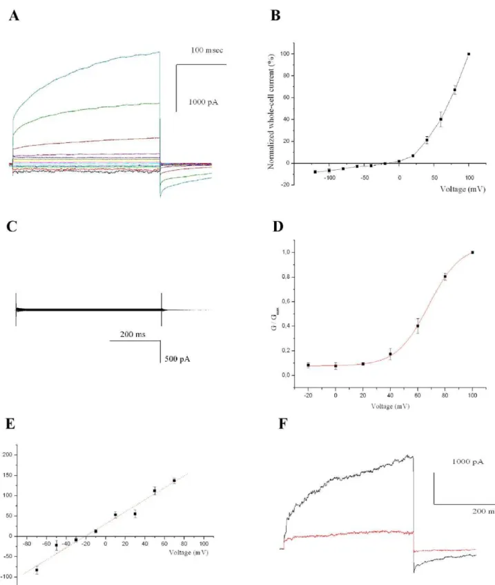
Transfected CHO cells were identified by green fluorescence and analyzed by patch clamping in whole-cell configuration. SynK gave rise to an outwardly rectifying current (Figure 3A and B) (n = 32). Cells either left untransfected or transfected with control plasmids never displayed such a current (Figure 3C) (n = 40). The SynK current had an instantaneous and a slowly activating component (Figure 3A), the latter having an activation voltage of+67 mV as determined from the Boltzman fit of the G/ Gmaxcurve (Figure 3D). SynK activity was selective for cations as
indicated by the fact that it was observed in the presence of potassium gluconate (Figure 3F, and not shown). Tail current analysis revealed a reversal potential (Erev) of22164 mV (n = 4)
which is consistent with potassium selectivity (the predicted Erev
for a perfectly selective channel in our ionic conditions is
223 mV) (Figure 3E). Furthermore, SynK was blocked by 15 mM cesium (Figure 3F) and could not be observed with solutions containing tetraethylammonium chloride (n = 10, not shown), a general potassium channel blocker [17]. To further prove that the activity observed was due to SynK, we also transfected CHO cells with SynK bearing a single point mutation in the selectivity filter GYGD (in the mutant tyrosine 181 was changed to alanine). K+
channels with GAGD sequence are known to be expressed, but are unable to conduct a current e.g. [27]. The mutant SynK was efficiently expressed and targeted to PM in CHO cells (Figure S3) but did not give rise to current (n = 6) (not shown). These data indicate that SynK does form a potassium selective channel.
SynK is located to both thylakoid and plasmamembrane in cyanobacteria
plasmamembrane (Figure 4D). As a positive control we used a specific antibody against CP43 protein of Photosystem II (Figure 4E), known to be located exclusively in the thylakoid membrane [29] and as negative control we used gold-coupled secondary IgG (Figure S7). Please note that the position of the anti-CP43-coupled gold particles with respect to the thylakoid membrane (white membraneous structure) is comparable to that obtained with anti-SynK antibody.
A homolog of SynK is present in the thylakoid membrane ofArabidopsis
The closest homolog of SynK in Arabidopsis is TPK3 (Score: 41,2; expect value: 3e-08, 36% identity, 51% positivities; Figure S8), which has a consensus prediction for localization in chloroplasts (http://aramemnon.botanik.uni-koeln.de/). TPK5 also shows some sequence similarity to SynK, and has a very strong predicted targeting for chloroplast according to several
Figure 2. Expression of SynK in Chinese Hamster Ovary cells. A) SynK-EGFP fusion protein expression in CHO cell plasma membrane, revealed by fluorescence microscopy. Fusion protein (left image) and PM-specific Vybrant DiI dye (central image) co-located as indicated by overlapping image (right). Representative images are shown. Bars: 10mm. Unequal distribution of Vybrant DiI may be due to preferential concentration of dye in rafts or to rapid vesicular uptake.B) SynK-EGFP is expressed with predicted molecular weight in CHO cells. Untransfected cells (lane 1) and CHO cells transfected with pEGFP-N1 (lane 2) or pSynK-EGFP (lanes 3, 4) were lysed 72 h after transfection, and 50mg (lanes 1, 2, 4) or 100mg (lanes 3) total proteins were loaded. Membranes were developed with anti-GFP (lanes 1–3) or anti-SynK (lane 4) primary antibodies. Arrows: positions of EGFP (28 kDa), SynK (27 kDa) and SynK-EGFP (54 kDa) proteins.C) SynK fusion protein is revealed in membraneous fraction. The purity of soluble and membrane fractions obtained from transfected CHO cells was checked by antibodies against marker proteins of the plasmamembrane (PMCA) (140 kDa), endoplasmatic reticulum (SERCA) (110 kDa) and cytosol (actin) (42 kDa) (upper panels). Actin is found also in the membraneous fraction because it is in part associated to organelles and cytoskeletron. SynK-EGFP fusion protein is present in the membraneous fraction (lower panels). Equal volumes of pellet and supernatant fractions, obtained as described in the Material and Method section, were loaded on SDS-PAGE (25ml for samples developed with anti-SynK and anti-KPORE and 15ml for those developed with anti-GFP antibody).
Figure 3. SynK functions as a potassium channel in CHO cells. A) Representative whole-cell currents in a pSynK-EGFP-transfected fluorescent cell, elicited by application of voltage steps of 300 ms duration, from2140 to+100 mV in 20-mV steps, from a holding potential of250 mV. Pulses were applied every 45 seconds, allowing complete deactivation of the channel. Different colours refer to different applied voltages.B) Current-voltage relationship. Peak currents normalized to current measured at+100 mV (n = 6, SEM values are reported).C) as in A), but from a control, pEGFP-N1-transfected cell.D) Boltzman fit of G/Gmax(n = 6).E) Determination of selectivity from tail currents, elicited by stepping voltage for 400 ms to+60 mV, followed by application of2100 to +100 mV in 20-mV voltage steps for 400 ms. Tail currents are reported as function of voltage. Reversal potential is22164 mV (n = 4). In A) to E) bath and pipette solutions contained 150 mM NaCl, 70 mM KCl and 134 mM KCl, respectively.F) Current recorded in K+
-gluconate solution at+100 mV, before (black) and after (red) addition of 15 mM Cs+
to bath. Results are representative of 4 experiments.
doi:10.1371/journal.pone.0010118.g003
algorithms. Although electrophysiological and biochemical evi-dence suggest the presence of potassium-conducting channel(s) in higher plant thylakoid membrane, the molecular nature of this(ese) protein(s) is unknown. Given that the SynK antibody was developed against the first 144 amino acids of the protein, i.e. a region comprising stretches of amino acid sequences which are conserved also in TPK5 and TPK3, we predicted thata priori, the anti-SynK antibody might recognize both proteins in Arabidopsis
thylakoids, if these proteins were located in that membrane system. Anti-SynK antibody revealed a protein with an apparent MW of 54 kDa in thylakoids isolated from Arabidopsis (Figure 5A). Membrane proteins often display a migration resulting in different MW from that predicted. Since an MW of 54 kDa is somewhat higher than that predicted for TPK5 and TPK3 (46,3 and 48,7 kDa, respectively), we developed a monoclonal antibody (3A8) against a region conserved inArabidopsisTPK3/5 but not in other members of the TPK family. 3A8 gave visible reaction already with 100 ng of the immunogenic peptide in dot blot (not shown). The 54 kDa band was recognized by both anti-SynK and 3A8 (Figure 5A) and also by other two monoclonal antibodies developed against the same peptide and by anti-KPORE (not shown). The specificity of the recognition by 3A8 is indicated by the significant decrease of the intensity of the band when the
antibody was pre-incubated with its immunogenic peptide prior to blot development (Figure 5B). The identified protein is an integral membrane protein (Figure S9). Furthermore, the 54 kDa protein, pulled down by anti-SynK antibody fromArabidopsisthylakoid, was recognized by the monoclonal anti-TPK3/5 antibody (Figure 5C). To further prove the nature of the 54 kDa band, we performed Western blots on thylakoids isolated from TPK5-knock-out
Arabidopsismutant (Figure 5D). The intensity of the 54 kDa band was not significantly altered in the thylakoid membrane isolated from the knock-out plant with respect to that observed in WT thylakoids. Given that in the TPK5-knock-out plants transcripts of TPK5 were absent (not shown), the 54 kDa band in the mutant plant was attributed to TPK3. Therefore we checked for the presence of this band in plants with a t-DNA insertion in the TPK3-encoding gene. t-DNA insertion mutants are only available in the UTR or in the promoter regions for TPK3. UTR (untranslated regions) may affect efficiency of translation and the lifetime of transcripts. The transcript level of TPK3 was slightly reduced in the UTR-insertion mutant with respect to that found in wild-type (not shown). In thylakoids isolated from these plants there was a decrease of the intensity of the 54 kDa band, but complete disappearance could not be observed, being compatible with the presence of a reduced amount of TPK3. Given that most
Figure 4. Localization of SynK inSynechocystis. A) Whole-cell cyanobacterial lysates containing 0.1mg chlorophyll/lane were loaded on SDS-PAGE without urea and blotted with anti-SynK (1:2500 dilution) (lane 1) and anti-KPORE (1:10000) (lane 2) polyclonal antibodies. Apparent MWs of monomer, SDS-resistant dimer trimer and tetramer forms correspond to 26, 52, 76 and ca. 110 kDa. The anti-KPORE antibody, as expected, given the predicted presence of various potassium channels in this organism, recognized other proteins as well (lane 2).B) Plasmamembrane (PM), soluble (SOL) and thylakoid membrane (THYL) fractions were isolated fromSynechocystis. The resulting fractions were checked for purity by using antibodies against markers of the plasmamembrane (NrtA), of the soluble fraction (PBS: allophycocyanin; LSU: large subunit of Rubisco) and of Thylakoid (ATP-ase and CP43). Cross-contamination to small extenct can be observed. 20mg of proteins/lane.C) The obtained fractions were assayed for SynK content by using anti-SynK (left panel) and anti-KPORE (right panel) antibodies. 20mg of proteins loaded/lane. The apparent MWs of the observed bands are 26 kDa (arrow) in the PM fraction and 26 and 24.5 kDa in the THYL fraction.D) Anti-SynK antibody used for immunogold electron microscopy confirms location of SynK protein in thylakoids (white membraneous structures). Arrows emphasize some of the gold particles. Bar: 200 nm.E) As control, anti-CP43 was used. Bar: 500 nm.
TPK channels, including TPK1, have been proposed to be located in the membrane around the vacuole, i.e. in tonoplast in plant cells [30], we checked for contamination of our thylakoid preparation by tonoplast. In Figure 5E the anti-TIP1.1 antibody raised against an aquaporin located to tonoplast [31], recognized a 28 kDa band in isolated tonoplasts, but not in thylakoids. As a further control, the localization of TPK1 inArabidopsiscells was assayed by using a specific anti-TPK1 monoclonal antibody. Western blot analysis of vacuolar and thylakoid fractions revealed the presence of a 51 kDa band only in vacuoles isolated from WT but not in those obtained from TPK1 knock-out plants, confirming tonoplast location of TPK1 and indicating that TPK proteins might migrate with a higher than predicted MW (Figure S10).
Discussion
In the present work we report cloning and functional characterization of a novel potassium channel of cyanobacteria. The SynK protein, identified as putative potassium channel by bioinformatics, was shown to mediate potassium transport when expressed in E.coli LB2003 and gave rise to potassium-selective current when studied in Chinese Hamster Ovary cells. Specific anti-SynK antibody localized the channel protein both in thylakoid and in plasmamembrane inSynechocystiscyanobacteria. SynK is thus the first potassium channel identified in the thylakoid membrane from molecular point of view. Furthermore, SynK seems to be the ancestor of a TPK family member in
Arabidopsis, which we show to be located in thylakoids of higher plants.
SynK is shown here to function as potassium-conducting channel when expressed in heterologous systems (Figures 1–3), although structural determinants of voltage sensitivity in SynK and
factors determining the instantaneous component remain to be clarified. Data of Figure 4 indicate SynK to be located in both plasma and thylakoid membranes in Synechocystis. Recently, we have identified another ion-conducting pathway, a sodium/proton antiporter, in the thylakoid membrane of the same organism [32]. Dual localization of several proteins and ion channels have been described in eukaryotic systems e.g. [33–35]. The targeting mechanisms are not well known in cyanobacteria, but according to one model, proteins may be initially targeted to either membrane and sorted afterwards, possibly by vesicle transport [29]. Recently, the Tat protein transport system was described to function in both membrane systems [36]. In the thylakoid membrane fraction the anti-SynK antibody detected two bands, one with a slightly lower MW than that predicted (Figure 4C). Whether this lower MW band corresponds to a mature form of the thylakoid-targeted protein or to a partially degraded protein remains to be determined.
Chloroplasts are descendents of an ancestral endosymbiont of cyanobacterial origin e.g. [37,38]. Nuclear genes coding for chloroplast proteins involved in photosynthesis and organelle biogenesis have been identified. A recent work identified other nuclear-encoded chloroplast proteins of endosymbiont origin by using functional orthogenomics [35]. Our data suggest that SynK may be an ancestor of TPK3 which is a member of the two-pore potassium channel family in Arabidopsis [39]. When BLAST analysis is performed, TPK3 is the closest homolog of SynK in the whole Arabidopsis genome and vice versa, according to Aramemnon. The evolutionary origin of eukaryotic tandem-pore channels is still elusive but according to one hypothesis, 6TM prokaryotic PNBD-less potassium channels (like SynK) might have given origin to TPK channels [40]. A conserved pore region feature (presence of YF residues) in both SynK and plant TPK
Figure 5. SynK homolog TPK3 is located in the thylakoid membrane ofArabidopsis. A) SynK and the monoclonal antibody 3A8 against TPK3/5 recognize the same, 54 kDa band inArabidopsiswild-type thylakoids (proteins corresponding to 30mg chorophyll were loaded).B) Intensity of the 54 kDa band decreased when the antibody was preincubated with 300mM immunogenic peptide. The two lanes (30mg Chl/lane) are from the same blot and were processed together.C) Thylakoids isolated from WTArabidopsisplants were immunoprecipitated with anti-SynK antibody and blotted with 3A8 monoclonal antibody.D) Thylakoids (30mg Chl/lane) isolated from wild type and TPK5-knock-out (left panel) and TPK3-knock-down (right panel) plants were loaded and assayed with the monoclonal antibody. The same membranes were stripped and reblotted with anti-ATP-ase to check for equal loading.E) Tonoplast and thylakoid fractions (20mg of total protein of each) were loaded and developed with anti-TIP1.1 antibody (TIP1.1 is indicated by arrow at 28 kDa). In A, C and E nitrocellulose membranes and the BCIP/NBT (Sigma) development system, while in B and D PVDF membrane and ECL system was used.
doi:10.1371/journal.pone.0010118.g005
channels further point to an evolutionary link between the two proteins (Figure S11).
Our findings indicate the presence of TPK3 protein in the thylakoid membrane (Figure 5). Independently of whether SynK is the precursor of TPK3 or not, this is the first thylakoid-located cation channel identified from molecular point of view in higher plants (in addition to proton-conducting F0/F1 ATP-ase). Given
that the electrophysiological activity of TPK3 has not been described up to now, it is difficult to predict which of the previously described electrophysiological activities [12–15] can be assigned to TPK3 protein. In any case, the thylakoid localization of this protein opens the way to functional characterization of this still putative channel. Despite a consensus prediction for chloroplast localization of TPK1, TPK2 TPK5 and TPK3 (see Aramemnon site), these proteins have previously been shown to be targeted to the vacuolar membrane of protoplasts from Arabidopsis cultured cells that transiently expressed AtTPK in fusion with GFP or YFP under the control of the cauliflower mosaic virus (CaMV) 35S promoter [30]. Interestingly, AtTPK3 fusion protein accumulated also in additional, non-identified internal membranes when using this system (Figure 2b of ref. 30). We would like to point out that we detect AtTPK3, shown to exhibit high transcript level [30], in thylakoids obtained from genetically non-manipulated Arabidopsis
plants, by using a specific monoclonal antibody. Thus, observation of the protein in thylakoids due to possible overexpression-induced mistargeting can be excluded. Our results do not exclude localization of TPK3 in other membranes as well, nor they exclude the presence of other channels as well in thylakoids. SynK and TPK3 might be involved counterbalancing cation fluxes from the lumen towards the stroma during photosynthesis, which would permit dissipation of the transmembrane potential but not that of the pH gradient [12,15,41]. Presuming the same orientation of SynK in the CHO plasma membrane and in thylakoids, at positive voltages of the thylakoid (proposed to reach +70 mV on the lumenal side during proton flux into the lumen [42]) SynK could permit the quick exit of potassium from the lumen. Direct genetic proof in favour of the ‘‘counterbalance’’ hypothesis is still missing, due also to the fact that cation channels have not been identified from a molecular point of view neither in cyanobacterial thylakoid nor in that of higher plants.
In summary, we report the molecular identification of two thylakoid-located potassium channels, SynK in cyanobacteria and TPK3 inArabidopsis. SynK represents the first cyanobacterial core-only type potassium channel, and seems to be the anchestor of TPK3 of the two-pore potassium channel family. Our results open the way for understanding the physiological roles of these thylakoid channels and for determining their role, if any, in the regulation of photosynthesis.
Materials and Methods
Strains and growth conditions are described in supplementary Text S1. Expression of SynK in E.coli and measurement of K+ uptake was performed according to [21] and [43]. Expression of SynK in CHO cells was performed according to [44]. DNA constructs and transformation ofSynechocystissp. PCC 6803 as well as plant growth, genotyping and transcript analysis ofArabidopsis
are detailed in the suplementary material. Thylakoids from plants were isolated as described [45]. Membrane fractionations of CHO cells, cyanobacteria and Arabidopsiswere performed according to [46], [47] and [48], respectively. Immunoprecipitation, electron microscopy and immunogold labelling were performed according to [49] and [50], respectively. Patch clamp analysis is according to [34,44] and is detailed in supplementary Text S1.
Supporting Information
Text S1
Found at: doi:10.1371/journal.pone.0010118.s001 (0.04 MB DOC)
Figure S1 Closest homologues of SynK are found in cyanobac-teria. A) The closest homologues of SynK (Syn, Synechocystissp. PCC 6803; gi:16331771) are found in other cyanobacteria species. Sequence alignment (ClustalW (1.83) algorithm) of SynK, of a hypothetical protein (Lyng,Lyngbyasp. PCC 8106; gi:119457762) and K+
channel pore region (Croco, Crocosphaera watsonii WH 8501; gi:46119130). ‘‘*’’ - identical residues in all aligned sequences; ‘‘:’’ - conserved and ‘‘.’’ - semi-conserved substitutions. BLAST analysis revealed E values (number of hits expected to be found by chance) of 2610–24 and 4610–19 and positivity over length of aligned sequence of 55% (223 amino acids) and 56% (207) when compared SynK withLyngbyaandCrocosphaera watsonii
proteins, respectively. Typical selectivity filter for potassium is in green. Glycine in S6, important for gating is in yellow.
Found at: doi:10.1371/journal.pone.0010118.s002 (0.02 MB DOC)
Figure S2 Potassium uptake by K+-depleted E.coli containing SynK or empty vector. Net potassium uptake measurements by K+
-depleted E. coli cells in the presence of 10 to 80 mM KCl revealed Vmax values of 553 and 460 nmol min21 g21 dry weight for SynK-expressing cells and for the control cells, respectively Lineweaver-Burk plot of K+
uptake data obtained from four independent experiments is shown.
Found at: doi:10.1371/journal.pone.0010118.s003 (0.02 MB PDF)
Figure S3 Expression of SynK and SynK mutant in Chinese Hamster Ovary cells. SynK-EGFP WT and mutant (non-conducting mutant with GAGD instead of GYGD in the pore region) fusion protein expression in CHO cell plasma membrane was revealed by confocal microscopy. Images with GFP fusion proteins (left images) and FM4-64 dye (central images) and merged signals (right images) are shown for WT SynK-GFP (upper panels) and mutant SynK-GFP (lower panels). Graphics shown beside the merged images represent profile plots of GFP (green) and FM4-64 (red) fluorescence intensity as a function of the distance for a particular region of interest (ROI), from inside the cell (in) to outside (out). Peaks falling in the same region correspond to co-localization.
Found at: doi:10.1371/journal.pone.0010118.s004 (0.48 MB PDF)
Figure S4 Anti-SynK antibody recognizes recombinant and native SynK. Recombinant protein (144 N-terminal amino acids of SynK fused with a 6 His-tag at C-terminus) was expressed inE. coliand purified as described in Materials and Methods. Protein was purified as a 30-kDa dimer (see lane 2). 30-kDa protein, recognized by anti-His antibody (not shown), was used for antibody production. Pre-immune antiserum did not recognize either purified 30 kDa protein (lane 3) or proteins in cyanobacteria whole-cell lysate (lane 4); serum from immunized rabbit clearly reacted with the recombinant protein (lane 5) and recognized SynK of 26 kDa in whole-cell lysate (in cells containing 0.1mg
chlorophyll) even at 1:5000 dilution (lane 6).
Found at: doi:10.1371/journal.pone.0010118.s005 (0.16 MB DOC)
channel with apparent MW of 65 kDa (Magic Marks loaded on lane 1). Same bands were recognized by anti-KPORE (lane 2) and by a specific antibody against Kv1.3 (1:200) (lane 3) in SDS-PAGE with 6 M urea. 50mg total proteins were loaded. Anti-KPORE antibody also recognized purified GST-Kv1.3 protein (lane 4, 10mg loaded, predicted MW 87 kDa) (production of GST-Kv1.3
is described in Gulbins et al, Biochim. Biophys. Acta, in press). Anti-KPORE antibody also recognized KCa3.1 in HCT116 colon cancer cell line (not shown), and monomeric as well as multimeric forms of the purified Kcv viral potassium channel (not shown) and of purified KvAP (kindly provided by P.Facci, not shown). Found at: doi:10.1371/journal.pone.0010118.s006 (0.07 MB DOC)
Figure S6 Anti-SynK antibody efficiently recognizes SynK in whole-cell lysate of cyanobacteria. Cells corresponding to the O.D. (at 730 nm) shown on the figure were solubilized in SB and loaded on SDS-PAGE. The blot was first developed with anti-SynK antibody and after re-stripping with anti-ATP-ase antibody (Agrisera). Efficiency of anti-Synk and anti-ATP-ase antibodies is comparable.
Found at: doi:10.1371/journal.pone.0010118.s007 (3.76 MB PDF)
Figure S7 Secondary antibody does not label cyanobacteria in immunogold electron microscopy. As control, only secondary IgG was used. Bar: 500 nm.
Found at: doi:10.1371/journal.pone.0010118.s008 (0.05 MB PDF)
Figure S8 Sequence homology between cyanobacterial SynK and Arabidopsis TPK3 (At4g18160). Aminoacid sequence align-ments obtained by T-COFFEE algorithm. ‘‘*’’ - identical residues in all aligned sequences; ‘‘:’’ - conserved, ‘‘.’’ - semi-conserved substitutions.
Found at: doi:10.1371/journal.pone.0010118.s009 (0.21 MB PDF)
Figure S9 The 54 kDa protein is an integral membrane protein. Thylakoids (100 mg total proteins) were subjected to alkaline extraction (0.2 M Na2CO3 for 30 minutes), pelleted and both pellet and supernatants were loaded. The 54 kDa band is not present in the supernatant fraction indicating that it is an integral membrane protein. Blots were developed with the indicated antibodies. Found at: doi:10.1371/journal.pone.0010118.s010 (0.10 MB PDF)
Figure S10 TPK1 locates to tonoplast inArabidopsis. A specific monclonal antibody was used to reveal location of TPK1 in WT and atkco1 plants. Cells were fractionated and loaded on continuous sucrose gradient. Fractions positive for tonoplast
TIP1 (VAC) or for thylakoid membrane D2 (THYL) were loaded. TPK1 is visible only in the vacuolar fraction of WT cells (at 50 kDa). An aspecific recognition is seen at approx. 35 kDa in thylakoids in both WT and mutant organisms.
Found at: doi:10.1371/journal.pone.0010118.s011 (0.06 MB PDF)
Figure S11 Pore region and YF residues are highly conserved between SynK and TPK channels ofArabidopsis. Voltage-gated Kv and KCNQ channels are characterized by a conserved pore region feature, namely, the presence of two tryptophans in tandem (W67 and W68 in KcsA) (Minor DL (2001) Potassium channels: life in the post-structural world. Current Opinion in Structural Biology, 11: 408–414). In plant shaker-like inward rectifier channels, the second tryptophan is highly conserved and the first is replaced by a tyrosine. These same positions are strongly conserved within other families of potassium channels, however, as different residues. Animal Kir channels harbour LF or SF residues in the same position (Minor 2001). Instead, in animal two-pore channels, in viral Kcv as well as in all plant two-pore channels the same positions are occupied by tyrosine and phenylalanine (YF). SynK has the same YF aminoacids in the corresponding position, further suggesting that SynK might have given origin to two-pore channels during evolution. Interestingly, GORK and SKOR outwardly rectifying voltage-dependent channels, also harbour YF residues in the corresponding position but, in contrast to TPK3, do not show significant homology with SynK. Aminoacid sequence alignments obtained by TCOFFEE algorithm. ‘‘*’’ identical residues in all aligned sequences; ‘‘:’’ conserved, ‘‘.’’ -semi-conserved substitutions. YF residues, typical of Kcv, animal and plant two-pore potassium channels are indicated. At4g01840: TPK5; Atg1g02510: TPK4; At4g18160: TPK3; At5g46370: TPK2; At5g55630: TPK1.
Found at: doi:10.1371/journal.pone.0010118.s012 (0.03 MB PDF)
Acknowledgments
The authors are grateful to Drs. M. Zoratti, A. Moroni, F. Tombola, A. Accardi and F. Rigoni for useful discussions. They thank very much Drs. A. Costa and E. Formentin for the confocal microscopy analysis. They thank G. Zambolin for help with construction of the patch clamp set-up and G. Walton for revision of the English text.
Author Contributions
Conceived and designed the experiments: MZ ET GMG IS. Performed the experiments: MZ ET NLR LZ VC TS YS IS. Analyzed the data: MZ ET NLR LZ VC TS YS GMG NU EB IS. Wrote the paper: NU EB IS.
References
1. Herrero A, Flores E (2008) The cyanobacteria: molecular biology, genetics and evolution. Caister Academic Press.
2. Kaneko T, Sato S, Kotani H, Tanaka A, Asamizu E, et al. (1996) Sequence analysis of the genome of the unicellular cyanobacteriumSynechocystis sp. strain PCC6803. II. Sequence determination of the entire genome and assignment of potential protein-coding regions. DNA Res 3: 109–136.
3. Kuo M-M-C, Haynes W-J, Loukin S-H, Kung C, Saimi Y (2005) Prokaryotic K+
channels: from crystal structures to diversity. FEMS Microbiology Reviews 29: 961–985.
4. Matsuda N, Uozumi N (2006) Ktr-mediated potassium transport, a major pathway for potassium uptake, is coupled to a proton gradient across the membrane inSynechocystis sp. PCC 6803. Biosci Biotechnol Biochem 70: 273–275. 5. Chen G-Q, Ciu C, Mayer M-L, Gouaux E (1999) Functional characterization of
a potassium-selective prokaryotic glutamate receptor. Nature 402: 817–821. 6. Bocquet N, Prado de Carvalho L, Cartaud J, Neyton J, Le Poupon C, et al.
(2007) A prokaryotic proton-gated ion channel from the nicotinic acetylcholine receptor family. Nature 445: 116–119.
7. Iyer R, Iverson T-M, Accardi A, Miller C (2002) A biological role for prokaryotic ClC chloride channels. Nature 419: 715–718.
8. Martinac B (2004) Mechanosensitive ion channels: molecules of mechanotrans-duction. Journal of Cell Science 117: 2449–2460.
9. Stingl K, Brandt S, Uhlemann EM, Schmid R, Altendorf K, et al. (2007) Channel-mediated potassium uptake inHelicobacter pyloriis essential for gastric colonization. EMBO J 26: 232–241.
10. Epstein W (2003) The roles and regulation of potassium in bacteria. Prog Nucleic Acid Res Mol Biol 75: 293–320.
11. Berry S, Esper B, Karandashova I, Teuber M, Elanskaya I, et al. (2003) Potassium uptake in the unicellular cyanobacterium Synechocystis sp. Strain PCC6803 mainly depends on a Ktr-like system encoded by slr1509 (ntpJ). FEBS Lett 548: 53–58.
12. Tester M, Blatt MR (1989) Direct measurement of K+
channels in thylakoid membranes by incorporation of vesicles into planar lipid bilayers. Plant Physiol 91: 249–252.
13. Enz C, Steinkamp T, Wagner R (1993) Ion channels in the thylakoid membrane (a patch clamp study). Biochim Biophys Acta 1143: 67–76.
14. Pottosin II, Schonknecht G (1996) Ion channel permeable for divalent and monovalent cations in native spinach thylakoid membranes. J Membr Biol 152: 223–233.
15. Hinnah SC, Wagner R (1998) Thylakoid membranes contain a high-conductance channel. Eur J Biochem 253: 606–613.
16. Fang Z, Mi F, Berkowitz G (1995) Molecular and physiological analysis of a thylakoid K+
channel protein. Plant Physiol 108: 1725–1734.
17. Hille B (2003) Chapter 5. In Ion channels of excitable membranes Sinauer Ed. Sundreland, USA, Third edition.
18. Jan L-Y, Jan Y-N (1997) Cloned potassium channels from eukaryotes and prokaryotes. Annu Rev Neurosci 20: 91–123.
19. Ruta V, Jiang Y, Lee A, Chen J, MacKinnon R (2003) Functional analysis of an archaebacterial voltage-dependent K+
channel. Nature 422: 180–184. 20. Santos J-S, Lundby A, Zazueta C, Montal M (2006) Molecular template for a
voltage sensor in a novel K+
channel. Identification and functional character-ization of KvLm, a voltage-gated K+
channel fromListeria monocytogenes. J Gen Physiol 128: 283–300.
21. Uozumi N (2001) Escherichia coli as an expression system for K+ transport systems from plants. Am J Physiol Cell Physiol 281: C733–C739.
22. Matsuda N, Kobayashi H, Katoh H, Ogawa T, Futatsugi L, et al. (2004) Na+ -dependent K+
uptake Ktr system from the cyanobacteriumSynechocystis sp. PCC 6803 and its role in the early phases of cell adaptation to hyperosmotic shock. J Biological Chemistry 279: 54952–54962.
23. Stumpe S, Bakker E-P (1997) Requirement of a large K+
-uptake capacity and of extracytoplasmic protease activity for protamine resistance ofEscherichia coli. Arch Microbiol 167: 126–136.
24. Kuo M-M-C, Saimi Y, Kung C, Choe S (2007) Patch clamp and phenotypic analyses of a prokaryotic cyclic nucleotide-gated K+
channel usingEscherichia coli as a host. J Biol Chem 282: 24294–24301.
25. Schrempf H, Schmidt O, Ku¨mmerlen R, Hinnah S, Mu¨ller D, et al. (1995) A prokaryotic potassium ion channel with two predicted transmembrane segments fromStreptomyces lividans. EMBO J 14: 5170–5178.
26. Gazzarrini S, Severino M, Lombardi M, Morandi M, DiFrancesco D, et al. (2003) The viral potassium channel Kcv: structural and functional features. FEBS Letters 552: 12–16.
27. Heginbotham L, Lu Z, Abramson T, MacKinnon R (1994) Mutations in the K+ channel signature sequence. Biophysical Journal 66: 1061–1067.
28. Cortes D-M, Perozo E (1997) Structural dynamics of theStreptomyces lividansK+ channel (SKC1): oligomeric stoichiometry and stability. Biochemistry 36: 10343–10352.
29. Zak E, Norling B, Maitra R, Huang F, Andersson B, et al. (2001) The initial steps of biogenesis of cyanobacterial photosystems occur in plasma membranes. Proc Natl Acad Sci USA 98: 13443–13448.
30. Voelker C, Schmidt D, Mueller-Roeber B, Czempinski K (2006) Members of the ArabidopsisAtTPK/KCO family form homomeric vacuolar channels in planta. Plant J 48: 296–306.
31. Ma S, Quist T-M, Ulanov A, Joly R, Bohnert H-J (2004) Loss of TIP1;1 aquaporin inArabidopsisleads to cell and plant death. Plant J 40: 845–59. 32. Tsunekawa K, Shijuku T, Hayashimoto M, Kojima Y, Onai K, et al. (2009)
Identification and Characterization of the Na+ /H+
Antiporter Nhas3 from the Thylakoid Membrane ofSynechocystissp. PCC 6803. J Biol Chem 284: 16513–21. 33. Karniely S, Pines O (2005) Single translation-dual destination. EMBO Reports
6: 420–425.
34. Szabo I, Bock J, Grassme´ H, Soddemann M, Wilker B, et al. (2008) Mitochondrial potassium channel Kv1.3 mediates Bax-induced apoptosis in lymphocytes. Proc Natl Acad Sci USA 105: 14861–14866.
35. Ishikawa M, Fujiwara M, Sonoike K, Sato N (2009) Orthogenomics of photosynthetic organisms: bioinformatic and experimental analysis of chloro-plast proteins of endosymbiont origin inArabidopsisand their counterparts in Synechocystis.Plant Cell Physiol 50: 773–788.
36. Aldridge C, Spence E, Kirkilionis MA, Frigerio L, Robinson C (2008) Tat-dependent targeting of Rieske iron-sulphur proteins to both the plasma and thylakoid membranes in the cyanobacterium Synechocystis PCC6803. Mol Microbiol 70: 140–150.
37. Martin W, Rujan T, Richly E, Hansen A, Cornelsen S, et al. (2002) Evolutionary analysis of Arabidopsis, cyanobacterial, and chloroplast genomes reveals plastid phylogeny and thousands of cyanobacterial genes in the nucleus. Proc Natl Acad Sci USA 99: 12246–12251.
38. Sato N (2006) Origin and evolution of plastids: genomic view on the unification and diversity of plastids. In The Structure and Function of Plastids. Edited by Wise, R.R. and Hoober, J.K. pp. 75–102. Springer, Dordrecht.
39. Maser P, Thomine S, Schroeder JI, Ward JM, Hirschi K, et al. (2001) Phylogenetic relationships within cation transporter families ofArabidopsis. Plant Physiol 126: 1646–67.
40. Derst C, Karschin A (1998) Evolutionary link between prokaryotic and eukaryotic K+
channels. J Exp Biol 201: 2791–2799.
41. Schonknecht G, Hedrich R, Junge W, Raschke K (1988) A voltage dependent chloride channel in the photosynthetic membrane of higher plant. Nature 336: 589–592.
42. Remis D, Bulychev AA, Kurella GA (1986) The electrical and chemical components of the proton motive force in chloroplasts as measured with capillary and pH-sensitive electrodes. Biochim Biophys Acta 852: 68–73. 43. Tholema N, Bakker EP, Suzuki A, Nakamura T (1999) Change to alanine of one
out ot four selectivity filter glycines in KtrB causes a two magnitude decrease in the affinities for both K+
and Na+
of the Na+
dependent K+
-uptake system KtrAB from Vibrio alginolyticus. FEBS Lett 450: 217–220.
44. Downey P, Szabo` I, Ivashinika N, Negro A, Guzzo F, et al. (2000) KDC1, a Novel Carrot Root Hair K+
Channel. Cloning, characterization and expression in mammalian cells. J Biological Chemistry 275: 39420–39426.
45. Bergantino E, Segalla A, Brunetta A, Teardo E, Rigoni F, et al. (2003) Light-and pH-dependent conformational change of the PsbS subunit of photosystem II. Proc Natl Acad Sci USA 100: 15265–15270.
46. Pulina MW, Rizzuto R, Brini M, Carafoli E (2006) Inhibitory interaction of the plasma membrane Na+
/Ca2+
exchangers with the 14-3-3 proteins. J Biol Chem 281: 19645–19654.
47. Bolter B, Soll J, Schulz A, Hinnah S, Wagner R (1998) Origin of a chloroplast protein importer. Proc Natl Acad Sci USA 95: 15831–15836.
48. Ishikawa F, Suga S, Uemura T, Sato MH, Maeshima M (2005) Novel type aquaporin SIPs are mainly localized to the ER membrane and show cell-specific expression inArabidopsis thaliana. FEBS Lett 579: 5814–5820.
49. Teardo E, de Laureto PP, Bergantino E, Dalla Vecchia F, Rigoni F, et al. (2007) Evidences for interaction of PsbS with photosynthetic complexes in maize thylakoids. Biochim Biophys Acta Bioenergetics 1767: 703–11.