Original Article
3 2 Arq Bras Oftalmol. 2015;78(1):32-5 http://dx.doi.org/10.5935/0004-2749.20150009
INTRODUCTION
The intraocular application of antiangiogenic drugs through in-travitreal injection (IVI) has changed the natural progression of many retinal diseases during the past 10 years. The treatment of retinal diseases began with the use of pegaptanib (Macugen®, Valeant, Canada) and was followed by the use of bevacizumab (Avastin®, Roche, Switzerland), as reported by Phillip Rosenfeld (2005)(1,2). At present, two antiangiogenic drugs have been approved by the Brazi-lian National Health Surveillance Agency (ANVISA) for intravitreal use: ranibizumab (Lucentis®, Novartis Switzerland) and aflibercept (Eylia®, Bayer HealthCare Pharmaceuticals, Germany).
The IVI of antiangiogenic drugs is intended to transiently decrea-se the amount of vascular endothelial growth factor (VEGF). VEGF is responsible for the alterations observed during diseases such as age-related macular degeneration (AMD), diabetic macular edema, and central retinal vein occlusions (CRVO) or branch retinal vein occlusions (BRVO). The pain experienced by patients is considered to be mild(3), and the procedure can be performed under topical anes-thesia or nerve block, with or without venous sedation.
The technique, operative precautions, and the area where the procedure is performed differ according to each surgeon and according to different standards adopted in each country. Green-Simms et al.
Survey: technique of performing intravitreal injection among members of the
Brazilian Retina and Vitreous Society (SBRV)
Survey: técnica para realização de injeção intravítrea pelos membros da
Sociedade Brasileira de Retina e Vítreo (SBRV)
Helio F. SHiroma1, micHel e. FaraH1, Walter Y. takaHaSHi2, andre m. V. GomeS2, mauro Goldbaum2, eduardo bucHele rodriGueS1
Submitted: September 8, 2014 Accepted: November 11, 2014
1 Department of Ophthalmology and Visual Science, Paulista School of Medicine (EPM). Federal
University of São Paulo (UNIFESP), São Paulo, Brazil.
2 Division of Ophthalmology, Medical School, University of São Paulo (USP), São Paulo, SP, Brazil.
Funding: No specific financial support was available for this study.
Disclosure of potential conflicts of interest: None of the authors have any potential conflict of interest to disclose.
Corresponding author: Helio Shiroma. Rua Pastor William Richard Schisler, 900 - Apto 1.011 - Florianópolis, SC - 88034-100 - Brazil - Email: helioshiroma@hotmail.com
Approved by the following Research Ethics Committee: Research Ethics Committee of UNIFESP under protocol No 132244. Brazil Research Platform No 24089513.3.0000.5505.
ABSTRACT
Objective: To evaluate and describe the precautions involved in the technique of intravitreal injection of antiangiogenic drugs adopted by the ophthalmologists who are members of the Brazilian Society of Retina and Vitreous (SBRV).
Method: A questionnaire containing 22 questions related to precautions taken before, during, and after intravitreal injection was sent electronically to 920 mem-bers of SBRV between November 15, 2013 and April 31, 2014.
Results: 352 responses (38%) were obtained. There was a predominance of men (76%) from the southwest region of Brazil (51%). The professional experience varied between 6 and 15 years after medical specialization (50%). Most professionals (76%) performed an average of 1 to 10 intravitreal injections a week, and 88% of the procedures were performed in the operating room using povidone iodine (99%), sterile gloves, and blepharostat (94%). For inducing topical anesthesia, usage of anesthetic eye drops was the most used technique (65%). Ranibizumab (Lucentis®) was the most common drug (55%), and age-related macular degeneration (AMD) was the most treated disease (57%). Regarding the complications treated, 6% of the ophthalmologists had treated at least one case of retinal detachment, 20% had treated cases of endophthalmitis, 9% had treated cases of vitreous hemorrhage, and 12% had encountered cases of crystalline lens touch.
Conclusion: Intravitreal injection is a procedure routinely performed by retina specialists and has a low incidence of complications. Performing the procedure in the operating room using an aseptic technique was preferred by most of the respondents. Ranibizumab was the most used drug, and AMD was the most treated disease.
Keywords: Intravitreal injections; Retinal diseases; Angiogenesis inhibitors; Topical anesthesia
RESUMO
Objetivo: Avaliar e descrever os cuidados envolvidos durante o procedimento de in jeção intravítrea de drogas antiangiogênicas realizado pelos oftalmologistas membros da Sociedade Brasileira de Retina e Vítreo (SBRV).
Método: Foi enviado um questionário aos 920 membros da SBRV, por meio de correio eletrônico, entre o período de 15/11/2013 a 31/04/2014, contendo 22 questões, rela-cionado aos cuidados pré, intra e pós-operatório da injeção intravítrea.
Resultados: Foram obtidas 352 respostas (38% dos sócios). Houve um predomínio do sexo masculino (76%), procedentes da região Sudeste (51%). O tempo de experiên-cia profissional se concentrou entre 6 a 15 anos após o término da espeexperiên-cialização (50%). A maioria dos participantes tem média semanal de 1 a 10 (76%), sendo 88% das vezes realizado dentro do centro cirúrgico, utilizando iodopovidona (99%), luvas e blefarostato estéreis (94%). A anestesia tópica com colírio anestésico foi a técnica
mais utilizada (65%). Entre os participantes, ranibizumabe (Lucentis®) é a droga mais
utilizada (55%) e a degeneração macular relacionada a idade (DMRI) é a doença mais tratada (57%). Das complicações citadas pelos oftalmologistas, 6% já vivenciaram pelo menos um caso de descolamento de retina, 20% endoftalmite, 9% hemorragia vítrea e 12% toque cristaliniano.
Conclusão: A injeção intravítrea é um procedimento realizado rotineiramente por retinólogos, com baixo índice de complicações. A realização do procedimento no centro cirúrgico com técnica asséptica é preferida pelos pesquisados. A droga mais utilizada foi o ranibizumabe e a doença mais tratada foi a DMRI.
Shiroma HF, et al.
3 3 Arq Bras Oftalmol. 2015;78(1):32-5 described the technique used for IVI in the United States(4). In June
2014, retina specialists described the technique used for IVI in Cana-da(5). However, we have not found any study describing the technique used for IVI in Brazil.
The objective of this study was to assess and describe the precau-tions, techniques, and materials used by ophthalmologists who are members of the Brazilian Society of Retina and Vitreous (Sociedade Brasileira de Retina e Vítreo; SBRV) for IVI.
METHODS
A descriptive questionnaire was created for specialists in the field of the retina and vitreous. This questionnaire was approved by the Research Ethics Committee of UNIFESP under protocol No. 132244.
Data were collected using a questionnaire containing 22 queries regarding epidemiological data; the anesthetic technique used; precautions adopted before, during, and after surgery; and the cal-culation of the incidence of complications. Questionnaires were sent electronically to 920 SBRV members between November 15, 2013 and April 31, 2014 and were sent at two other occasions to profes-sionals who did not respond to the first invitation. The answers were compiled and analyzed using the Survey Monkey method(6) (Palo Alto, California, USA). The variable results were expressed as relative frequencies. Only questionnaires answered in full were evaluated. The confidentiality of the participants was maintained during all stages of the study, and it was not possible to match the participants with their responses.
RESULTS
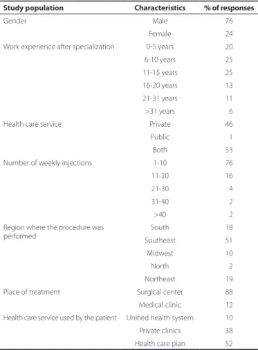
A total of 352 SBRV members (38%) responded to the question-naire and herein will be referred to as the study participants. There was a predominance of men (76%) responding to this questionnaire. With regard to the period of professional experience after retina spe-cialization, 20% of the respondents had ≤5 years of experience, 25% had 6-10 years of experience, 25% had 11-15 years of experience, 13% had 16-20 years of experience, 11% had 21-30 years of experience, and 6% had >31 years of experience.
Of the study participants, 51% were located in the southeast of Brazil, 18% in the northeast, 18% in the south, 10% in the midwest, and 2% in the north. Moreover, 46.02% of the participants worked ex-clusively in the private sector, 1.42% in the public sector, and 52.56% in both sectors. With regard to the type of health care provided to the patients, 38% of the patients were assisted in private clinics, 52% used health care plans, and 10% used the Unified Health System (SUS) (Table 1).
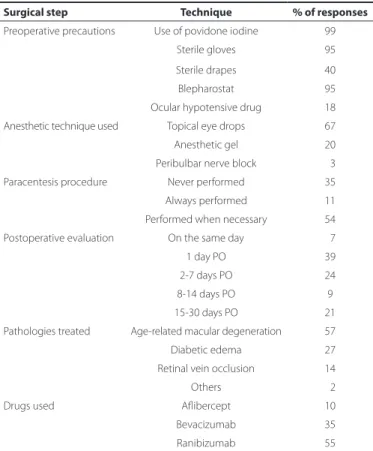
The location chosen to perform the IVI procedures was the ope-rating room (88.10%), ophthalmic clinics (5.10%), and in minor pro-cedure rooms (6.80%). The mean number of IVI propro-cedures per week performed by the study participants was as follows: 1-10 injections (76.10%), 11-20 injections (16.40%), 21-30 injections (4.20%), 31-40 injections (1.70%), and >40 injections (1.40%). Povidone iodine was used in 99.10% of the cases whereas sterile gloves, drapes, and ble-pharostat were used in 94.80% of the cases. Sterile drapes were used by 39.40% of the professionals.
For anesthesia, topical anesthesia with anesthetic eye drops was the most widely used technique (used by 67% of the participants), followed by anesthetic eye drops with anesthetic gel (20%), and pe-ribulbar nerve block (3%). Approximately 12% of the respondents reported having used intravenous sedation.
Most retina specialists (80%) used a compass to measure the distance of the limbus. Moreover, tunneling and conjunctival displa -cement were performed by 30% and 58% of the professionals, res-pectively. Intravitreal injection in both eyes was performed on the same day by 36% of the respondents when necessary. With regard to the type of needle/syringe used, 43% used the syringe that came
with ranibizumab whereas 48% used a syringe attached to an ultra-fine needle (BD ultraultra-fine® needle, Becton, Dickinson, and Company, New Jersey, USA).
Furthermore, 5% of the participants used hypotensive eye drops, 10% used acetazolamide, and 3% used compression of the eyeball with a Honan balloon in the preoperative period; 30% of the parti-cipants had never performed paracentesis whereas 54% reported having performed the procedure when necessary.
AMD was the most common disease treated (57%), followed by diabetic macular edema (27%), retinal vein occlusion (14%), and other pathologies (2%). Ranibizumab was the most widely used drug among the participants (55%), followed by bevacizumab (35%), and afliber-cept (10%) (Table 2).
With regard to the AMD treatment protocols, 30% of the partici-pants preferred the Pro Re Nata (PRN) approach (7), 26% preferred the “treat and extend” approach(8), and 12% preferred the monthly appli-cations. One-third of the participants used one of these treatment regimens, depending on individual patients.
Postoperative evaluation was conducted on the same day by 7% of the participants, on the first day postoperatively (PO) by 39%, within 3-7 days PO by 24%, within 7-14 days PO by 9%, and within 14-30 days PO by 21%. The use of antibiotic eye drops for 3-7 days PO was prescribed by 89.21% of the professionals.
In the postoperative period, mild punctate keratitis was the most frequent complication, which was reported to be encountered by 62% of the participants. Most (65%) of the participants reported having encountered no serious complications, 20% reported having treated at least one case of endophthalmitis, 6% had treated cases
Table 1. Proile of the respondents
Study population Characteristics % of responses
Gender Male 76
Female 24 Work experience after specialization 0-5 years 20 6-10 years 25 11-15 years 25 16-20 years 13 21-31 years 11 >31 years 06 Health care service Private 46 Public 01
Both 53
Number of weekly injections 1-10 76 11-20 16 21-30 04 31-40 02 >40 02 Region where the procedure was
performed
Survey: technique of performing intravitreal injection among members of the Brazilian Retina and Vitreous Society (SBRV)
3 4 Arq Bras Oftalmol. 2015;78(1):32-5
of retinal detachment, 9% had treated cases of vitreous hemorrhage, and 12% had treated cases of crystalline lens touch (Table 3).
DISCUSSION
The subspecialty of retina and vitreous care has attracted the in-terest of young ophthalmologists because of considerable advances in diagnostic and surgical equipment and because of novel therapies against diseases that would invariably have progressed to blindness. Although the questionnaires were sent repeatedly to all SBRV members, only 38% responded, and this low response rate introdu-ced a source of bias that precluded the generalization and interpre-tation of data. However, a great merit of the present study was the description of the IVI technique adopted by a representative sample of the ophthalmologists, and the responders may have included the most active members of the SBRV.
Men predominated (76%) among the study participants, and most questionnaires came from the southeast region, which has the lar-gest concentration of specialists. Specialists with various levels of professional experience participated, including recently graduated doctors (20%) and professionals with >30 years experience (6%), with an average of 6-15 years of experience.
Most retina specialists performed an average of 1-10 IVIs a week, and 88% of the procedures were performed in the operating room. The medical clinic was chosen for the procedure by only 12% of the respondents, in contrast to the results of large studies such as those conducted by the Comparison of Age-related Macular Degeneration Treatments Trials (CATT), involving 12,886 injection procedures and the retrospective study by Cheung et al., involving 14,895 injection procedures, wherein all procedures were performed in the clinic (9). Sterile surgical materials, povidone iodine, and topical eye drops during patient preparation were used in approximately 99% of the cases. Until date, there is no consensus on the use of sterile drapes. In the present study, it was used in <40% of the cases in an attempt to decrease the risk of endophthalmitis. In the U.S., gloves are not mandatory, and syringes do not need to be sterile, but the tip of any surgical instrument should remain sterile until it touches the pa tient’s eye(4).
The most used anesthetic technique was topical anesthesia with eye drops or gels, with or without compression using cotton swabs. However, intravenous sedation conducted by anesthesiologists can provide comfort and reassurance to patients and the surgeon. On the other hand, the disadvantages of intravenous sedation include the risks associated with CNS depressant drugs, high cost of the proce-dure, and increased length of hospitalization(10).
The application site was demarcated using a compass in 80% of the cases. The most used syringe was BD ultrafine® in 48% of the cases. One reason for preferring the use of small-volume syringes is the accuracy in adjusting the drug volume, which is directly related to treatment efficacy and response variability(11).
Precautions involving intraocular pressure (IOP) during the posto-perative period were not very relevant among the physicians, con-sidering that 48% of the respondents did not measure IOP after the procedure. A study by Yannuzzi et al. (2014) based on a questionnaire sent to retina specialists suggested that the sustained increase in IOP after IVI was directly associated with the volume and rate of injection of the drug. Accordingly, patients who received volumes of >0.05 cm3 injected in less than one s showed 5.56 times more likelihood of ha-ving ocular hypertension due to potential damage of the trabecular meshwork(12). The use of oral or ocular hypotensive eye drops was reported by <10% of the respondents. Previous studies have shown that scleral compression using cotton swabs containing anesthetic(13) or the use of a Honan balloon(14) can dehydrate the vitreous and lower IOP, thereby avoiding peaks during the immediate postopera-tive period. In the present study, paracentesis was performed when necessary by 54% of the respondents, whereas 31% reported never having used this procedure. The Diabetic Retinopathy Clinical Research Network (DRCR.net) was founded in 2002 by the National Institute of Health with the aim of performing multicenter studies in 200 cities across the U.S., Europe, and Asia and defining the best treatment for diabetic retinopathy and macular edema using IVI. However, DRCR. net did not require the measurement of IOP and only assessed visual acuity and optic nerve perfusion using indirect ophthalmoscopy(14). In our study, 25% of the respondents reported having assessed the optic nerve perfusion, and 25% measured IOP after the procedure.
With regard to postoperative complications, 65% of the profes-sionals reported not having encountered any serious complications related to IVIs during their professional career. Among the ophthal-mologists who encountered at least one case with complications, retinal detachment and crystalline lens touch may have been asso-ciated with the inadvertent movement of the patient during the pro cedure. Endophthalmitis is another serious complication, and 20%
Table 2. Techniques and precautions adopted during intravitreal injection
Surgical step Technique % of responses
Preoperative precautions Use of povidone iodine 99 Sterile gloves 95 Sterile drapes 40 Blepharostat 95 Ocular hypotensive drug 18 Anesthetic technique used Topical eye drops 67 Anesthetic gel 20 Peribulbar nerve block 03 Paracentesis procedure Never performed 35 Always performed 11 Performed when necessary 54 Postoperative evaluation On the same day 07 1 day PO 39 2-7 days PO 24 8-14 days PO 9 15-30 days PO 21 Pathologies treated Age-related macular degeneration 57 Diabetic edema 27 Retinal vein occlusion 14 Others 02 Drugs used Aflibercept 10 Bevacizumab 35 Ranibizumab 55
PO= postoperatively.
Table 3. Percentage of participants who encountered complications associated with intravitreal injection during their professional career
Complication Frequency
Endophthalmitis 20.2%
Shiroma HF, et al.
35 Arq Bras Oftalmol. 2015;78(1):32-5 of the respondents had encountered this situation. However, the
reported incidence of this complication is <0.10% (15,16) and cannot be compared with the benefits provided by the treatment(17).
The most used drugs were ranibizumab (55%), followed by be-vacizumab (35%), and aflibercept (10%). Ranibizumab was the first antiangiogenic drug registered by ANVISA for retinal diseases. Bevaci-zumab is still used off-label in Brazil but less frequently than in the U.S. In the present study, it was used in 61% of the cases of AMD, in 62% of the cases of CRVO, and in 64% of the cases of BRVO(18). Aflibercept is being used increasingly by ophthalmologists and was released in Brazil in early 2013. Further studies should indicate possible changes in the pattern of use of antiangiogenic drugs in Brazil.
The use of antibiotics in the postoperative period is prevalent (89%) in Brazil, although several studies have reported the low incidence of endophthalmitis together with possible selection of resistant pathogens(19). The fact that the indication for endophthalmitis is mentioned on the package leaflet as well as legal aspects may explain their exaggerated use. The DRCR.net does not impose the use of antibiotics(20) in the postoperative period, and they should be used at the discretion of each doctor. In 2011, a study reported 5 cases of en-dophthalmitis after 6251 applications of ranibizumab in the form of eye drops. Furthermore, a clinical study published in 2009 evaluated the incidence of endophthalmitis after the injection of ranibizumab or triamcinolone in patients with diabetic retinopathy and found a low incidence of endophthalmitis (0.09% with ranibizumab and no incidence with triamcinolone) along with blepharostat and povido-ne iodipovido-ne without the povido-need for topical eye drops, antibiotics, sterile gloves, or sterile drapes(21).
In summary, despite the limitations of our study with respect to the moderate participation of retina specialists who are SBRV members, we can conclude that IVI is a common and standardized procedure with low risk of complications. The performance of IVI in surgical centers and the precautions involving sterile equipment are evident in Brazil. Among the respondents, ranibizumab was the most used drug, and AMD was the most treated disease.
REFERENCES
1. Michels S, Rosenfeld PJ, Puliafito CA, Marcus EN, Venkatraman AS. Systemic beva-cizumab (Avastin) therapy for neovascular age-related macular degeneration twelve-week results of an uncontrolled open-label clinical study. Ophthalmology. 2005; 112(6):1035-47.
2. Fung AE, Rosenfeld PJ, Reichel E. The International Intravitreal Bevacizumab Safety Survey: using the internet to assess drug safety worldwide. Br J Ophthalmol. 2006; 90(11):1344-9.
3. Rifkin L, Schaal S. Factors affecting patient’s pain intensity during in office intravitreal procedure. Retina. 2012:32(4):696-700.
4. Green-Simms AE, Ekdawi NS, Bakri SJ. Survey of intravitreal injection techniques among retinal specialists in the United States. Am J Ophthalmol. 2011;151(2):329-32. 5. Xing L, Dorrepaal SJ, Gale J. Survey of intravitreal injection techniques and treatment protocols among retina specialists of Canada. Can J Ophthalmol. 2014;49(3):261-6. 6. Disponível em:<http://www.surveymonkey.com>
7. Lalwani GA, Rosenfeld PJ, Fung AE, Dubovy SR, Michels S, Feuer W, et al. A variable--dosing regimen with intraviteral ranibizumab for neovascular age-related macular degeneration: year 2 of the PrONTO Study. Am J Ophthalmol. 2009;148(1):43-58. 8. Engelbert M, Zweifel SA, Freund KB. “Treat and extend” dosing of intravitreal
anti-vascular endothelial growth factor therapy for type 3 neoanti-vascularization/retinal an giomatous proliferation. Retina. 2009;29(10):1424-31.
9. Fagan XJ, Al-Qureshi S. Intravitreal injections: a review of the evidence for best practi-ce. Clin Experiment Ophthalmol. 2013;41(15):500-7.
10. Tesniere A, Servin F. Intravenous techniques in ambulatory aesthesia. Anesthesiol Clin North America. 2003;21(2):273-88.
11. Sampat KM, Wolfe JD, Shh MK, Gang SJ. Accuracy and reproducibility of seven brands of small-volume syringes used for intraocular drug delivery. Ophthalmic Surg Lasers Imaging Retina. 2003;44(4):385-9.
12. Yannuzzi NA, Patel SN, Bhavsar KV, Sugiguchi F, Freund KB. Predictors of sustained intraocular pressure elevation in eyes receiving intravitreal anti-vascular endothelial grownth factor therapy. Am J Ophthalmol. 2014;6(14):233-5.
13. Gregori NZ, Weiss MJ, Goldhardt R, Schiffman JC, Vega E, Mattis CA, et al. Ocular de compression with cotton swabs lowers intraocular pressure elevation after intra-vitreal injection. J Glaucoma. 2014;23(8):508-12.
14. Kim KS, Jee D. Effect of the Honan intraocular pressure reducer on intraocular pressu-re incpressu-rease following intravitpressu-real injection using the tunneled scleral technique. Jpn J Ophthalmol. 2011;55(6):632-7.
15. CATT Research Group. Martin DF, Maguire MG, Ying GS, Grunwald JE, Fine SL, Jaffe GJ. Ranibizumab and bevacizumab for neovascular age-related macular degeneration. N Engl J Med. 2011;364(20):1897-908.
16. Solomon SD, Lindsley K, Vedula SS, Krzystolik MG, Hawkins BS. Anti-vascular endothe-lial growth factor for neovascular age-related macular degeneration. Cochrane Database Syst Rev. 2014;29.8:CD005139.doi: 10.1002/14651858.CD005139.pub3. 17. Sampat KM, Garg SJ. Complications of intravitreal injections. Curr Opin Ophthalmol.
2010;21(3):178-83.
18. Stone TW, Mitra RA, Raef S. Preferences and trends (PAT) survey 2013 [Internet]. Ame -rican Society of Retina Specialists. [cited 2014 Nov 17]. Available from: http://www. asrs. org/content/documents/_2013asrspatsurveyresults.pdf
19. Storey P, Dollin M, Pitcher J, Reddy S, Vojtko J, Vander J, et al. The role of topical antibiotic prophylaxis to prevent endophthalmitis after intravitreal injection. Ophthal-mo logy. 2014;121(1):283-9.
20. Aiello LP, Beck RW, Bressler NM, Browning DJ, Chalam KV, Davis M, Ferris FL 3rd, Glassman
AR, Maturi RK, Stockdale CR, Topping TM. Rationale for the diabetic retinopathy clinical research network treatment protocol for center-involved diabetic macular edema. Ophthalmology. 2011;118(12):e5-e14.