Ciência Rural, v.46, n.9, set, 2016.
Laparoscopic inguinoiliac lymphadenectomy following
staining using different lymphatic markers in healthy dogs
Linfadenectomia inguinoilíaca laparoscópica, após impregnação linfática com diferentes marcadores em cadelas hígidas
Fernando Wiecheteck de SouzaI* Maurício Veloso BrunI João Pedro Scussel FerantiI
Marília Teresa de OliveiraI Bruna CopatI Sabrina BaumerI Priscila Natasha KasperI
Bernardo SchimittI Rafael Oliveira ChavesI Juliana Speroto BrumI ISSN 1678-4596
Received 08.11.15 Approved 01.23.16 Returned by the author 05.25.16 ABSTRACT
The lymphatic mammary system in healthy and neoplastic animals has been poorly studied. There is sparse information regarding the communication pattern along the mammary lymphatic chain and abdominal and pelvic lymph nodes in healthy animals or in those bearing mammary neoplasms. Thus, the purpose of this study was to evaluate the occurrence of such intra-abdominal lymphatic communications, as well as the feasibility of laparoscopic assessment of the inguinoiliac lymphatic chain. Animals were randomly divided into three groups: dogs that underwent lymphatic staining with sterile methylene blue solution (G1); lymphatic staining with indocyanine green dye (G2); placebo subjects were submitted to the same injection technique, but using sterile normal saline (G3). In all groups, intradermal administration of dye or saline around the right inguinal mammary gland was performed 12 hours or 30 minutes before skin incision. Afterwards, two areas containing adipose tissue were sampled laparoscopically, lateral to the external and internal iliac arteries, and the number of lymph nodes retrieved was assessed. Animals were submitted to ovariohysterectomy using the same laparoscopic approach, following adipose tissue/lymph node excision. Other assessments included operative time, intra and postoperative complications and technical difficulties. The proposed technique for intra-abdominal iliac lymphadenectomy was not successful in recovering lymph nodes. The lymphatic staining technique using either methylene blue or indocyanine green provided no intraoperative staining. Regarding the complications and technical difficulties, all animals presented mild hypersensitivity reactions regardless of the dye used. Laparoscopy for inguinoiliac lymph node exploration presented no other technical difficulties or postoperative complications.
Key words: laparoscopy, metastasis, lymphatic drainage, sentinel lymph node, dogs.
RESUMO
O sistema linfático mamário de animais saudáveis e neoplásicos é pouco estudado. Inexistem respostas detalhadas quanto ao padrão de comunicação entre as cadeias linfáticas mamárias com as abdominais e pélvicas em animais sadios ou com neoplasmas na referida glândula. Por essa razão, pretendeu-se avaliar a ocorrência dessa comunicação linfática intrabdominal,
verificando também se a cadeia linfática inguinoilíaca poderia
ser adequadamente abordada via cirurgia laparoscópica. Para
tanto, os animais foram distribuídos, de forma randômica, em três
grupos cirúrgicos: pacientes submetidos à técnica de coloração linfática com o corante azul de metileno estéril (G1); animais submetidos à técnica de impregnação linfática com o corante verde de indocianina (G2); animais submetidos à técnica demarcação
linfática, utilizando solução fisiológica de NaCl a 0,9% (G3). Em
todos os grupos, foi realizada a aplicação intradérmica, do corante
ou solução fisiológica, ao redor da mama inguinal direita, em
tempos distintos de 12h ou 30min, antes da incisão de pele. Após, foram coletadas, por videolaparoscopia, duas regiões contendo
tecido adiposo, laterais às artérias ilíacas interna e externas,
buscando avaliar a presença e o número de linfonodos extirpardos. Por último, realizou-se a ovário-histerectomia eletiva pelos mesmos portais de acesso. Foram considerados ainda, o tempo
de cirurgia e as complicações e dificuldades técnicas trans e pós-operatórias. A técnica proposta de linfadenectomia inginoilíaca
intrabdominal não obteve êxito na coleta de linfonodos. As técnicas de marcação linfática realizadas, utilizando tanto o
azul de metileno a 1%, como o verde de indocianina a 1%, não
evidenciaram nenhuma marcação transoperatória nos tempos
estudados. Em relação às complicações e dificuldades técnicas
trans e pós-operatórias, observou-se, em todos os animais, reações de hipersensibilidade cutânea ao uso dos corantes. As técnicas
propostas não apresentaram maiores dificuldades de execução ou
complicações transoperatórias.
Palavras-chave: laparoscopia, metástase, drenagem linfática, linfonodo sentinela, cães.
IPrograma de Pós-graduação em Medicina Veterinária, Universidade Federal de Santa Maria (UFSM), 97105-190, Santa Maria, RS, Brasil.
E-mail: wiecheteck@hotmail.com. *Corresponding author.
INTRODUCTION
The study of lymphatic drainage targeting the therapeutic planning of breast neoplasms in medicine began with Henry François Le Dran in the eighteenth century, who described the possible spread of disease by the lymphatic system. William Stewart Halsted, in the nineteenth century, developed the full mastectomy technique by resection of the chest muscles and lymphatic dissection of axillary lymph
nodes. This was the first treatment that led to a cure
for this cancer (MOULIN, 1983).
Mammary tumors in female dogs are mainly treated by surgery because of the possibility of malignancy and the occurrence of subsequent metastases to other organs. Radical mastectomy is the main treatment choice; however, the removal of sentinel lymph nodes (axillary and inguinal) is recommended in order to prevent the spread of tumor cells and subsequent metastases (SCHRENK et al., 2007). In these situations, the lymphatic system plays an important role in local tumor control as well as in cellular transport mechanisms, associated with the onset of metastasis in many types of cancer (HAIGH & GIULIANO, 2000). The formation of new lymphatic vessels resulting from lymphangiogenesis is also related to tumorigenesis (SOUZA et al., 2014).
There are some studies, in both human and veterinary medicine, which show that the regional lymphatic system associated with mammary glands can be observed by impregnating lymph nodes with dyes (PINE et al., 2003; SUGA et al., 2007; PINE et al., 2009; MIYASHIRO et al., 2010). Different types of markers have been tested, of which sterile methylene blue is the most common (PINE et al., 2003). Additionally, iopamidol (SUCKS et al., 2007), hemosiderin (PINHEIRO et al., 2009) and indocyanine green (MIYASHIRO et al., 2010) have been tested. Assessment of the breast lymphatic system in healthy and cancerous dogs with the use of sterile methylene blue has demonstrated neovascularization and lymphatic sites (PINE et al., 2003). Thus, a lymphatic tag becomes very important in cases of mammary tumors in dogs, in order to
check metastases in body tissues, to define lymphatic
vessels and regional lymph nodes, and to make their extirpation more accurate.
In medicine, laparoscopic lymphadenectomy is a technique that has been used frequently in patients with malignancies, when the tumor is large and in cases of target lymph node involvement (LAVERY et al., 2010). Laparoscopy also has a prognostic role using biopsies to
determine the progression of the disease, as well as a therapeutic role when the surgical removal of lymph nodes and lymphatic vessels is performed (LAVERY et al, 2010). Still, there is the possibility of performing exploratory laparoscopy, also aiding in the diagnosis of other possible tumor foci (FREEMAN & POTTER, 1999).
This study aimed to evaluate the efficacy of
the laparoscopic inguinoiliac laparoscopic technique in healthy dogs, focusing on two regions adjacent to the iliac vessels (internal an external iliac) after lymphatic targeting using 1% methylene blue and 1% indocyanine green different time points.
MATERIALS AND METHODS
Fifteen female mongrel dogs were selected for this study, obtained from shelters in the city of Santa Maria - RS. Subjects were healthy in terms of clinical and hematological tests and were not neutered, with an average weight of 14±3.45kg. Dogs were evaluated clinically and blood samples were collected for complete blood counts and serum biochemistry in order to assess the health condition of the subjects. Animals were housed in individual cages for 24 hours before the procedures and received a commercial diet and water ad libitum.
The animals were distributed randomly into three surgical groups: six undergoing lymphatic staining with sterile methylene blue (G1); six undergoing lymphatic impregnation with indocyanine green (G2) and three control animals that received an intradermal injection of saline solution (0.9% NaCl). The subjects in G1 and G2 were further subdivided into two groups of three subjects, related to time point of lymphatic staining, i.e. 12 hours or 30 hours before the start of the surgical procedure. To perform lymphatic staining, the aforementioned team, a concentration of 1% was used with a volume of 0.5mL for bitches up to 15kg and 1mL when above 15kg. The animals were placed in a dorsal recumbent position and clipped in the right inguinal region. Breast surgical antisepsis was performed with 0.5% chlorhexidine then, using a 1mL syringe with a 13x4.5mm needle, being the volume of the
corresponding dye applied intradermally in five
equispaced points surrounding the gland.
the techniques of pelvic lymphadenectomy ovariohysterectomy, which were not included in post-operative evaluations of the study. Pre-anesthetic medication included acepromazine 0.2% (0.1mg kg-1,
IM) and tramadol 5% (3mg kg-1, IM). Twenty minutes
later, the subjects were induced with propofol 1% (6mg kg-1, IV) and maintained with inhaled isoflurane
vaporized in 100% oxygen, using a semi-closed and
assisted ventilation circuit. All subjects received fluid
therapy with saline solution (10mg kg-1 min-1) and
received ampicillin (20mg kg-1, IV) as antibacterial
prophylaxis, both applied 30 minutes before incision. Surgical times were divided as it follows:
T1 - from the first incision to the establishment
of access to the peritoneal cavity and initial inspection of the abdomen, with the three portals for abdominal access; T2 - from the start to the
finish of pelvic lymphadenectomy on the right side (right internal iliac); T3 - from the start to the finish
of pelvic lymphadenectomy on the left side (left internal iliac); T4 - from the beginning to the end of the laparoscopic ovariohysterectomy technique. The
entire surgical period was measured from the first
skin incision until the last skin suture.
A 12mm ventral midline incision was
made in the umbilical region, inserting the first 10mm trocar by the open technique and insufflating medical
CO2 (1.5L min-1) until an intracavitary pressure of
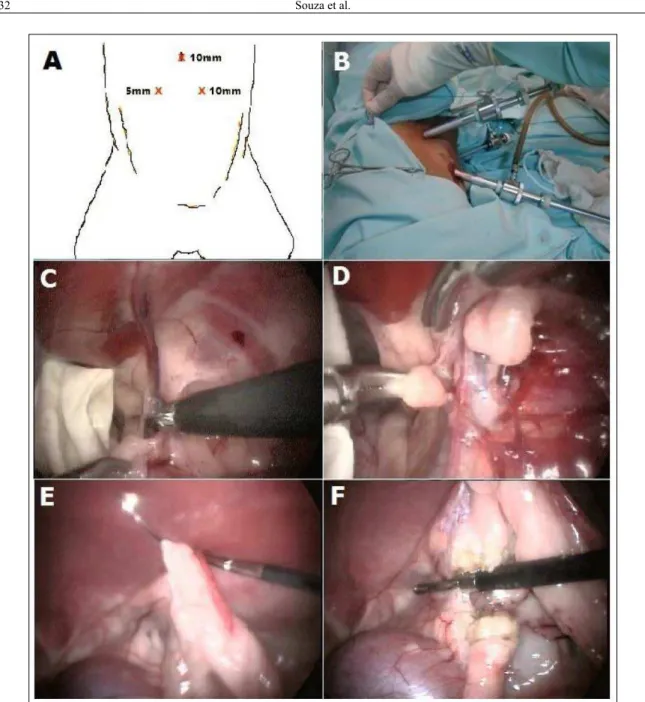
12mmHg was reached. Under endoscopic vision using a 0° and 10mm endoscope, the two other portal positions were selected (Figure 1A). The second puncture was performed on the left abdominal wall, using a 5mm trocar. The third approach was performed on the right abdominal wall with a second 10mm portal. After inspection of the abdominal cavity, the right inguinoiliac lymphadenectomy procedure began laterally to the deep epigastric vessels (Figure 1C). Two standardized areas of 2cm2 were cleared of fat
tissue surrounding the arteriovenous inguinoiliac complex. Regarding the depth of the dissection plane, a dissection 1cm into the femoral canal on the left side of the iliac vessels was performed. Another sample was collected 1cm from these vessels, toward the right inguinal canal (Figure 1D). These samples were collected to be sent for histopathological examination, to evaluate the presence or absence of lymphatic tissue and also to quantify the average number of micro lymph nodes in each region. To the right of the internal iliac artery at the beginning of its junction with the ventral peritoneum, the fat tissue was dissected using a Maryland forceps and Metzenbaum scissors. To release the dissected tissue, a 5mm tripolar forceps (PowerBlade TM®) was used
through a bipolar cautery section followed by a cutting blade. With the aid of the Maryland grasping forceps, tissue was removed through the 10mm trocar
in a retrieval bag made with glove fingers. Using this
same technique, the tissue from the left quadrant to the deep iliac vessels was also dissected and collected.
Using the same portal accesses, ovariohysterectomy began with the elevation of the cranial portion of the cervix, using a Kelly forceps, then two openings were made in the uterus broad ligament, parallel to the two uterine arteries. Bipolar hemostasis and sectioning of the junction between the cervix and the beginning of the body of the uterus was achieved using the same tripolar forceps used before. After this step, the animals were placed in a left lateral recumbent position (Figure 1B) to promote the displacement of the viscera to the contralateral side and facilitate access to the right mesovarium region. With the aid of the Maryland forceps, the ovarian proper ligament area was grasped and raised along the lateral abdominal wall, then a temporary trans-parietal suture using two polypropylene threads
with a 4 cm circular needle was made to fix the
corresponding ovary (Figure 1E). With the tripolar forceps, cauterization and sectioning caudal to the ovarian arteriovenous complex until the ovarian proper ligament was performed (Figure 1F). The same steps were performed in the contralateral ovary region by changing to a right recumbent position. After complete hemostasis and sectioning of the left ovarian artery and vein, animals were repositioned in a supine recumbent position and the area close to the suspensory ovarian ligament was grasped using the Kelly forceps. Then, the right ovary was removed through the 10mm portal located on the right abdominal wall, followed by the right uterine horn, uterine body, left uterine horn and left ovary. After
confirmation of the absence of abdominal bleeding, the abdominal cavity was deflated and the accesses
wounds were sutured in the muscle layer using a Sultan pattern and 2-0 polyglact in 910 thread. Skin sutures were made with a Wolff pattern and 4-0 nylon
monofilament. At the end of all surgical procedures, the wounds were dressed and anti-inflammatory
therapy with meloxicam (0.2mg kg-1, SC, SID) and
tramadol (3mg kg-1, SC, TID) was given for three
days. Immediately after surgery, the animals were
kept under observation, receiving fluid therapy until
full anesthetic recovery.
In all subjects, hypersensitivity reactions were noted 12 hours after application of the dye intradermally. Ice was applied to the site of
ointment based on escin (10mg g-1) and diethylamine
salicylate (50mg g-1) was applied twice daily to help
resolve signs of hypersensitivity. One animal had necrosis two days after the application of indocyanine green dye 1%. In this case, antimicrobial therapy
was given with enrofloxacin (5mg kg-1, PO, BID)
for seven days, as well as anti-inflammatory therapy
with prednisone (0.5mg kg-1, PO, BID) for five days.
Wound management was performed using saline solution and the same ointment described before.
Excised adipose tissue was placed in 10% formalin and it was referred for histopathological evaluation. For adipose tissue histology of the pelvic
region, the sample was first evaluated macroscopically.
Thicker segments were sectioned into two equal parts for better use. The samples were processed routinely Figure 1 - Laparoscopic inguinoiliac lymphadenectomy andovariohysterectomy in dogs. In “A”, disposition of the
for histology and embedded in paraffin blocks. Serial
sections were made at a thickness 3.0mm, using most of the fragment at intervals of about 500mm (around three sections from each piece). Slides were stained with hematoxylin and eosin (H&E) and observed by optical microscopy.
RESULTS AND DISCUSSION
According to the histopathological evaluation, pelvic adipose tissue fragments from
15 bitches (right and left sides, specifically)
weighed between 0.1g and 0.38g, with dimensions between 0.5x0.2x0.2cm and 3.5x2.0x1.5cm. Tissues were predominantly made up of pale yellow and white tissue, interspersed with small whitish areas, sometimes reddish (interpreted as blood vessels). Structures similar to lymph nodes were not observed during palpation and visual inspection. Histologically, the sections were made up predominantly of a large and well-differentiated cell mantle, with abundant and intensely vacuolated cytoplasm with distinct boundaries. Nuclei of these cells were shifted to the periphery, round in shape and consisting of aggregated chromatin (adipocytes). Blood vessels of various calibers were found within the adipose tissue. The edges of most cuts showed a loss of tissue architecture with
the distortion of adipocytes (defined as an artifact
resulting from the manipulation of the sample, with
no clinical significance). There were no lymph nodes
or lymphocytic accumulations in any of the samples. SOUZA et al. (2013) performed the same technique of pelvic lymphadenectomy; however, this was performed only on the left side of a dog presenting mammary tumors in the abdominal and inguinal glands. Intradermal application of methylene blue dye 1% was performed on the largest tumor located in the inguinal mammary gland. Two micro lymph nodes were isolated from the excised area, but there was no sign of the dye in those structures. In the present study, no lymph nodes were isolated in this area. This condition may have occurred because the laparoscopic technique was performed on the contralateral pelvic chain in animals without neoplastic mammary glands, unlike the procedure performed in the report above. Another factor may
have influenced this situation, since the dissection
plane adjacent to the deep iliac vessels did not include areas containing lymph nodes, making it necessary to further extend lymphadenectomy. Another possibility is that there are micro lymph nodes, but as they were not reactive or were located for inclusion in
the histological sections. These results emphasize
the importance of further studies to better define the
regions referred for collection, and the comparison of the lymphatic drainage mechanisms of healthy animals and those with mammary neoplasms, since the existence of micro lymph nodes in this region has been previously demonstrated (SOUZA et al., 2013).
As for the smaller sample volume on the right side, this was likely due to the anatomical
position, making it difficult to collect samples;
this region lies between the inguinal and femoral rings and is demarcated by the presence of rich vascularity. However, the use of Power blade tripolar forceps improved the safety of the procedure with bipolar cauterization of blood vessels in the lymphadenectomy areas as well as cauterization of the ovarian venous arterial complex and uterine arteries, similar to the degree of safety described by MACHADO-SILVA et al. (2012).
The use of methylene blue dye and indocyanine green demonstrated some toxicity around the application site in all subjects, since there was no surgical removal of the stained tissue, unlike what is done in cases of mastectomy. Such signs included skin necrosis in two animals from the group where indocyanine green 1% was used. Reactions manifested as edema and erythema of the skin, consistent with type IV hypersensitivity, in all other animals in which dye application was performed,
similar to the findings of PINHEIRO et al. (2003) and
CAMPOS et al. (2007). The dose and concentration used for sterile methylene blue and indocyanine green dye needs to be adjusted and better studied to minimize the side effects in dogs, particularly in cases where the impregnated lymphatic tissue is not surgically removed. It is also necessary to assess the safety of such dyes under these conditions.
Means and standard deviations of surgical times in minutes were 18.06±5.40 for surgical access (T1); 19.33±5.47 for right pelvic lymphadenectomy (T2); 26.53±6.36 for left pelvic lymphadenectomy (T3); 35.06±5.70 ovariohysterectomy (T4) and 98.88±7.82 as the average sum of the procedures. It is noteworthy that the surgical time in laparoscopic surgery tends to decrease as the technical learning curve is achieved (DAVIDSON et al., 2004; MALM et al., 2004). The surgeon who performed this study has solid experience related to peritoneal cavity access and laparoscopic ovariohysterectomy, but was in the initial phase of the learning curve for right and left lymphadenectomy techniques for pelvic lymph nodes, a condition that certainly
CONCLUSION
The proposed model of laparoscopic lymphadenectomy did not allow the collection of lymph nodes of the areas (left and right) lateral to the deep epigastric vessels (internal and external iliac artery) in healthy animals.
The lymphatic staining technique performed 12 hours and 30 minutes before the start of the surgical procedure using sterile methylene blue 1% and indocyanine green 1% did not allowed visual demarcation of lymphatic vessels and intra abdominal lymph nodes from abdominal and pelvic cavities.
The procedures performed by the laparoscopic approach showed no major technical difficulties and were found to be safe in their implementation.
BIOETHICS AND BIOSSECURITY COMMITTEE APPROVAL
Ethics Committee for the Use of Animals of Universidade Federal de Santa Maria (UFSM) has approved the methods employed in this study (protocol 043/2013).
ACKNOWLEDGMENTS
The authors would like to thanks to the WEM Equipamentos Eletrônicos (Ribeirão Preto, SP, Brasil) for providing the bipolar forceps Tripol PowerBlade TM® used in this study.
REFERENCES
CAMPOS, M.L.C. et al. Pesquisa do linfonodo sentinela (LNS) através da administração de corante azul de metileno em cães portadores de neoplasias. Revista Nosso Clínico, v.10, n.56, p.18-34, 2007.
DAVIDSON, E.B. et al. Comparison of laparoscopic ovariohysterectomy and ovariohysterectomy in dogs. Veterinary Surgery, v.33, p.62-69, 2004.
FREEMAN, L.J. Minimally Invasive Surgery of the hemolymphatic system. Veterinary Endosurgery. In: FREEMAN, L.J.; HENDRICKSON, D.A. (Eds.), St Louis: Mosby, 1999. V.1. p.192-204.
HAIGH, P.I.; GIULIANO, A.E. Role of sentinel lymph node dissection in breast cancer. Annals of Medicine, v.32, p.51-56, 2000. Available from: <http://informahealthcare.com/doi/ abs/10.3109/07853890008995910>. Accessed: May 30, 2012. doi: 10.3109/07853890008995910.
LAVERY, H.J. et al. Robotic extended pelvic lymphadenectomy for bladder cancer with increased nodal yield. British Journal of Urology, v.107, p.1802-1805, 2010. Available from: <http:// www.ncbi.nlm.nih.gov/pubmed?term=Robotic%20extended%20 pelvic%20lymphadenectomy%20for%20bladder%20cancer%20 with%20increased%20nodal%20yield>. Accessed: May 30, 2012. doi: 10.1111/j.1464-410X.2010.09789.x.
MALM, C. et. al. Ovário-histerectomia: estudo experimental comparativo entre as abordagens laparoscópica e aberta na espécie canina. Intra-operatório I. Arquivo Brasileiro de Medicina Veterinária e Zootecnia, v.56, n.4, p.457-466, 2004.
MOULIN, D. The short history of breast cancer. Kluwer: Academic Publishers; p.124, 1983. doi: 10.1007/978-94-017-0601-8.
MIYASHIRO, I. et al. Laparoscopic detection of sentinel node in gastric cancer surgery by indocyanine green fluorescence imaging.
Surgical Endoscopy. 2010. Available from: <http://www.ncbi. nlm.nih.gov/pubmed/20976497>. Accessed: May 30, 2012. doi: 10.1007/s00464-010-1405-3.
PINHEIRO, L.G.P. et al. Estudo Experimental de linfonodo sentinela na mama da cadela com azul patente e tecnécio Tc99. Acta Cirúrgica Brasileira, v.18, p.545-552, 2003. Available from: <http://www.scielo.br/pdf/acb/v18n6/ a12v18n6.pdf>. Accessed: May 30, 2012.
PINHEIRO, L.G.P. et al. Hemosiderin: a new marker for sentinel lymph node identification. Acta Cirúrgica Brasileira, v.24, p.432-436, 2009. Available from: <http://www.scielo.br/scielo. php?script=sci_arttext&pid=S0102-86502009000600002>. Accessed: May 30, 2012. doi: 10.1590/S0102-865020009000600002.
SCHRENK, P. et al. Morbidity following sentinel lymph node biopsy versus axillary lymph node dissection for patients with breast carcinoma. Cancer, v.88, p.608-614, 2000. Available from: <http://onlinelibrary.wiley.com/ doi/10.1002/(SICI)10970142 (20000201)88:3%3C608::AID CNCR17%3E3.0.CO;2-K/full>. Accessed: May 30, 2012. doi: 0.1002/(SICI)1097-0142(20000201)88.