Rev Bras Anestesiol SCIENTIFIC ARTICLE 2010; 60: 4: 376-382
376 Revista Brasileira de Anestesiologia
Vol. 60, No 4, July-August, 2010
Evaluating the Depth of the Epidural Space with the Use of
Ultrasound
Pablo Escovedo Helayel, TSA
1, Diogo Bruggemann da Conceição, TSA
2, Gustavo Meurer
3,
Claudia Swarovsky
3, Getúlio Rodrigues de Oliveira Filho, TSA
4Summary: Helayel PE, Conceição DB, Meurer G, Swarovsky C, Oliveira Filho GR – Evaluating the Depth of the Epidural Space with the Use of
Ultrasound.
Background and objectives: The objective of the present study was to evaluate the use of the ultrasound on the determination of the depth of
the epidural space.
Methods: Sixty patients were included in this prospective study; the L3-L4 space was initially identified by palpation followed by the ultrasound
me-asuring the depth of the epidural space (PU). After the epidural puncture the measurements o the depth (PA) were recorded. The data underwent descriptive statistics, and the concordance correlation coefficient and Bland-Altman analysis, with 95% confidence interval were calculated.
Results: Analysis of concordance between the palpation and ultrasound methods was 86.6%. Mean values of PU obtained were 4.97 ± 0.51 cm
and PA 4.97 ± 0.71 cm, and Pearson correlation coefficient of 0.66 while Bland-Altman analysis revealed a mean difference of 0.0035 ± 0.53 cm with 95% confidence interval between -0.228 and 0.221.
Conclusions: The ultrasound is a precise tool to determine the depth of the epidural space.
Keywords: ANESTHETIC TECHNIQUES, Regional: epidural; EQUIPMENT, Ultrasound; METHODOLOGY: validation studies.
[Rev Bras Anestesiol 2010;60(4): 376-382] ©Elsevier Editora Ltda.
The objective of the present study was to validate the use of the ultrasound as a tool to determine the depth of the epi-dural space as well as to evaluate its precision on identifying the L3-L4 intervertebral space.
METHODS
After approval by the Ethics on Research Committee of the Hospital Governador Celso Ramos and signing of the infor-med consent, 60 patients, physical status ASA I and II, ages between 18 and 65 years, scheduled for elective surgeries under epidural block in the field of general, urologic, vascular, and orthopedic surgeries were enrolled in this study. Patients with neurological diseases, history of spinal surgery, defor-mities of the spine, infection at the puncture site, coagulopa-thies, and any other contraindication to neuroaxis block were exclude from this study. The physical status (ASA), age, wei-ght, heiwei-ght, and body mass index (BMI) of all patients were recorded.
All patients were monitored with cardioscope, pulse oxime-ter, and non-invasive blood pressure. A line was inserted with an 18G catheter for administration of midazolam IV (0.05 mg.kg-1)
10 minutes before the blockade.
Patients were placed in the sitting position and the L3-L4
space was identified by palpation based on the Tuffier line (horizontal line between the iliac crests) and marked with a pen. This was followed by the ultrasound using a convex transducer of 2-5 MHz (Sonoace 8000SE®, Medison, South
Korea). Initially the intervertebral space determined by
pal-INTRODUCTION
The high variability of the distance between the skin and the epidural space and its surface anatomical references hinder its correct identification 1,2 demanding care when positioning
the patient and technical experience that could affect the suc-cess rate of epidural blocks 3-6. The depth of the epidural
spa-ce depends on the trajectory of the needle. Several attempts to relate this depth with patient-related parameters, such as weight and height, proved ineffective for clinical use 7. From
1980 on, a strong correlation between the depth of the epi-dural space visualized on ultrasound and the distance mea-sured by the needle was observed 8,9. Thus, the ultrasound
has been considered a useful tool to identify the depth of the epidural space and its anatomical structures 10,11.
Received from Hospital Governador Celso Ramos – CET/SBA aggregated of Secretaria de Estado da Saúde de Santa Catarina (SES-SC) and Núcleo de Ensino e Pesquisa em Anestesia Regional (NEPAR), Florianópolis, SC.
1. Anesthesiologist; Coordinator and Researcher of NEPAR, and Instructor Co-responsible for CET/SBA aggregated of SES-SC
2. Anesthesiologist; Researcher of NEPAR and Instructor Co-responsible for CET/SBA ag-gregated of SES-SC
3. Anesthesiology Resident
4. Anesthesiologist; PhD in Anesthesiology; Researcher of NEPAR and Responsible for CET/SBA aggregated of SES-SC
Submitted on January 8, 2010 Approved on March 1, 2010
Correspondence to: Dr. Pablo Escovedo Helayel
Av. Governador Irineu Bornhausen, 3440/204 Agronômica
EVALUATING THE DEPTH OF THE EPIDURAL SPACE WITH THE USE OF ULTRASOUND
Revista Brasileira de Anestesiologia 377
Vol. 60, No 4, July-August, 2010
pation was confirmed with the transducer on the longitudinal position identifying the sacrum when it was slowly moved to the cephalad direction until the L4-L4 space. Once the
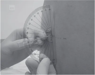
inter-vertebral space was identified, the transducer was moved 90° to obtain the transverse position, and, inclining slightly on the cephalad or caudal direction to obtain a better image of the in-tervertebral space, which was frozen for the measurement of the depth of the epidural space (Figure 1). With the transducer on the same position the skin was marked with a pen on the center of the horizontal surface of the transducer (middle line), and another coinciding with the meddle point of the right late-ral surface of the transducer 11, the inclination angle in relation
to the inferior apophysis was measured with the help of a pro-tractor (Figure 2). The puncture site was determined by the intersection of both marks; the angle provided the inclination
of the needle in relation to the skin. The epidural block was performed maintaining the patient on the same position after antisepsis, placement of sterile surgical fields, and local anes-thesia of the skin and deeper planes with 5 ml of 1% lidocai-ne. A 17G Tuohy needle (8.89 cm) marked at 1-cm intervals was inserted in a point and angulation determined previously, and it was redirected if necessary. The number of repositions (change in angle) and puncture attempts (different puncture area) were recorded. After identification of the epidural space by the loss of resistance technique with saline, the angulation of the needle in relation to the skin was determined by a sterile protractor (Figure 3) and the needle was marked close to the skin with a pen to measure the depth.
After collection of the data, the ultrasound images of the intervertebral spaces were stored for posterior evaluation by two investigators regarding the quality of the images of the following structures: spinous, transverse, and articular apo-physis, posterior border of the vertebral body, and yellow liga-ment and dura-mater (visualized as a single line) similar to the description of other studies 11. An independent evaluation was
performed and the degree of concordance was determined. Calculation of the sample size was based on the following parameters, according to previous findings 3: distance from
the skin to the epidural space = 50.9 ± 12 mm, and depth of the epidural space = 6.9 mm. This was considered the maxi-mal accepted difference between the two measurements of this study. Thus, it was estimated that 60 patients would be necessary with a type I probability error of 5%, and type II probability error of 10%. Descriptive analysis of the data was undertaken using means and standard deviations, for conti-nuous parameters, and percentages for nominal parameters. The Spearmen correlation coefficient was calculated to esti-mate the degree of concordance between both methods of evaluation, ultrasound depth of the epidural space (PU) ver-sus the depth of the needle introduced until the epidural space (PA), and ultrasound angulation in relation to the skin (AU) Erector spinae muscle
CINE
Vertebral body
Spinous apophysis + accoustic shade
Articular apophysis
Transverse apophysis
Dura mater + Yellow ligament
Depth of the epidural space
Figure 1 – Ultrasound of the L3-L4 Intervertebral Space on the
Trans-versal Approach, Showing Structures of the Neuroaxis and Measu-rement of the Distance between the Skin until the Epidural Space in Centimeters.
Figure 2 – Convex Transducer Positioned Transversally to the L3
-L4 Intervertebral Space to Measure the Distance between the Skin and the Epidural Space and the Inclination Angle in Relation to the Inferior Apophysis with the Aid of a Sterile Protractor.
Figure 3 – Determination of the Angle of the Tuohy Needle in
HELAYEL, CONCEIÇÃO, MEURER ET AL.
378 Revista Brasileira de Anestesiologia
Vol. 60, No 4, July-August, 2010 versus the angle of the needle in relation to the skin (AA).
Concordance of both methods of measuring the depth of the epidural space was estimated by the Bland-Altman method
12. The remaining data related to patient characteristics were
submitted to multiple linear regression in order to evaluate the possible significant association with the differences of depth and angulation.
RESULTS
Sixty patients with mean age of 45 ± 14 years, of which 41 (68%) were males, participated in this study. They had a mean height of 167 ± 8 cm; mean weight of 71 ± 12 kg; and mean BMI 25 ± 4 kg.m2. According to the criteria of the ASA, 29 (48%) were
classified as ASA I, 27 (45%) as ASA II, and 4 (7%) as ASA III. Considering the type of procedure, 27 (45%) underwent vascular surgery, 22 (37%), urologic surgery, 7 (12%), general surgery, and 4 (6%), orthopedic surgery.
Mean PU values were 4.97 ± 0.51 cm, and PA 4.97 ± 0.71 cm, with Pearson correlation coefficient of 0.66, while Bland-Altman analysis revealed a mean difference of 0.0035 ± 0.11 cm, with 95% confidence limits between -0.228 and 0.221 (Chart 1). Mean AU values were 84.45 ± 5.14° and AA 80.68 ± 7.39°, with Pearson correlation coefficient of 0.41, while Bland-Altman analysis revealed a mean difference of 3.76 ± 1.15°, with a 95% confidence limit between 1.47 and 6.05. Repositioning of the needle did not show a correlation with the difference observed in the angles (Spearman coe-fficient of 0.043 and p = 0.74). On multiple linear regression analysis, only the weight showed a significant association with the difference between measurements (R coefficient = 0.3 and p = 0.019). However, only 9.1% variance in the differen-ces among measurements was explained by weight variation, since the adjusted determination coefficient (R2) was 0.091.
Analysis of the concordance between the palpation and
ultrasound methods on the identification of the L3-L4 space
was 86.6%. In all cases, only one puncture on the skin at the point determined previously was necessary, and, in 34 (55%), redirection of the needle was not necessary. On the remaining cases, 1 (14.5%) to 5 (3.2%) changes in the angle of insertion of the needle were necessary.
DISCUSSION
This study showed a high correlation between the measu-rements of the distance between the skin and the epidural space by palpation and ultrasound. Similarly to other studies, the findings of the present study revealed that ultrasound is capable of generating a fairly precise estimate of the depth of the epidural space 8,9.
As observed previously 3,13, the depth of the epidural
space had a close relationship with the weight and BMI of patients. The values of those variables observed in this study were comparable to those observed in previous studies3,4,9 done in patients with equally lower body mass
index. The concordance between PU and PA was not affec-ted by the BMI, which has also been demonstraaffec-ted 11,13,14.
Besides estimating the depth the ultrasound can facilitate the epidural puncture before it is performed by evaluating the anatomy of the spine and providing greater accuracy on the identification of the site of puncture on the skin 7,15,16.
Only one attempt was necessary in all patients, and in 55% of the cases it was not necessary to redirect the needle. This indicates that the ultrasound can help define the best place of entry on the skin and the ideal direction of the needle. The usefulness of measuring the angle between the transducer and the skin to make the relationship PU/PA more precise and reduce the need of repositioning the needle did not show a good correlation with the difference obser-ved among angles. Thus, the measurement of those angles does not seem to facilitate epidural punctures, it only increa-ses the time of the ultrasound.
Identification of the L3-L4 space by palpation showed a high
correlation with the ultrasound (86.6%). Those levels of accu-racy are much higher than those observed by other authors 2,
who reported accuracy lower than 30%. Although this result shows a better identification of the L3-L4 space, it does not
guarantee the correct position of the needle in the desired in-tervertebral space by palpation.
To conclude, the ultrasound is a precise tool to determine the depth of the epidural space. Besides, it facilitates epidural puncture by the correct identification of the intervertebral spa-ce and spinal anatomy; however, it does not exclude the need to use the loss of resistance technique.
1.5
4.075 4.3 4.42 4.455 4.455 4.57 4.63 4.685 4.735 4.78 4.915 4.955 5.025 5.12 5.255 5.375 5.41 5.5 5.93 6.25
0.5
Mean
Difference Dif. Mean +2DP –2DP
Diff
erence–0.5
–1.5 –1
–2 0 1
Chart 1 – Bland-Altman Chart of the Measurements of the Depth of
382 Revista Brasileira de Anestesiologia Vol. 60, No 4, Julho-Agosto, 2010 HELAYEL, CONCEIÇÃO, MEURER E COL.
REFERÊNCIAS / REFERENCES
01. Furness G, Reilly MP, Kuchi S. An evaluation of ultrasound imaging for identification of lumbar intervertebral level. Anaesthesia, 2002;57:277-280.
02. Broadbent C. Ability of anaesthetists to identify a marked lumbar inter-space. Pain Practice, 2001;1:199.
03. Bevacqua BK, Haas T, Brand F. A clinical measure of the posterior epidural space depth. Reg Anesth, 1996;21:456-460.
04. Sutton DN, Linter SP. Depth of extradural space and dural puncture. Anaesthesia, 1991;46:97-98.
05. Hamza J, Benhamou D. Dural puncture and depth of the extradural space. Anaesthesia, 1992;47:169-170.
06. Oliveira Filho GR, Gomes HP, Fonseca MHZ et al. Predictors of suc-cessful neuraxial block: a prospective study. Eur J Anaesthesiol, 2002;19:447-451.
07. Grau T, Leipold RW, Conradi R et al. Ultrasound control for presumed difficult epidural puncture. Acta Anaesthesiol Scand, 2001;45:766-771. 08. Cork RC, Kryc JJ, Vaughan RW. Ultrasonic localization of the lumbar
epidural space. Anesthesiology, 1980;52:513-516.
09. Currie JM. Measurement of the depth to the extradural space using ultrasound. Br J Anaesth, 1984;56:345-347.
10. Helayel PE, Conceição DB, Oliveira Filho GR. Bloqueios nervosos guiados por ultrassom. Rev Bras Anestesiol, 2007;57:106-123. 11. Arzola C, Davies S, Rofaeel A et al. Ultrasound using the transverse
approach to the lumbar spine provides reliable landmarks for labor epidurals. Anesth Analg, 2007;104:1188-1192.
12. Bland JM, Altman DG. Measuring agreement in method comparison studies. Stat Methods Med Res, 1999;8:135-160.
13. Balki M, Lee Y, Halpern S et al. Ultrasound imaging of the lumbar spine in the transverse plane: the correlation between estimated and actual depth to the epidural space in obese parturients. Anesth Analg, 2009;108:1876-1881.
14. Oliveira Filho GR, Boso AL, Benedetti RH. Distância da pele ao espaço subaracnoideo em pacientes geriátricos: comparação entre os acessos mediano e paramediano. Rev Bras Anestesiol, 1997;47:226-230.
15. Grau T, Leipold R, Conradi R et al.Ultraschall und Periduralanasthe-sie. Technische Moglichkeiten und Grenzen einer diagnostischen Un-tersuchung des Periduralraums. Anaesthesist, 2001;50:94-101. 16. Grau T, Leipold RW, Conradi R et al. Ultrasound imaging facilitates
localization of the epidural space during combined spinal and epidural anesthesia. Reg Anesth Pain Med, 2001;26:64-67.
Resumen: Helayel PE, Conceição DB, Meurer G, Swarovsky C,
Oli-veira Filho GR – Evaluación de la Profundidad del Espacio Epidural con el Uso del Ultrasonido.
Justificativa y objetivos: El objetivo de este estudio fue evaluar el
uso del ultrasonido para la determinación de la profundidad del es-pacio epidural.
Método: Sesenta pacientes fueron ubicados, prospectivamente
te-niendo la identificación del espacio intervertebral L3-L4
inicialmen-te realizada por el método de palpación. Posinicialmen-teriormeninicialmen-te se usó el método de ultrasonido, y se realizó la medida de la profundidad del espacio epidural (PU). Después de la punción epidural, se anotaron las medidas de la profundidad (PA). Se midieron las estadísticas des-criptivas de los datos y se calculó el coeficiente de correlación de concordancia y análisis de Bland-Altman, con un intervalo de un 95% de confianza para las medidas de profundidad.
Resultados: El análisis de concordancia entre el método de
palpaci-ón y el ultrasonido fue de un 86,6%. Se obtuvieron valores promedios de PU 4,97 ± 0,51 cm y PA 4,97 ± 0,71 cm y un coeficiente de correla-ción de Pearson de 0,66, mientras el análisis Bland-Altman arrojó una diferencia promedio de 0,0035 ± 0,53 cm, con un límite de un 95% de confianza entre -0,228 a 0,221.
Conclusiones: El ultrasonido es un instrumento preciso para la