MOLECULAR AND CELL BIOTECHNOLOGIES
UDC 616.13/.14 : 616.126.3-06 : (617-089.843 : 547.962.9)
Tissue engineering of vascular/valvular equivalents
on the base of the xenogeneic decellularized matrix
M. V. Savchuk
State Instituton «Institute of urgent and recovery surgery named after V. K. Gusak National Academy of Medical Science of Ukraine» 47, Leninskyi Ave., Donetsk, Ukraine, 83045
According to the WHO, in 2008 cardiovascular diseases claimed the lives of 17.5 million people (30 % of all diseases). Often the only option to save a patient’s life is a replacing the injured part of an organ by the prosthe-sis.Aim. This research was aimed to produce biomodificated cardiovascular graft by decellularisation of por-cine heart valve.Methods. Our method of decellularization permits to make morphologically and physically non-modified decellularised extracellular matrix.Results. The analysis of matrix shows a decrease of the total number of cells, preservation of the collagen and elastin fibers structure, and safety of physiological adhesion.
Conclusions. The matrix can be used as a framework for the vessel-valvular tissue-engineering prosthesis after its recellularization by the recipient’s autologous cells.
Keywords: tissue engineering, extracellular matrix, decellularization.
Introduction. Each year, cardiovascular diseases claim the lives of millions of people, and are the prevailing cause of death for last two decades. Often the only op-tion to save a patient’s life is a replacing the injured part of an organ by the prosthesis. Annually in the world about 275,000 of such surgeries have been performed. Surgical replacement by either mechanical or bioprosthetic heart valves is the most common treatment for the end-stage valvular diseases [1, 2].
Mechanical valves display good structural durabili-ty but are associated with some problems for recipients. The mechanical prostheses are unable to provide full adaptation to physiological environment such as the pressure change and strength characteristics, since the non-living materials are used for their production [3]. For the same reason, they are not capable of growth and development in the body, which makes them unsuitable in pediatric cardiac surgery. Furthermore, the use of me-chanical prostheses can cause the thromboembolic di-sease which needs lifelong anticoagulant therapy [4–6].
Bioprosthetic heart valve replacements are either of animal origin (xenografts), such as porcine aortic
val-ves and bovine pericardial valval-ves, or taken from human donors (homografts). Usage of xenografts makes it pos-sible to have the valves of different sizes stored and available off-the-shelf. Moreover they enable to avoid anticoagulative therapy and have a potential of remode-ling in recipient’s organism [7]. Several attempts have been made to create functional heart valve replacements able to grow as a patient grows older, to be repaired and remodeled. With the approach of tissue engineering, the patient’s own cells are isolated,e. g. from a bone mar-row, and seeded onto prepared matrices, in the three-di-mensional structure (for example, extracellular matrix (ECM) of xenografts previously decellularized) [4].
All tissues and organs are made up of cells and an associated extracellular matrix – a secreted product of the resident cells consisting of a unique, tissue-specific three-dimensional environment of structural and func-tional molecules. They are usually regarded as cells with a supporting stroma.
So, the success in creating viable autologous cardio-vascular graft depends on three main elements: autolo-gous cells suitable phenotypically and functionally; a matrix as a temporary scaffold that gives the tissue strength as long as the new extracellular matrix with
autologous cells is being synthesized; the tissue forma-tion and development under thein vitroconditions clo-se to the physiological ones [3].
The extracellular matrix is viewed in terms of its ro-le in structural maintenance and three-dimensional sha-pe of the ressha-pective tissue or organ. However, the extra-cellular matrix is the microenvironmental niche, and it is in the dynamic reciprocity with the resident cell po-pulation. That is why the phenotype of the resident cells, including their active genetic profile, proteome, and functionality, is influenced by the conditions of their microenvironmental niche. Thus, the native extracellu-lar matrix is a logical and ideal scaffold for the organ and tissue reconstruction.
Moreover, structural and functional molecules in the extracellular matrix (glycosaminoglycans, collagen, ela-stin, fibronectin, laminin, and vitronectin) are highly conserved proteins in eukaryotic organisms, which lar-gely explains the absence of an adverse immune respon-se after xenotransplantation [8].
There are evidences that grafts failure, at least in part, is a result of immunological reactions, caused by cellular elements remaining in the homograft’s matrix, that trigger the immune reaction by the receiver. The en-dothelial cells and fibroblasts are able to express Class I and II histocompatibility complex that are recognized by the receiver’s immune system and can be a cause of tissue degeneration and graft failure [9].
The decellularization of xenogeneic matrices is a possibility to prevent the graft failure induced by the im-mune response. Moreover, it is a chance to create a pati-ent specific viable heart valve with tissue engineering (TE) techniques. There are the studies suggesting that decellularized heterografts lead to a reduced inflam-matory response of the receiver and are able to be pro-gressively repopulated by the autologous cells [9].
The main purpose of decellularization is to remove all cellular components, by maintaining intact the struc-tural elements of the extracellular matrix. Of today, se-veral methods of the pre-transplant treatment of trans-plants have been developed. They provide a donor cell death without using crosslinking agents, and a subsequ-ent colonization of the recipisubsequ-ent cells prior to implanta-tion [10].
Of now, a variety of tissue decellularization methods have been developed in the world. All of them can be
assembled into two main groups: methods, suggesting the use of substances that cause necrosis; and methods allowing decellularization by inducing apoptotic cell death. Both groups have their pros and cons [10–15].
The method of decellularization with the calcium-free solution of ethylenediaminetetraacetic acid (EDTA) belongs to the apoptosis-inducing methods. EDTA is a chelate, which is able to bind calcium ions, so that the in-teraction between cadherins becomes broken, which re-sults in cells dissociation. At the same time, according to the published data, it is possible that EDTA in high concentration initiates the apoptosis processes [12, 15]. However, it should be noted that the method of de-cellularization with the EDTA does not allow achie-ving a complete removal of dead cells from the depth of the matrix. Perhaps, this is due to the fact that cells can migrate from the surface of the tissue to its depth be-cause of the negative chemotaxis. On the other hand, it is common knowledge that apoptosis can be realized till the end in vivo with macrophages [15].
The aim of research is to assess the effective con-ditions of apoptosis-inducing decellularization of xeno-geneic valves, and to evaluate the integrity and physio-logical intactness of the obtained extracellular matrix during decellularization of heart valves.
Materials and methods. The research was perfor-med with cardiac valves of 6-month-old pigs (n = 7). Heart sampling was carried out in the operating room conditions in accordance with the requirements of Eu-ropean Convention for the Protection of Vertebrate Ani-mals Used for Experimental and Other Scientific Pur-poses (Strasbourg, 1986) and the statements of the 1st National Congress on Bioethics (Kyiv, 2001). The val-ves were derived under sterile conditions on the avera-ge four hours after heart sampling. The obtained samp-les were exposed in apoptosis-inducing solution of 10 mM EDTA («Sigma», USA) for two days. After expo-sure the samples were carefully washed in the medium with salts of concentration close to physiological one.
We supposed that the medium Eagle MEM without Ca2+
is a better solvent for EDTA as a chelate of calci-um ions. Due to the usage of this modification of me-dium we could reduce the washing time after decellula-risation solution to two days. Moreover, we permanent-ly mixed up the medium with the decellularised graft by roller, which helps us to increase the efficiency of de-cellularisation. Our modification of the Akatov’s decel-lularisation method, is protected by the Ukrainian pa-tent N 85240.
To examine the efficiency of apoptosis initiation, the tissue integrity and cell removing samples were analyzed histologically. Slices of each conduit were embedded in paraffin. For general morphology, three sections (5
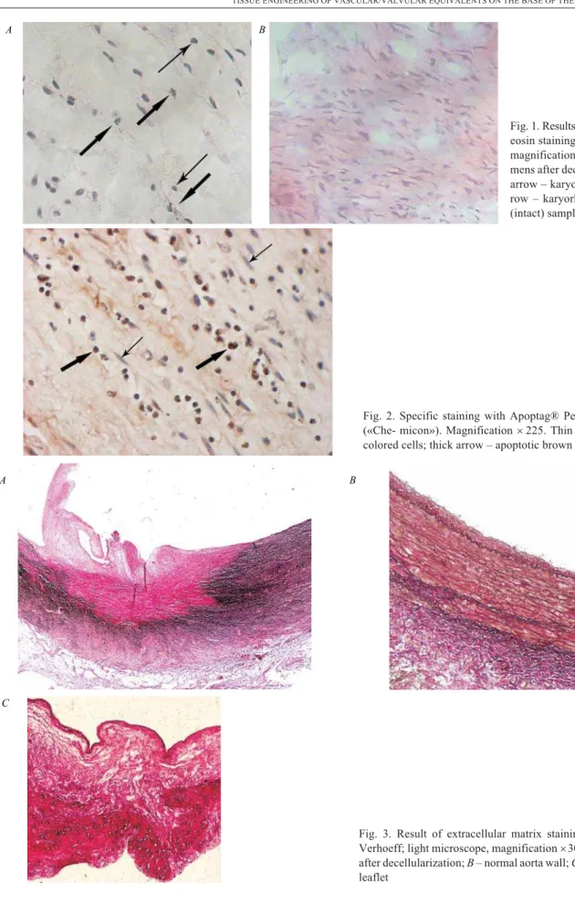
mm) of each slice were stained with hematoxylin and eo-sin [17]; underwent special staining (Apoptag® Peroxi-dase ISOL Kit, «Chemicon», USA) [www. millipore. com/catalogue/item/s7200] to allow the identification of breaks in DNA at initial stages of apoptosis [18]; and stained according to Verhoeff to evaluate the state of elastic and collagen fibers for assessment of safety of extracellular matrix strength and flexibility [17].
To assess adhesive properties of matrix the decellu-larized grafts were incubated with human fetal fibro-blasts derived and cultured according to the standard method [19]. Before incubation the culture of fibro-blasts was stained with vital fluorescent dye PKH 67 Green («Sigma») and resuspended in cultural medium. The adhesion rate of decellularized tissue of cardiac val-ves was estimated by the fifth day of incubation using fluorescent microscopy.
Results and discussion. Specimens of decellulari-zed porcine matrix were not cell-free in the leaflets and the conduit wall. Histological analysis revealed just a decrease in the total number of cells, appearance of pro-nounced changes in nuclei morphology in most of them (karyopyknosis, karyorhexis) testifying to the success-ful initiation of apoptosis (Fig. 1,A). The cells of control (intact) samples on the contrary had normal morphologi-cal characteristic for fibroblasts: extended spindlesha-ped cells with oval shaspindlesha-ped nucleus (Fig. 1,B) [20, 21].
Specific staining with Apoptag®
peroxidase showed different content of cells at the apoptosis state in various structures of valve (Fig. 2). The highest number of the apoptotic cells was found in the leaflet. This may be as-sociated with its relative thinness if compared with the
vessel wall or the annulus fibrosis region that could simplify the penetration of treating solution deep into the structure and following apoptosis induction. Additio-nally, a larger number of viable cells in tissue depth, if compared with superficial zones in regions with comp-licated structure, is also associated with the cell migra-tion due to the negative chemotaxis [21, 22].
Histological analysis of the sections after specific staining for extracellular matrix showed good preserva-tion of the matrix architecture [23, 24], the structure and orientation of collagen and elastin fibers after decellula-rization are safe. The staining of extracellular matrix fi-bers according Verhoeff was successful in both aorta wall area (maroon color) and valve leaflet area (crimson color) (Fig. 3).
A fluorescent analysis of supposed transplants revea-led the bright fluorescencing cells adhered to connective tissue matrix under violet blue excitation, which shows the preservation of adhesive properties of the extracellu-lar matrix and its availability for further colonization with tissue-forming autologous cells of the recipient.
The present study provides evidence of a successful in vitroproducing of tissue engineered grafts, fits for the reconstruction of heart valve. The described method of tissue engineering is based on decellularization of por-cine heart valves by inducing apoptotic cell death with the calcium-free solution with EDTA. According to the obtained data we may suggest that EDTA solution of 10 mM as well as the proposed scheme of grafts treat-ment are effective in terms of decellularization. As we expected this type of tissue treatment induces apoptotic cell death in the donor grafts, total number of cells de-creases, the changes in nuclei morphology (karyopyk-nosis, karyorhexis) in most part of remaining cells ap-pear, but the total decellularization of tissue is not rea-chedin vitro. The initiation of apoptosis in valve leaflet as well as on the surface of the valve vascular region was efficient. The initiation of apoptosis was not uniform in different structures of xenograft.
colo-A B
Fig. 1. Results of hematoxylin and eosin staining (light microscope, magnification´400):A– speci-mens after decellularization (thin arrow – karyopyknosis; thick ar-row – karyorhexis);B– control (intact) samples
Fig. 2. Specific staining with Apoptag® Peroxidase ISOL Kit («Che- micon»). Magnification´225. Thin arrow – alive blue colored cells; thick arrow – apoptotic brown colored cells
A B
C
nization by autologous recipient cells (to realize the re-vitalization) and use as a tissue-engineered cardiovascu-lar prosthesis.
Òêàíèííî-³íæåíåðíèé ñóäèííî-êëàïàííèé åêâ³âàëåíò íà îñíîâ³ êñåíîãåííîãî äåöåëþëüîâàíîãî ìàòðèêñó
Ì. Â. Ñàâ÷óê
Ðåçþìå
Çã³äíî ç äàíèìè ÂÎÎÇ, ó 2008 ðîö³ â ñâ³ò³ â³ä ñåðöåâî-ñóäèííèõ çà-õâîðþâàíü ïîìåðëî 17,5 ìëí ëþäåé (30 % óñ³õ çàçà-õâîðþâàíü). ×àñ-òî ºäèíî ìîæëèâèì âàð³àí×àñ-òîì ñïàñ³ííÿ æèòòÿ ïàö³ºíòà º çàì³-íà ïîøêîäæåíî¿ ä³ëÿíêè îðãàçàì³-íà ïðîòåçîì.Ìåòà. Îòðèìàííÿ á³î-ìîäèô³êîâàíîãî ñåðäöåâî-ñóäèííîãî ãðàôòà çà ðàõóíîê äåöåëþëÿ-ðèçàö³¿ ñåðöåâîãî êëàïàíà ñâèí³.Ìåòîäè. Òåõíîëîã³ÿ îäåðæàííÿ ìîðôîëîã³÷íî ³ ô³çè÷íî íå çì³íåíîãî äåöåëþëÿðèçîâàíèé åêñòðà-öåëþëÿðíîãî ìàòðèêñó.Ðåçóëüòàòè. Àíàë³ç ìàòðèêñó äåìîíñò-ðóº çíèæåííÿ çàãàëüíî¿ ê³ëüêîñò³ êë³òèí, çáåðåæå- í³ñòü ñòðóêòó-ðè êîëàãåíîâèõ òà åëàñòèíîâèõ âîëîêîí, à òàêîæ ô³ç³îëîã³÷íî¿ àäãåçèâíîñò³ ìàòðèêñó.Âèñíîâêè. Ìàòðèêñ ï³ñëÿ äåöåëþëÿðè-çàö³¿ º ïðèäàòíèì äëÿ âèêîðèñòàííÿ ÿê êàðêàñà äëÿ ñóäèííî-êëà-ïàííîãî òêàíèííî-³íæåíåðíîãî ïðîòåçó ï³ñëÿ éîãî ðåöåëþëÿðè-çàö³¿ àóòîëîã³÷íèìè êë³òèíàìè ðåöèﳺíòà.
Êëþ÷îâ³ ñëîâà: òêàíèííà ³íæåíåð³ÿ, åêñòðàöåëþëÿðíèé ìàòðèêñ, äåöåëþëÿðèçàö³ÿ.
Òêàíåèíæåíåðíûé ñîñóäèñòî-êëàïàííûé ýêâèâàëåíò íà îñíîâå êñåíîãåííîãî äåöåëëþëèðîâàííîãî ìàòðèêñà
Ì. Â. Ñàâ÷óê
Ðåçþìå
Ñîãëàñíî äàííûì ÂÎÇ, â 2008 ãîäó â ìèðå îò ñåðäå÷íî-ñîñóäèñ-òûõ çàáîëåâàíèé óìåðëè 17,5 ìëí ÷åëîâåê (30 % âñåõ çàáîëåâàíèé). ×àñòî åäèíñòâåííî âîçìîæíûì âàðèàíòîì ñïàñåíèÿ æèçíè ïà-öèåíòà ÿâëÿåòñÿ çàìåíà ïîâðåæäåííîãî ó÷àñòêà îðãàíà ïðîòå-çîì.Öåëü. Ïîëó÷åíèå áèîìîäèôèöèðîâàííîãî ñåðäå÷íî-ñîñóäèñ-òîãî ãðàôòà âñëåäñòâèå äåöåëëþëèðîâàíèÿ ñâèíîãî ñåðäå÷íîãî êëàïàíà.Ìåòîäû. Íàøà òåõíîëîãèÿ ïîçâîëÿåò ïîëó÷èòü ìîðôî-ëîãè÷åñêè è ôèçè÷åñêè íå èçìåíåííûé äåöåëëþëèðîâàííûé ýêñòðà-öåëëþëÿðíûé ìàòðèêñ.Ðåçóëüòàòû. Àíàëèç ìàòðèêñà äåìîíñò-ðèðóåò ñíèæåíèå îáùåãî êîëè÷åñòâà êëåòîê, ñîõðàííîñòü ñòðóê-òóðû êîëëàãåíîâûõ è ýëàñòèíîâûõ âîëîêîí, à òàêæå ôèçèîëîãè-÷åñêîé àäãåçèâíîñòè ìàòðèêñà.Âûâîäû. Òàêèì îáðàçîì, ìàò-ðèêñ ÿâëÿåòñÿ ïðèãîäíûì äëÿ èñïîëüçîâàíèÿ åãî â êà÷åñòâå êàð-êàñà ñîñóäèñòî-êëàïàííîãî òêàíå-èíæåíåðíîãî ïðîòåçà ïîñëå ðåöåëëþëÿðèçàöèè àóòîëîãè÷íûìè êëåòêàìè ðåöèïèåíòà.
Êëþ÷åâûå ñëîâà: òêàíåâàÿ èíæåíåðèÿ, ýêñòðàöåëëþëÿðíûé ìàòðèêñ, äåöåëëþëÿðèçàöèÿ.
REFERENCES
1.Hoerstrup SP, Sodian R, Daebritz S, Wang J, Bacha EA, Martin DP, Moran AM, Guleserian KJ, Sperling JS, Kaushal S, Vacanti JP, Schoen FJ, Mayer JE Jr. Functional living trileaflet heart val-ves grownin vitro.Circulation. 2000;102(19 Suppl 3):III44–9. 2.Schmidt CE, Baier JM.Acellular vascular tissues: natural bioma-terials for tissue repair and tissue engineering.Biomaterials. 2000;21(22):2215–31.
3.Schmidt D, Hoerstrup SP. Tissue engineered heart valves based on human cells.Swiss Med Wkly.2006;136(39–40):618–23. 4.Schmidt D, Stock UA, Hoerstrup SP. Tissue engineering of heart
valves using decellularized xenogeneic or polymeric starter matri ces.Philos Trans R Soc Lond B Biol Sci. 2007;362(1484): 1505–12.
5.Kasimir MT, Rieder E, Seebacher G, Nigisch A, Dekan B, Wolner E, Weigel G, Simon P. Decellularization does not eliminate throm-bogenicity and inflammatory stimulation in tissue-engineered porcine heart valves.J Heart Valve Dis. 2006;15(2):278–86 6.Klopsch C, Steinhoff G.Tissue-engineered devices in
cardiovas-cular surgery.Eur Surg Res. 2012;49(1):44–52.
7.Steinhoff G, Stock U, Karim N, Mertsching H, Timke A, Meliss RR, Pethig K, Haverich A, Bader A. Tissue engineering of pulmona-ry heart valves on allogenic acellular matrix conduits:in vivo res-toration of valve tissue.Circulation. 2000;102(19 Suppl 3):III50–5. 8.Badylak SF, Weiss DJ, Caplan A, Macchiarini P. Engineered
whole organs and complex tissues. Lancet. 2012;379(9819): 943–52.
9.Lopes SA, Costa FD, Paula JB, Dhomen P, Phol F, Vilani R, Ro-derjan JG, Vieira ED. Decellularized heterografts versus cryo-preserved homografts: experimental study in sheep model.Rev Bras Cir Cardiovasc. 2009;24(1):15–22.
10.Van Nooten G, Somers P, Cornelissen M, Bouchez S, Gasthuys F, Cox E, Sparks L, Narine K. Acellular porcine and kangaroo aortic valve scaffolds show more intense immune-mediated calcification than cross-linked Toronto SPV valves in the sheep model.Interact Cardiovasc Thorac Surg.2006;5(5):544–9. 11.Popandopulo AG, Petrova MV.Acellular matrix as a substrate
for tissue-engineered graft of heart valve.Cell and Organ Trans-plantology. 2013;1(1):52–55.
12.Grauss RW, Hazekamp MG, van Vliet S, Gittenberger-de Groot AC, DeRuiter MC.Decellularization of rat aortic valve allografts reduces leaflet destruction and extracellular matrix remodeling. J Thorac Cardiovasc Surg. 2003;126(6):2003–10.
13.Rieder E, Seebacher G, Kasimir MT, Eichmair E, Winter B, De-kan B, Wolner E, Simon P, Weigel G.Tissue engineering of heart valves: decellularized porcine and human valve scaffolds differ importantly in residual potential to attract monocytic cells. Circulation. 2005;111(21):2792–7.
14.Tomazic BB, Edwards WD, Schoen FJ. Physicochemical cha-racterization of natural and bioprosthetic heart valve calcific de-posits: implications for prevention.Ann Thorac Surg. 1995;60(2 Suppl):S322–7.
15.Rosanova IB, Mischenko BP, Zaitsev VV, Vasin SL, Sevastianov VI.The effect of cells on biomaterial calcification: experiments within vivodiffusion chambers.J Biomed Mater Res. 1991; 25(2):277–80.
16.Akatov BC, Fesenko NI, Solov''ev VV, Fadeeva IE, Chekanov AV, Muratov RM, Britikov DV, Sachkov AS. Inhibition of calcifica-tion of heart valves’ transplants by their devitalizacalcifica-tion. Kletoch-naja transplantalogija i tkanevaja inzhenerija. 2010;5(1):41–46. 17.Korzhevskij D, Giljarov A. Fundamentals of histological
techni-ques. St. Petersburg, SpecLit, 2010; 95 p.
18.EducationGuide: Immunohistochemical Staining Methods: Pa-thology. Eds GL Kumar, L Rudbeck. Carpinteria, 2009;162 p. 19.Popandopulo AG, Ignatov DJu, Slipchenko IO, Vasil'ev RG, Merkulova EV, Gerasimov IG. Effect of culturing factors on viability of fetal human fibroblasts. Vestnyk Neotlozhnoy i Vos-stanovitelnoy Meditsyny. 2003;4(2): 323–325.
20.Eghbali M. Cardiac fibroblasts: function, regulation of gene ex-pression, and phenotypic modulation.Basic Res Cardiol.1992; 87Suppl 2:183–9.
21.Hannun YA, Obeid LM. Ceramide and the eukaryotic stress res-ponse.Biochem Soc Trans. 1997;25(4):1171–5.
22.Weedon D, Searle J, Kerr JF. Apoptosis. Its nature and implica-tions for dermatopathology.Am J Dermatopathol. 1979;1(2): 133–44.
23.Robinson KA, Li J, Mathison M, Redkar A, Cui J, Chronos NA, Matheny RG, Badylak SF.Extracellular matrix scaffold for car-diac repair.Circulation. 2005;112(9 Suppl):I135–43.
24.Ross M, Wojciech P. Histology: A Text and Atlas. Lippincott, Williams & Wilkins, 2010; 235–241.