Tipping the Scale from Disorder to
Alpha-helix: Folding of Amphiphilic Peptides in the
Presence of Macroscopic and Molecular
Interfaces
Cahit Dalgicdir1, Christoph Globisch2, Christine Peter2*, Mehmet Sayar1*
1College of Engineering, Koç University, Istanbul, Turkey,2Theoretical Chemistry, University of Konstanz, Konstanz, Germany
*christine.peter@uni-konstanz.de(CP);msayar@ku.edu.tr(MS)
Abstract
Secondary amphiphilicity is inherent to the secondary structural elements of proteins. By forming energetically favorable contacts with each other these amphiphilic building blocks give rise to the formation of a tertiary structure. Small proteins and peptides, on the other hand, are usually too short to form multiple structural elements and cannot stabilize them internally. Therefore, these molecules are often found to be structurally ambiguous up to the point of a large degree of intrinsic disorder in solution. Consequently, their conforma-tional preference is particularly susceptible to environmental conditions such as pH, salts, or presence of interfaces. In this study we use molecular dynamics simulations to analyze the conformational behavior of two synthetic peptides, LKKLLKLLKKLLKL (LK) and EAA LAEALAEALAE (EALA), with built-in secondary amphiphilicity upon forming an alpha-helix. We use these model peptides to systematically study their aggregation and the influence of macroscopic and molecular interfaces on their conformational preferences. We show that the peptides are neither random coils in bulk water nor fully formed alpha helices, but adopt multiple conformations and secondary structure elements with short lifetimes. These pro-vide a basis for conformation-selection and population-shift upon environmental changes. Differences in these peptides’response to macroscopic and molecular interfaces (pre-sented by an aggregation partner) can be linked to their inherent alpha-helical tendencies in bulk water. We find that the peptides’aggregation behavior is also strongly affected by pres-ence or abspres-ence of an interface, and rather subtly depends on their surface charge and hydrophobicity.
Author Summary
Small proteins and peptides have important biomedical implications with their ability to penetrate cells, disrupt cell membranes, or form—potentially neurotoxic—fibrillar aggre-gates. It is rather intriguing that the same peptide can take part in all these processes
a11111
OPEN ACCESS
Citation:Dalgicdir C, Globisch C, Peter C, Sayar M (2015) Tipping the Scale from Disorder to Alpha-helix: Folding of Amphiphilic Peptides in the Presence of Macroscopic and Molecular Interfaces. PLoS Comput Biol 11(8): e1004328. doi:10.1371/journal. pcbi.1004328
Editor:Avner Schlessinger, Icahn School of Medicine at Mount Sinai, UNITED STATES
Received:October 2, 2014
Accepted:May 6, 2015
Published:August 21, 2015
Copyright:© 2015 Dalgicdir et al. This is an open access article distributed under the terms of the
Creative Commons Attribution License, which permits unrestricted use, distribution, and reproduction in any medium, provided the original author and source are credited.
Data Availability Statement:All relevant data are within the paper and its Supporting Information files.
depending on its environment. Hence it is important to understand how the environment guides the peptide from a disordered state to an alpha-helix or beta-sheet. By studying two different peptides with computer simulations, we find that one clue to this puzzle is the state of the allegedly disordered molecule in water, which is in fact characterized by a vast number of partially folded short-lived conformations. We investigate how interaction with macroscopic interfaces affects this multi-state conformational ensemble by promoting an alpha-helix over the other conformers. Furthermore, we show that association of two pep-tides can also be viewed as an interaction with an interface—a molecular interface. We show that the impact of macroscopic and molecular interfaces strongly depends on their ability to enforce partitioning of hydrophobic and hydrophilic residues. The effect gets more subtle as soon as macroscopic partitioning is coupled with peptide association where two interfaces compete: the relative balance of opposing forces determines aggregate sta-bility and structure.
Introduction
The partitioning, folding and aggregation properties of soluble peptides and small proteins at hydrophobic/hydrophilic interfaces (HHI) play a crucial role in biomedically important pro-cesses. One example that has been extensively investigated over the last years are amyloid-fibril forming peptides which are responsible for many human disorders, among them severe neuro-degenerative diseases [1–3]. The amphiphilic character of the involved amyloidogenic proteins and peptides, such as islet amyloid polypeptide (IAPP), amyloid-β(Aβ) peptide orα-synuclein is responsible both for their aggregation propensity as well as for their strong interface activity. For several of the amyloidogenic peptides not only interaction with biomembrane surfaces but also membrane penetrating, membrane rupturing, and pore-forming mechanisms are dis-cussed [4]. This aspect of partitioning and penetration through hydrophobic barriers like cell membranes links them to a related class of natural and synthetic peptides, so called cell pene-trating peptides (CPP). They have gained importance as a tool for the targeted delivery of non-permeable therapeutics like other peptides, proteins, oligonucleotides or nanoparticles into the cell. [5,6]
The folding and aggregation equilibria of these systems are usually complex and very sensi-tive to external factors (ion strength, pH, etc.). In particular, folding patterns are very diverse, spanning from different helical structures to variousβ-structure-rich arrangements [4], and
the transitions between these states are coupled to changes in peptide/protein concentration, the presence of initial aggregates as seeds or the presence of membranes, interfaces or surfaces. Aggregate formation appears to occur both in theα-helical and in theβ-sheet state, and in sev-eral systems a partially helical structure has been reported prior to fibril formation, while the fibrils themselves typically showβ-structure-rich regions [4,7]. For many of these systems an intrinsically disordered state has been found at low concentrations in solution, and it is specu-lated that this state is an essential intermediate and of great importance for self assembly and aggregation [8].
In particular, very little is known about the low concentration species that are crucial for the early stages of aggregation and that are folding/refolding intermediates. Their molecular state is largely unknown since they are very difficult to detect and characterize experimentally at low concentrations. In addition, their ill-defined fold and their highly dynamic behavior with con-formations that remain stable only for short timescales contribute to the challenges in Competing Interests:The authors have declared
detection. Yet these states are of incredibly high interest as they may well be the actually toxic species in neurodegenerative diseases.
Due to the amphiphilic nature of the peptides/proteins, folding into more defined conforma-tions is observed at interfaces such as the air/water interface or at a biomembrane. In addition to this stabilization of conformations, the interface propensity also leads to an increase in local pep-tide/protein concentration. Both factors presumably further promote aggregation and fibril for-mation (see [9–12]). The effect of the interface on the folding process of these—in bulk mostly disordered—peptides/proteins and their aggregation is not understood yet in its entirety [13–16].
Molecular simulation can shed light on the above-described disordered, half-folded, and highly-dynamic states at low concentration. This information can be used to interpret ensem-ble data from spectroscopic experiments such as NMR, IR or other. In addition, a mechanistic understanding of intrinsically disordered states and the driving forces that trigger folding and aggregation can be obtained. Atomistic insight into unfolded or partially folded intermediates can help to identify the role of different contributions, such as polar and hydrophobic interac-tions, hydrogen bonding, conformational propensities of amino acid sequences, etc.
In the present study, we use classical atomistic molecular dynamics simulations to investi-gate the environment-dependent folding equilibrium of two (synthetic) model peptide
systems—with a special focus on the influence of soft hydrophobic interfaces and oligomer for-mation on this equilibrium. The two chosen systems—a cationic model peptide based on LK repeats [17] and EALA, a pH-switchable anionic peptide based on the so-called GALA system [18]—are both designed to display secondary amphiphilicity in theα-helical conformation
(Fig 1). Both systems are reported to have an affinity to biomembranes and to oligomerize in solution (and in/on the membranes) [17–19]. Peptides composed of leucine and lysine residues have been analyzed in recent studies for their adsorption behavior on hard surfaces with enhanced sampling methods [20], as well as for their role as templates for biomineralization via coarse grained simulations [21]. As we will demonstrate in the course of this paper, both systems display a highly dynamic conformational behavior, with a sizeable amount of intrinsic disorder in solution and an ability to formα-helices under certain conditions.
Thus, our synthetic peptides share the key features of their conformational behavior with natural systems such asα-synuclein. With their highly repetitive designed sequences they are good model systems to analyze the contribution of different interactions and driving forces to the coupled equilibria of folding, aggregation and partitioning at (macroscopic) hydrophobic/
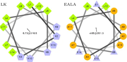
Fig 1. Helical wheel representation of LK and EALA.Both peptides are designed to display secondary amphiphilicity when they adopt theα-helix conformation.
hydrophilic interfaces (seeFig 2), and to characterize typical microscopic intermediates and pathways. The significant motifs in biological systems such asα-synuclein may not be as
regu-lar as in the model systems but the general features (such as repeats of charged and hydropho-bic elements—seeS1 Fig) are comparable.
In the present study we have chosen the most simple representation of a HHI, the (air)vac-uum/water interface. Experimentally, the air/water interface is used to study the folding equi-libria of various protein systems, and its importance for amyloidogenic intrinsically disordered proteins such as A-βorα-synuclein has recently been recognized [9,11,22,23]. Air/water
interfaces are abundantly present in many experimental systems due to agitation of the samples and they appear to affect fibril formation and growth. From a computational point of view the vacuum/water interface is an ideal soft hydrophobic/hydrophilic model interface that allows to systematically study generic effects and fundamental driving forces.
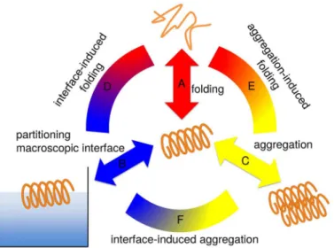
In the course of the paper we will first characterize the folding equilibrium of individual LKKLLKLLKKLLKL (LK) and EAALAEALAEALAE (EALA) molecules in bulk water (equilib-rium A inFig 2), in particular with respect to potential intrinsic disorder of the individual mol-ecule in bulk, i.e. without a further ordering environmental effect. Then we will subsequently study, how mesoscopic interfaces (in the form of the air/water interface; indicated as connect-ing arc D inFig 2), as well as nanoscale interfaces (in the form of a second molecule, as a repre-sentative of early stages of oligomerization; indicated as connecting arc E inFig 2) influence the folding equilibrium of the two molecules. As a last step we will show, how the presence of a HHI affects aggregate formation and possibly further influences the conformational behavior (arc F inFig 2).
Results
Single Peptide in Bulk Water
LK and EALA are both designed to display secondary amphiphilicity [24] inα-helix
conforma-tion as shown in the helical wheel representaconforma-tion inFig 1. The hydrophobic/hydrophilic Fig 2. Three separate contributions govern the dynamic conformational equilibrium of an amphiphilic peptide.The folding of the individual molecule in solution (A); the partitioning of hydrophobic/hydrophilic residues induced by macroscopic interfaces (B) or molecular interfaces upon aggregation (C). Combinations of these effects (connecting arcs D, E and F) determine the preferred secondary structure in a given environment.
partitioning of the residues in theα-helical state plays a major role in determining these pep-tides’conformational behavior.
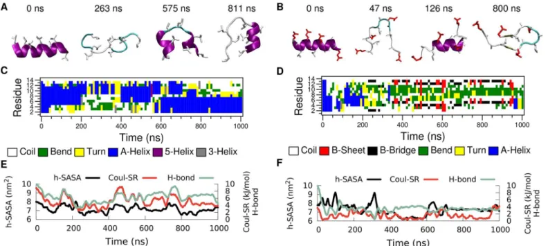
In order to explore the folding-unfolding equilibrium of individual peptides in bulk water (Fig 2path A) we performed molecular dynamics simulations of LK and EALA.Fig 3shows the time evolution of the secondary structure for both molecules. The simulations were started from an idealα-helix conformation of the peptides. Analysis of the secondary structural
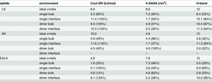
changes via DSSP [25] (Fig 3C and 3D) reveals that for both molecules theα-helix conforma-tion is not stable in bulk water. They adopt a variety of different conformaconforma-tions, where a few representative snapshots are shown for LK and EALA inFig 3A and 3B, respectively. The struc-tural instability of LK and EALA can be assessed based on the competition between three main factors: Coulomb repulsion between the charged sidechains, hydrophobic forces acting on the hydrophobic residues, and backbone-backbone or backbone-sidechain hydrogen bond forma-tion. We discuss the role of these three main factors by comparing their running averages with an idealα-helix conformation inTable 1.
When LK is in idealα-helix conformation all six positively charged lysine sidechains are
located on the same side of the helix. The electrostatic self-energy of the peptide can be approx-imated by the (screened) short range Coulomb repulsion between the charged sidechains (see
Methods).Fig 3EandTable 1show that when the molecule unfolds the Coulomb energy of LK drops to roughly 60% of the value for theα-helix. As expected, Coulomb repulsion drives the
molecule towards conformations which allow larger distances between the charged sidechains. With a total of eight leucine residues LK exhibits a rather large hydrophobic surface (8.6 nm2), which is completely exposed to water when in idealα-helix conformation in bulk water.
Hydro-phobic forces drive the molecule towards conformations where the leucine sidechains are less exposed to solvent and form a hydrophobic core. However, this is rather difficult to achieve when LK is isolated on its own. By measuring the SASA for hydrophobic leucine sidechains of Fig 3. Time evolution of secondary structure for LK (left) and EALA (right) when isolated in bulk water.Snapshots depicting various conformations adopted by these molecules (A and B), DSSP analysis of secondary structure (C and D), SASA for hydrophobic sidechains (h-SASA), number of intra-peptide backbone hydrogen bonds (H-bond) and short-range Coulomb interaction energies (Coul-SR) between the charged groups (E and F) are given for both of the molecules. SeeMethodssection for the color coding and representation of peptides in A and B.
LK, one can assess the degree of shielding from water achieved by any particular conformation (Fig 3E). The h-SASA drops on average only by 16% compared to the idealα-helix (Table 1).
The half-α-helix conformation, captured with the snapshot at 811 ns, is an attempt to form a hydrophobic core with just a single LK. This conformation is very similar to the TRP-cage structure [26,27], but it lacks the stability and specificity found for TRP-cage. LK can form a half-α-helix structure at either the C- or the N-terminus as observed between 400–550 ns and 700–1000 ns, respectively (see DSSP analysis inFig 3C). Reduction of the Coulomb repulsion and h-SASA via the loss of the idealα-helix conformation comes at a price: number of
back-bone hydrogen bonds is reduced by half (Fig 3EandTable 1).
In general all three of these contributions display a positive correlation. For the running averages given inFig 3Ethe correlation coefficients of h-SASA with H-bond, h-SASA with Coul-SR, and H-bond with Coul-SR are 0.44, 0.68, and 0.74, respectively. LK peptide is a frus-trated molecule, since there is no unique conformation, which is favorable for all three main contributions to free energy, when LK is isolated in bulk water. Experiments at low peptide and chloride concentrations suggest a random conformation for LK. [17] Hence our results at infi-nite dilution are in agreement with the experimental findings.
EALA, with 4 glutamic acid residues, has a slightly smaller total charge (−4) compared to
LK. As for the hydrophobic residues, in comparison to the eight leucine residues of LK, EALA has 3 leucine and 7 alanine residues. Despite the small size of the alanines, due to the increased total number of hydrophobic residues, the h-SASA value for EALA is only slightly smaller than LK, 7.8nm2, for the idealα-helix conformation LK (Table 1).
Similar to LK, EALA (with deprotonated glutamic acid sidechains at pH 7) lacks a well defined equilibrium structure in bulk water (seeFig 3B and 3D). However, EALA shows con-siderably less tendency towardsα-helix conformation. Qualitatively the individual secondary
structure elements found for EALA appear to have shorter lifetimes compared to LK. Table 1. Average short range Coulomb energy, hydrophobic SASA, and number of intra-mainchain hydrogen bonds for LK, AK and EALA.
peptide environment Coul-SR (kJ/mol) h-SASA (nm2) H-bond
LK idealα-helix 8.6 8.6 12
single bulk 5.2 (60%) 7.2 (84%) 6.4 (53%)
single interface 11.4 (133%) 1.7 (20%) 10.1 (84%)
dimer bulk 9.0 (105%) 4.9 (57%) 10.4 (87%)
dimer interface 12.3 (143%) 2.2 (26%) 11.3 (94%)
AK idealα-helix 10.0 4.6 12
single bulk 4.9 (49%) 4.4 (96%) 3.8 (32%)
single interface 11.6 (116%) 1.7 (37%) 11.3 (94%)
dimer bulk 4.5 (45%) 4.6 (100%) 2.6 (22%)
dimer interface – – –
EALA idealα-helix 4.9 7.8 12
single bulk 1.6 (33%) 7.3 (94%) 3.5 (29%)
single interface 5.1 (104%) 3.8 (49%) 9.9 (83%)
dimer bulk 0.6 (12%) 4.8 (62%) 2.8 (23%)
dimer interface 6.1 (124%) 2.2 (28%) 10.2 (85%)
The running averages are calculated for an idealα-helix in bulk water and under four different setups: a single peptide in bulk water and at the vacuum/ water interface, and two peptide molecules in bulk water and at the vacuum/water interface. For all four cases comparison with the idealα-helix is shown as percentages. For the dimers average of the two chains is reported. Running averages are calculated by discarding the equilibration period.
Given that under acidic conditions, when the sidechains are protonated, half GALA (the system from which EALA had been derived, see alsoS1 Fig) folds into anα-helix in bulk water [28], electrostatic repulsion can be listed as the dominant driving force in the unfolding of the helix. Upon unfolding the short range Coulomb interactions are reduced to 33% of the idealα -helix conformation (Table 1). h-SASA is not significantly changed, and hydrogen bonds are down to 29% of the idealα-helix conformation. Interestingly, no strong correlation between h-SASA, hydrogen bonds and electrostatic repulsion is observed (the correlation coefficient for h-SASA with H-bond, h-SASA with Coul-SR, and H-bond with Coul-SR are -0.27, 0.14, and 0.16, respectively.
Of course, MD results from a single simulation (even when it is one microsecond long) do not reveal the full picture, the system is not in full thermodynamic equilibrium and we are far away from having sampled states and transitions repeatedly and exhaustively. To get a better impression of the degree of sampling (or the lack thereof), we have performed additional simu-lations for the LK peptide with different initial velocities (results are given inS2 Fig). In these additional MD runs, the sequence of conformations adopted by LK differed considerably, how-ever the main conclusions did not change: None of the conformational changes, which take place over several hundred nanoseconds, lead to a single stable conformation of the molecule. Hence, both LK and EALA can be characterized as peptides with a high degree of intrinsic dis-order in bulk aqueous solution. However, it is important to note that these molecules do not behave like a random coil. LK displays a higher propensity towardsα-helical conformations, while EALA mostly adopts loop-like structures which transiently change intoβ-hairpins.
Single Peptide at the Vacuum/Water Interface
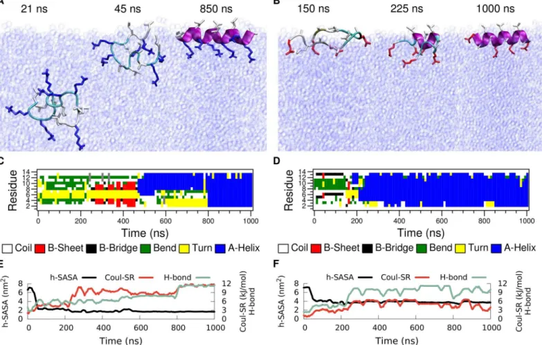
In this section we investigate LK and EALA at the vacuum/water interface, in particular we show the drastic effect that this interface has on their conformational preferences (Fig 2arc D). As in the previous section we first focus on the behavior of asinglepeptide molecule. Initially, the molecules were positioned at the center of the water phase in a random conformation (exemplary shown for LK inFig 4Asnapshot at 21 ns). After a short time (45 ns for LK and 50 ns for EALA) both molecules diffuse to the interface. In the particular simulations shown here, the peptides still lacked a well defined secondary structure when they reached the interface (Fig 4C and 4D) and folded subsequently. Since in bulk water the peptides adopt a large variety of transient conformations, molecules can in principle also make the first contact with the inter-face with a partially formed secondary structure.
The first structural change observed at the interface is the partitioning of the hydrophobic and hydrophilic residues as marked by the sharp drop in h-SASA (Fig 4E and 4F). Note that h-SASA calculation is performed such that only the surface of the hydrophobic residues that are actually in contact with the solvent is taken into account (seeMethodssection). Coupled with this reduction in h-SASA, the initial signs of secondary structure formation can be seen via an increase in the number of intra-peptide backbone hydrogen bonds (Fig 4E and 4F). At the same time, the partitioning effect leads to a locally increased density of charged sidechain groups in the water phase resulting in an increased electrostatic repulsion (green curve) which, however, is not strong enough to prevent folding.
Upon partitioning LK adopts aβ-hairpin structure which lasts up to 475 ns, at which point
a transition to theα-helix conformation starts. This halfα-helix structure persists for more than 300 ns, which finally transforms into a fullα-helix. Similarly, after going through a series
of conformational changes, EALA adopts a fullα-helix conformation. Separately, for both mol-ecules we simulated systems where the molecule was directly placed at the interface in anα
for more than one microsecond for both peptides. Hence, we can safely conclude that in the presence of a hydrophobic/hydrophilic interface both LK and EALA adopt anα-helix as their
preferred conformation. Note that the transition to theα-helical conformation does not
signifi-cantly reduce the h-SASA for both peptides compared to the other states at the interface (black lines inFig 4E and 4F). The hydrophobic sidechains are already desolvated as a result of parti-tioning, therefore the h-SASA is not strongly affected by folding. The remaining driving force for folding into anα-helix is the formation of hydrogen bonds. For those parts of the peptide
that are not exposed to water, intramolecular (α-helical) hydrogen bond formation will be very
favorable. Comparing the total values of h-SASA and electrostatic interactions between LK and EALA once the helix is formed at the interface, for EALA both the favorable reduction of h-SASA upon partitioning as well as the unfavorable Coulomb repulsion upon helix formation are smaller than for LK (Table 1). An analysis of the distribution of hydrophobic and hydro-philic sidechains at the interface can be found inS4 Fig. The data clearly show the preferential orientation of the LK and EALA helices as expected.
Fig 4. Simulations of a single peptide at the vacuum/water interface for LK (left) and EALA (right).Simulations are started with random conformations at the center of the water slab. Upon adsorption of peptides (after 45 ns for LK and 50 ns for EALA) partitioning of hydrophobic/hydrophilic residues and folding into theα-helix structure (after 800 ns for LK and 350 ns for EALA) is observed. Snapshots depicting the adsorption and adoption of theα-helix structure (A and B) and the DSSP analysis of the secondary structure evolution (C and D) are shown for both molecules. SASA, intra-peptide backbone hydrogen bonds and short-range Coulomb interaction energies between the charge groups (E and F) display the partitioning of hydrophobic/hydrophilic residues and secondary structure formation in the presence of the interface.
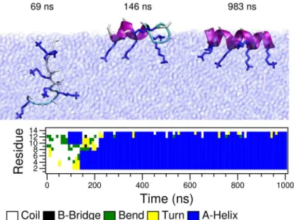
In order to better understand the contribution of the hydrophobic sidechains to the observed folding and partitioning equilibria, we have performed anin silicomutation of the
leucine residues to alanine in LK, resulting in an alanine-lysine peptide (from now on denoted as AK).Fig 5shows timeline and snapshots of the secondary structure formation of a single AK in the presence of the vacuum/water interface. Similar to LK and EALA, AK peptide, which was initially placed at the center of the water layer with a random conformation, moved to the interface in a short time (approx. 80 ns) and subsequently folded intoα-helix. The distribution
of alanine and lysine sidechains with respect to the interface are shown inS4 Fig. Notably, the decrease in sidechain hydrophobicity apparently neither prevents the partitioning nor the fold-ing at the interface.
In summary, the vacuum/water interface dictates a strong partitioning of the hydrophobic and hydrophilic residues in all three peptides considered. Once the partitioning takes place– since all three molecules are designed to display secondary amphiphilicity in theα-helix
con-formation–complete folding and stabilization of secondary structure is observed. All the pep-tides diffuse to the interface in an unfolded conformation and fold subsequently, with AK and EALA folding significantly faster than LK (see Figs4and5). This observation can be explained by the fact that folding at the interface requires intermediate conformations where hydropho-bic sidechains are (re-)solvated in water and hydrophilic sidechains are temporarily desolvated. This former step for example is more unfavorable for leucine than for alanine sidechains which may affect the height of the transition barriers in the folding process. The observations suggest that the folding times at the interface may be related to the hydrophobic moment [29] of the peptides, with LK being the slowest one to fold with the highest hydrophobic dipole. Further data collected from a variety of sequences will be needed to support this hypothesis.
Two Peptide Molecules in Bulk Water
As demonstrated in the previous sections, the conformation of these intrinsically disordered peptides strongly depends on the environment. In bulk water both LK and EALA display a Fig 5. Time evolution of secondary structure of a single AK (in-silico mutated form of LK) peptide at the vacuum/water interface.Initially the peptide is submerged in bulk water and moves to the interface in 80 ns and remains there for the rest of the simulation.
variety of secondary structures, which also include partialα-helices. However, in the presence
of a vacuum/water interface, all alternative secondary structures, except the fullα-helix are completely eliminated, supporting the conformation-selection & population-shift hypothesis for intrinsically disordered systems [30,31]. Interfaces are not limited to macroscopic ones. In the cell environment or in an experimental setup at finite concentration, proteins and peptides are surrounded by several other molecules. These surrounding molecules also present an inter-face, which we will refer to as a molecular interface. These nanoscale interfaces differ from the macroscopic ones (such as the vacuum/water interface) in two important ways. First, in terms of their size, they are of molecular scale and therefore force the guest molecule to fit to this area. Second, they are not permanent but rather transient structures and their stability is strongly influenced by the guest molecule.
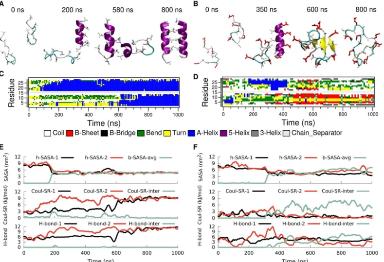
As a first example we have studied systems with two identical molecules in bulk water to see how the presence of such a molecular interface can affect the folding equilibrium (Fig 2arc E). Simulations for both LK and EALA were started with two molecules, each in random confor-mations and separated by several water layers (seeFig 6A and 6B). Snapshots depicting the
Fig 6. Folding and association of a pair of LK (left) and EALA (right) peptides in bulk water.Snapshots illustrating the aggregation process (A and B), DSSP secondary structure analysis (C and D), SASA, short range Coulombic interaction energies between the charged groups and the number of intra and inter-peptide backbone hydrogen bonds (E and F) are displayed as a function of simulation time. Association of peptides take place at 180 ns for LK and 300 ns for EALA, which can be observed via the sharp drop in SASA.
association of the peptides and time series of the secondary structures, the h-SASA and buried SASA values, the short range Coulomb energy between the charged sidechains, and the number of backbone hydrogen bonds are presented inFig 6for the course of one microsecond
simulation.
As shown before, neither LK nor EALA single peptides in bulk water adopt a unique confor-mation. Now, with two peptides, the behavior of LK and EALA is qualitatively different from each other. For LK (Fig 6left) the DSSP data show that initially one of the molecules starts to form anα-helix at around 80 ns (Fig 6C). Initial contact between the two separate LK mole-cules is made at around 200 ns via a pair of leucine sidechains (Fig 6A). From this initial con-tact on, a hydrophobic interface between the two molecules is formed rather rapidly. It appears that the intrinsically disordered nature of LK accelerates the aggregation process in good corre-spondence with a fly-casting mechanism [32], where initial contact is established via extension of unfolded tails (seeS5 Fig,S1andS2Videos). Although the kinetic effect due to the fly-cast-ing mechanism is reported to be less than 2-fold [32], it may nevertheless be significant for an intrinsically disordered system where multiple pathways exist, and small kinetic advantages may tip the balance, in particular in typical non-equilibrium experimental situations.
The formation of the hydrophobic interface can be observed in the SASA plot (Fig 6E). The h-SASA of each peptide chain declines rapidly at around 180 ns and the buried SASA corre-sponding to the contact area of the peptides increases. This overall behavior persists for a time-span of approximately 400 ns. Finally, at a simulation time of approximately 600 ns, also the second molecule folds into anα-helix. The increase of sidechain Coulomb repulsion within
each individual molecule uponα-helix formation which is perfectly correlated with the respec-tive increase in the number of backbone hydrogen bonds (Fig 6E) is completely analogous to the single peptide folding simulations discussed earlier. At the same time the arrangement of the two helices relative to each other allows for an inter-molecular separation of the charged NH3groups such that the inter-molecular Coulomb repulsion approaches zero (dropping lower than in the earlier stages of the aggregation between 100 and 600 ns where one of the peptides is still unfolded). This shows that the dimer interface consists mainly of hydrophobic residues. The fully formedα-helices remain stable for the rest of the simulation.
A separate run where the LK peptides were initially setup as aggregatedα-helices and remain stable throughout, supports these observations (S6 Fig). Hence, LK molecules can act as hydrophobic/hydrophilic molecular surfaces for each other, and the matching interface sta-bilizes the designedα-helix conformation. Comparing the molecular interface with the
macro-scopic one for LK, one can see that the h-SASA is only reduced to 57% of the value of an ideal
α-helix that is fully exposed to water. In comparison the vacuum/water interface reduces the
h-SASA to 20%. This suggests that the dimerization does not yet lead to a perfect shielding of hydrophobic sidechains from water, i.e. the aggregation is not necessarily limited to two pep-tides. This is in good agreement with the experimental findings that LK forms tetramers in aqueous solution [17]. Preliminary simulations of a preaggregated LK-tetramer have shown indeed, that this aggregate is stable in solution (seeS7 Fig). Further analysis to determine the equilibrium aggregate size and free energy profile are under way.
The picture is rather different for EALA. At first, similar to LK, the two molecules get in contact at roughly 300 ns and form a hydrophobic interface with their hydrophobic side chains which leads to a reduction in h-SASA. However, unlike LK, EALA molecules do not form or maintainα-helical conformations in the aggregate. In the dimer simulation shown inFig 6
(right panel), one of the EALA molecules displays aβ-hairpin structure, whereas the second
molecule does not adopt a well defined secondary structure (Fig 6D). Even if one starts with both EALA molecules inα-helix conformation the picture does not change (S8 Fig): the
mole-cules merge, however multiple times in the course of this 2 microsecond long simulation one of the two helices unfolds and adopts different secondary structure elements, such as turn or coil structures.
One key difference for EALA is that the glutamic acid residues play a dual role: due to their charge they repel the other EALA molecule while they can at the same time make contact with the backbone of the neighboring molecule because of their hydrogen bonding capacity. Indeed, the inter-molecular hydrogen bonds (Fig 6F) show that the two EALA molecules are connected via hydrogen bonds in addition to hydrophobic contacts. In the case of LK no inter-peptide hydrogen bond formation is observed (Fig 6E) and the dimer is held together purely by hydro-phobic attraction. It should be noted that these sidechain-backbone hydrogen bonds poten-tially destabilize theα-helices, and predominantly show up for EALA dimers withoutα-helical elements (S8 Fig).
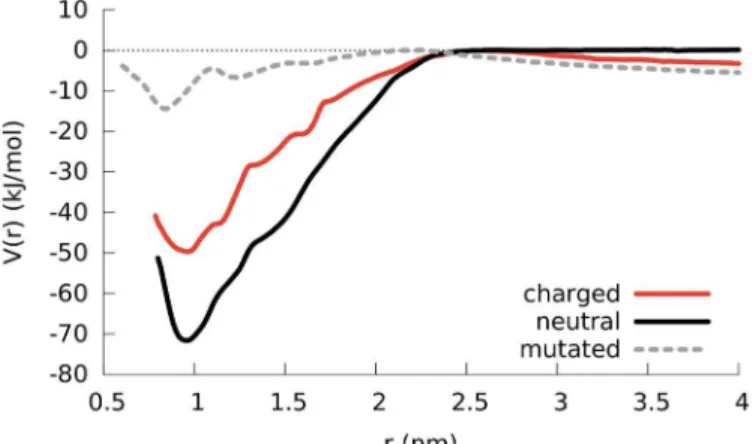
To quantify the strength of the dimerization, we employed umbrella sampling and com-puted the potential of mean force (PMF) as a function of the distance between two LK peptides.
Fig 7compares different PMFs to identify contributions from electrostatic and hydrophobic interactions: between two normal, i.e. charged, LK molecules (red line), between two neutral LK molecules where the lysines hold no charge (black line) and for the previously mentioned AK molecules (with leucine mutated to alanine; dashed grey line). The Coulombic repulsion between the positively charged lysine residues decreases the propensity of aggregation as evi-dent from the approximately 20 kJ/mol difference between the charged and neutral dimers at the PMF minimum (red and black curves, respectively). When the peptides are completely sep-arated (approximately above 2 nm), one observes a repulsion which can be seen from the slope in the PMF curves of charged LK and AK dimers. The fact that this slope does not exist for the
Fig 7. PMF results comparing the separation of a LK dimer in bulk water for the cases where lysine residues are positively charged, lysine residues are neutral and the leucine residues are in silico mutated to alanine residues.The curves are shifted so that the maximum points are zero. The distance refers to the distance between the center of mass of backbone atoms of the peptides. For the mutated AK dimer, when the peptides are in contact they maintain their helical structures. However when they are separated or when they make a loose contact theα-helical structure is not conserved.
PMF of the neutral LK dimer confirms that the repulsion is of electrostatic origin, resulting from the lysine charges. This repulsion acts as a barrier against aggregation where the probabil-ity of overcoming it can be increased by unfolded peptides, initiating contact through hydro-phobic residues. The hydrohydro-phobic attraction between the peptides for the AK dimer is smaller compared to the LK dimer because of the reduced volume of hydrophobic groups, supporting the hypothesis that the aggregation is driven by the formation of the hydrophobic core. Due to the reduced hydrophobic attraction in the AK dimer, the peptides unfold easily, yielding differ-ent PMF curves for the folded and the unfolded cases, which makes it harder to achieve conver-gence in a reasonable amount of time. As a result, the block analysis of the PMF yields different curves for different blocks (seeS9 Fig). However, the poor convergence does not change the fact that the aggregation of AK is significantly reduced compared to LK. This is further con-firmed by unconstrained simulations of two AK molecules (seeS10 Fig) which neither fold intoα-helices nor aggregate (as can be seen from the values for buried SASA and inter-peptide
hydrogen bonds). Even when the simulation starts with a pre-aggregatedα-helical AK dimer, the molecular interface is not sufficient to maintain the helical aggregate (seeS11 Fig) where, the peptides unfold and after one microsecond, the aggregate is weakly held by inter-chain hydrogen bonds. These data show that the mutation from leucine to alanine completely removes aggregation propensity from the LK peptides, as well as the tendency to form transient partial helical elements. In this sense AK is the most drastically disordered peptide investigated here. Remarkably it does formα-helices at the air/water interface, though.
Given the previously observed instability of secondary structures for EALA in solution and in aggregation (which hinders convergence), no PMF calculation were performed for two EALA molecules.
Two Peptide Molecules at the Interface
Considering the strong influence of the interface on the conformation of peptides, we finally investigated the combined effect: two peptides at the vacuum/water interface (Fig 2arc F). As we had observed that the interface induces theα-helix conformation in both molecules, the
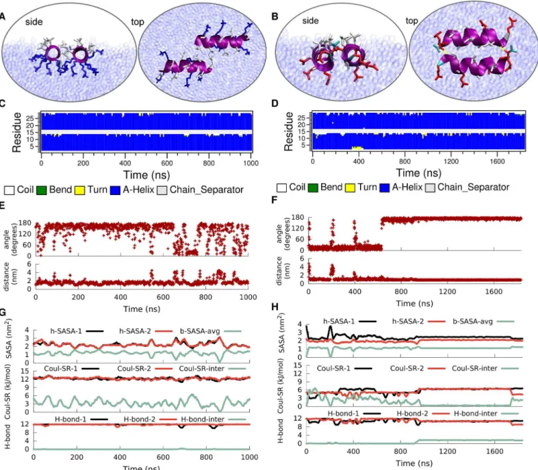
simulations were started from pre-foldedα-helices. For EALA, the two peptides were aligned at the interface, initially well-separated from each other by several layers of water molecules. For LK, the peptides were initially aggregated, but this starting configuration does not intro-duce any bias on the system, as the molecules display multiple aggregation/dissociation events during the simulation.
Both LK peptides retain theirα-helical structure for the entire simulation as characterized
This reduction in dimer stability for LK at the vacuum/water interface compared to bulk is a direct consequence of the disruption of the hydrophobic core. In bulk water hydrophobic leu-cine residues of the two peptides face each other and form a hydrophobic contact interface, whereas at the vacuum/water interface the leucine sidechains preferentially extend into vacuum instead of towards the leucine sidechains of the neighboring peptide. Furthermore, the align-ment of the sidechains of both peptides at the vacuum/water interface causes the charged Fig 8. Time evolution of the secondary structure of a dimer of LK (left) and EALA (right) at the vacuum/water interface.Typical snapshots when the peptides are associated at the interface (A and B), DSSP structural analysis (C and D), angle between the helix axis for the peptides and center-to-center distance (E and F), the h-SASA for each peptide along with the buried SASA for the whole peptide, the inter and intra molecular short-range Coulomb energies and the number of inter- and intra-molecular hydrogen bonds (G and H) are shown in figure.
lysines to come into closer contact compared to the dimer in bulk water (see snapshot in
Fig 8A), resulting in unfavorable electrostatic repulsion. Therefore, the inter-peptide short range Coulomb energy (Fig 8G) for the dimer at the interface is higher compared to the one for the dimer in bulk water (Fig 6E). Both of the peptides yield similar h-SASA values to the single LK at the interface due to the identical stability of theα-helix. One can follow the aggregation
of the dimers from both the inter-peptide distance (Fig 8E) and buried SASA (Fig 8G) which are inversely correlated. Moreover, the molecules do not make any inter-molecular hydrogen bonds.
Once again the picture is rather different for EALA. Under the strong influence of the inter-face both EALA peptides retain theirα-helical conformation (Fig 8D). In this conformation they strongly associate with each other by forming leucine-leucine contacts at the interface. Unlike LK, for EALA charged glutamic acid residues do not appear to prevent such an align-ment, presumably because they are differently distributed on the peptide surface. Differently from LK, the dimeric state of EALA at the interface appears to be more deeply submerged into the water phase compared to a single EALA molecule (S4 Fig). This suggests that dimerization and mutual shielding of hydrophobic sidechains leads to an overall structure where other parts of the molecule can be more favorably solvated. Initially EALA peptides orient in a parallel fashion, which switches to the anti-parallel alignment at 600 ns. Upon switching an additional stabilizing factor comes in to play: hydrogen bonds at the two ends of the aggregate.
Fig 8Hshows the intra and inter-peptide hydrogen bonding for EALA. Inter-peptide hydro-gen bonds that appear after ca. 900 ns, at the cost of intra-peptide ones, lead to the locking-in of the antiparallel orientation. Same trade-off can be observed for the electrostatic repulsion where the inter-chain value is minimized while the intra-chain repulsion is increased. The bur-ied SASA is in agreement with the enforced interaction and additionally shows that the orienta-tional changes, such as flipping of the peptide between anti-parallel and parallel orientations, are possible while the peptides stay in contact. The h-SASA of the aggregate is strongly reduced by a factor of 2 compared to a single peptide at the interface and keeps the hydrophobic area of the dimer at the same level as a single molecule. While h-SASA and Coulomb interaction describe the less explicit or long range interactions, the formation of the inter-chain hydrogen bonds locks in the angle and distance between the peptides for the rest of the simulation (Fig 8B, 8F, and 8H).
Discussion
In this paper, we have investigated the influence of interfaces on the conformational behavior of two synthetic peptides, EALA and LK, along with its alanine mutant AK, with built-in sec-ondaryα-helical amphiphilicity. First, we have characterized the behavior of individual
A change in environmental conditions typically changes the relative depths of the free-energy minima corresponding to these ensemble structures. This possibly explains why for many intrinsically disordered polypeptides certain environmental triggers induce the forma-tion of distinct structures. The formaforma-tion ofβ-sheet-rich amyloid fibrils upon aggregation of IDPs such as A-βorα-synuclein can be given as examples.
One important class of external stimuli that induce structure formation related to secondary amphiphilicity are interfaces—both macroscopic as well as molecular ones. We first investi-gated the conformational behavior of LK and EALA under the influence of the macroscopic vacuum/water interface. This interface leads to a strong partitioning of hydrophobic/hydro-philic residues, which enforces a unique conformation by stabilizing theα-helical form for both model systems. In the case of the LK peptide, the bulk water ensemble is already domi-nated by conformations where a large fraction of the residues are in anα-helical state. On the contrary, for EALA the probability of structures withα-helical elements in the bulk water
ensemble is low (but non-zero). Thus, the shift towards fully formedα-helices for both systems supports a conformational-selection & population-shift hypothesis, where the partitioning of the side chains at the interface shifts the existing equilibria and enhances the population of the
α-helical structures that are (at least partially) already present.
In their response to molecular interfaces presented by an aggregation partner in bulk water the peptide systems differ considerably. For LK, the molecular interface stabilizes theα-helical
state, even already at a dimer level. On the other hand, EALA dimers adopt a broad distribution of transient secondary structural elements, including (a very small fraction of)α-helical
ele-ments as well asβ-sheet structures. For AK the further reduced hydrophobicity of the side-chains destabilizes the dimer state entirely. Our MD simulations and PMF calculations have shown that the differences between LK, EALA and AK can be linked to the peptides’inherent
α-helical tendencies in bulk water, as well as to their different balance of hydrophobic forces,
electrostatic repulsion, and intra- and intermolecular hydrogen-bonding.
The simulations also allow an interesting glimpse on to the early stages of dimer formation, when the two molecules make their first contact. Not surprisingly, the first contact is estab-lished via the extended tail of one of the molecules in its disordered state as proposed in the fly-casting mechanism. This gives rise to the speculation, that a certain degree of disorder is kineti-cally advantageous for aggregation of these peptide systems, since it makes random encounters more likely. One might speculate that it is not entirely coincidental that so many amyloid form-ing peptides are intrinsically disordered in solution since (i) they are conformationally ambigu-ous and (ii) disorder may enhance aggregation.
We find that the peptides’aggregation behavior is also strongly affected by presence or absence of a macroscopic interface. While the LK peptides aggregate less strongly at the vac-uum/water interface compared to bulk water, two EALA molecules form very stableα-helical
dimers at the vacuum/water interface. The different behavior of EALA can be linked to the dis-tribution of surface charge and hydrophobicity, and again, strong intermolecular hydrogen bonds further stabilizes the dimer.
Methods
Setup
The amino acid sequences for LK and EALA are Ace-(LKKLLKL)2-Nme and Ace-(EAALAEA-LAEALAE)-Nme, respectively. All simulations were performed at neutral pH where the lysine sidechains in LK were protonated, and the glutamic acid sidechains in EALA were deproto-nated. The systems were neutralized by six chloride and four sodium ions for LK and EALA respectively.
Simulations
Molecular dynamics simulations were conducted using GROMACS version 4.5.6 [34] with the leap-frog integrator employing the GROMOS 54a7 force-field [35] and SPC/E water [36]. All bonds were constrained with LINCS algorithm [37] to enable a time-step of 2 fs. Coulomb interactions were calculated by particle mesh Ewald (PME) [38] method with a 1 nm cut-off. Lennard Jones interactions were calculated with a twin-range cut-off scheme of 1.0 and 1.4 nm with a neighbor list update at every 10 steps.
All systems were energy minimized with steepest-descent algorithm for 2000 steps, followed by three consecutive simulations for equilibration (100 ps each) where the heavy atoms, the backbone atoms and the Cαatoms were constrained. This was followed by a 100 ps simulation
for further equilibration. The actual simulations for data collection were at least oneμslong.
Simulations in bulk water and interface were performed at 298 K by using the Velocity-rescaling algorithm [39] with a coupling time of 1 ps. The bulk water systems were simulated in an isobaric-isothermal ensemble where the pressure was set to 1 atm using isotropic Berend-sen pressure coupling [40] with a pressure relaxation time of 1 ps for both the solvent and the peptide. The isothermal compressibility of 4.5×10⁻⁵bar−1which corresponds to that of pure water was used for the system.
The simulations for the single peptide in bulk water had 2919 and 2965 water molecules for LK and EALA respectively with cubic boxes of dimension of 4.5 nm on the average. The dimer simulation for LK in bulk water was started in a rhombic dodecahedron box of dimensions 7.27 nm in x and y and 5.14 nm in z-dimension with 8882 water molecules. After the LK mole-cules merge, to accelerate the simulation, the system size was reduced to 2984 molemole-cules in a box size of 5.12 × 5.12 × 3.62 nm. In order to verify that the box size does not change the out-come the original simulation with the larger box size was extended until folding (S13 Fig). In case of EALA, the simulation box had the dimensions of 8.03 × 8.03 × 5.68 nm and 12108 water molecules.
Vacuum/water interface simulations were performed with systems with cubic water slabs that have two interfaces to a vacuum slab in z-direction. These simulations were performed in the canonical (NVT) ensemble to maintain the shape of the box. For the single peptides the box sizes were 5.16 × 5.16 × 16.00 nm and 6.0348 × 6.0348 × 18.1043 nm, with 4467 and 7268 water molecules for LK and EALA respectively. The size of the vacuum in z-dimension was chosen to be twice the size of the z-dimension of the water slab. The dimer simulations at the vacuum/water interface contained 10258 (LK) and 10331 (EALA) water molecules in rectangu-lar boxes with average dimensions of 6.78 × 6.78 × 20.35 nm.
windows. A force constant of 1000 kJ mol−1nm−2was used to restrain the distance between the peptides. Each window for the charged and neutral cases was sampled for 200 ns whereas the in silico mutated case was run for 300 ns. Weighted histogram analysis [42] was applied to obtain the PMF curves using the g_wham tool [43] of GROMACS. The convergence of the PMF curves was checked with the block analysis method.
Analysis
Solvent accessible surface area (SASA) calculations were performed with a probe size of 0.24 nm using the g_sas tool of GROMACS. The relatively large probe was chosen to avoid inclu-sion of cavities in bulk water for the SASA calculations at the interface (see below). With few exceptions (which are separately indicated in the text) we report the SASA of the hydrophobic (alanine and leucine) sidechains of the peptides, denoted as h-SASA. Hence, for a single pep-tide in bulk water the g_sas tool is used by specifying the whole peppep-tide as calculation group (i.e. the specification of what is considered solute and what is solvent), but restricting the out-put to only the surface of the hydrophobic sidechains. At the vacuum/water interface the calcu-lations are done in two steps. First, by using the peptide as calculation and the hydrophobic sidechains as output group, the whole contact area of the hydrophobic sidechains is calculated. Next, by using water and peptide as calculation group and the hydrophobic sidechains as out-put group, the contact area of the hydrophobic sidechains facing vacuum is calculated. Taking the difference between the two values yields the h-SASA excluding the area exposed to vacuum. For the dimer state of peptides both peptides are given as calculation group, but only the hydrophobic sidechains of one of the peptides is given as output group. Finally, for two pep-tides at the interface, the same two step calculation is performed to calculate the h-SASA of the hydrophobic sidechains that are actually in contact with the solvent as described above for the single molecule at the interface. In addition, we compute also the buried (total) SASA between the two peptides. This is the difference between the sum of the SASA values of the individual peptides and the SASA of the aggregate (in the case of the interface simulation again account-ing for the fact that part of the surface is exposed to the vacuum).
Effective electrostatic energies of the solvated peptides were calculated using the“rerun” option of GROMACS’mdrun tool: the electrostatic interaction between the protein charges was re-calculated using a cutoff scheme with a relative permittivity of 80 to account for the screening by water. In Figs3,4,6the short range Coulomb interaction energies between the CE-NH3groups at the end of the lysine sidechains and the COO groups at the end of the glu-tamic acid sidechains are considered for LK and EALA respectively. Hydrogen bond analysis was performed using the g_hbond tool of GROMACS using a 30° cutoff for the acceptor-donor-hydrogen angle and a 3.5 Å cutoff for the distance between donor and acceptor. The reported inter-chain hydrogen bonds are between the main chain atoms unless otherwise stated. SASA, hydrogen bonds and the electrostatic energies are presented as running averages over 2000 frames with a data step size of 10 ps throughout the paper. The idealα-helix data in
Table 1are the average values from a 100 ns simulation where the single peptide is initially an idealα-helix with the backbone atoms position restrained throughout the simulation.
Supporting Information
S1 Fig. Sequences of LK, EALA and GALA and comparison withα-synuclein.The figure
shows the sequences of the peptides studied in the present paper (LK and EALA), and for refer-ence, also the experimental model peptide GALA is shown from which the EALA model peptide had been derived. GALA/1-30 refers to the full GALA sequence whereas the GALA05/1-16 rep-resents a shortened version as studied in [18]. The difference between GALA and EALA lies in the replacement of the tryptophane and histidine which are used as spectroscopic labels. GALA had been simulated at two different protonation states in [18]: At neutral pH the histidine side-chain is neutral and glutamic acid sideside-chains are deprotonated, the C-terminus is deprotonated and the N-terminus is protonated. At low pH, the histidine, glutamic acid sidechains as well as the C- and N- termini are protonated. The figure also shows a comparison with the part of the sequence ofα-synuclein where anα-helical state is formed upon membrane binding which relies on amphiphilic repeats. The alignment shows similarities in the hydrophobic profiles, and even though the sequence similarity betweenα-synuclein and EALA and LK is not high, there are repeating motifs in the hydrophobicity profile showing the secondary amphiphilicity. The colors range from blue to red for the hydrophilic to hydrophobic residues respectively. (TIFF)
S2 Fig. DSSP analysis for the additional runs of single LK peptide in bulk water.In order to test the dependence on the initial conditions we have run three additional MD simulations with different initial velocities. In two of these runs LK displayed a highα-helix content in its secondary structure, whereas in the second one the predominant structure is observed to be a
β-hairpin. These additional runs demonstrate that LK is an intrinsically disordered peptide, as it does not have a preferred secondary structure on its own in bulk water. The percentages of residues with defined secondary structures: 44%α-helix, 26% coil, and 17% bend & turn (top); 8%α-helix, 25%β-sheet, 36% coil, and 20% bend & turn, (middle); 41%α-helix, 24% coil, and
19% bend & turn (bottom); and finally 44%α-helix, 34% coil, 20% bend & turn for the simula-tion shown in the manuscript.
(TIFF)
S3 Fig. Single peptide at the vacuum/water interface withα-helical starting conformation.
DSSP analysis of time evolution of the secondary structure of a single LK (top) and EALA (bot-tom) at the vacuum/water interface color coded for each secondary structure.
(TIFF)
S4 Fig. Atom density plots comparing single peptides (solid lines) and dimers (dashed lines) at the vacuum/water interface: LK (top) and EALA (middle).The plot at the bottom compares the single LK with the single AK in the same environment. Vertical gray lines indi-cate the position of the interface where the interface is determined as the point as the water density is half of that of bulk water. Density plots are shown for backbone atoms (black), hydrophobic sidechains (red), and hydrophilic sidechains (green) separately. The calculated groups are as follows: iso-propyl group at the end of leucine sidechain (labelled as LEU(ip)), nitrogen atom at the end of lysine sidechain (labelled as NZ) and CO2group at the end of glu-tamic acid sidechain (labelled as GLU(CO2). Slices for computing densities are taken every 1 Å. The snapshot at the bottom is of a single AK at the vacuum/interface where similar to LK, the lysine sidechains (blue) extend into water and the alanines face vacuum.
(TIFF)
LK, the leucine residues are colored red and lysine residues are colored blue, for EALA the same coloring scheme in the manuscript is applied here. The ACE caps are shown with yellow CPK representation.
(TIFF)
S6 Fig. DSSP analysis of the simulation of two LK molecules initially inα-helix conforma-tion and pre-assembled into a dimer.Theα-helix conformation and the aggregate remain sta-ble for 1μs, in agreement with the simulation results presented for a pair of LK molecules in
bulk water with random starting conformations. (TIFF)
S7 Fig. Pre-aggregated LK tetramer in bulk water.The peptides remain aggregated andα -helical throughout the simulation time of 1 microseconds. The snapshot shows that the aggre-gate forms a hydrophobic core dehydrating the leucine sidechains. The ACE caps are shown with yellow CPK representation.
(TIFF)
S8 Fig. Additional EALA dimer simulation in bulk water where the peptides are initiallyα -helical and separated.Time evolution of secondary structure (top) and h-SASA, short range Coulombic interaction energies and the number of hydrogen bonds of the main chain (solid) along with the hydrogen bonds between the sidechain and the main chain (dashed) are shown (bottom). As can be seen from the buried SASA (green curve) the peptides aggregate approxi-mately in 50 ns. Although the dimer remains stable, the peptides do not maintain the initial full
α-helical secondary structure unlike the LK dimer in bulk water. After unfolding, the peptide can
make hydrogen bonds between the sidechain and the main chain at the cost of the ones between the main chains of the molecules. This is visible after 1800 ns and it illustrates the counterplay between the different inter and intra molecular driving forces acting in the folding and aggrega-tion processes. This conformaaggrega-tion increases both intra-peptide and inter-peptide electrostatic repulsion. The inter-peptide hydrogen bonding compensates for the intra-molecular hydrogen bonding as well as the electrostatic repulsion, while for SASA the effect is less pronounced. (TIFF)
S9 Fig. The block analysis for the PMF of separating an AK dimer.Because of the unfolding of the AK peptides the PMF blocks for the first 150 ns (purple) and the second 150 ns (yellow) yield different values. Although this behavior prevents proper sampling in a reasonable amount of time, it does not change the result that the aggregation energy of AK dimers is less than LK. Entropic correction is applied to the PMF curves and they are shifted so that the PMF equals zero at the moment of total separation at 2.1 nm.
(TIFF)
S10 Fig. Two AK peptides in bulk water initially separated and unfolded.DSSP analysis of secondary structure (top), SASA of the sidechains of hydrophobic residues and short-range Coulomb interaction energies between the charge groups are given. Similar to the EALA and unlike LK dimer in bulk water, the AK peptides remain unfolded and do not show any stable secondary structure. However, in contrast to EALA, the stronger charge repulsion (six charged lysine residues in AK versus four charged glutamic acid residues in EALA) together with the reduced hydrophobic attraction does not allow a stable aggregate as seen from the buried SASA.
(TIFF)
S11 Fig. Two AK peptides in bulk water pre-aggregated and pre-folded intoα-helix.The
(middle), hydrophobic SASA of the sidechains, short-range Coulomb interaction energies between the charge groups and number of hydrogen bonds (bottom) are given. Unlike the LK dimer the peptides unfold after approximately 350 ns. Although the peptides are unfolded, they are still in contact after one microsecond. The final structure is not stable and it is held weakly by inter-chain hydrogen bonds. As a result of the separation of the inter-residue charge groups the intra-Coulomb short range energies decrease, where at the same time the inter-dimer ones increase. The final h-SASA values are higher than the initial folded values, because the peptides cannot dehydrate their hydrophobic residues through a stable hydrophobic core.
(TIFF)
S12 Fig. Orientational analysis for dimer simulations.Inter-dimer distance vs inter-dimer angle correlation plots for LK (a and c) and EALA (b and d) in bulk water (top) and at the air/ water interface (middle). Part e shows the representative snapshots of the most populated dimer arrangements of LK peptides at the air/water interface (c). The DSSP plots can be seen inFig 6for the ones in bulk water (top), and inFig 8for the simulations at the vacuum/water interface (middle). The correlation plot for the LK dimer in bulk water contains the data after the peptides are aggregated and folded (first 800 ns of the simulation shown in part a was dis-carded).
(TIFF)
S13 Fig. Extension of the simulation of initially unfolded and separated LK peptides in bulk water.After the initial 500 ns the box size of the simulation in the manuscript was reduced to match the smaller aggregate size. The outcome is similar to the one presented in the manuscript as well as the results.
(TIFF)
S1 Video. Fly-casting mechanism observed for two LK dimers in bulk water.The leucine residues are colored red and lysine residues are colored blue. The ACE caps are shown with yel-low CPK representation.
(MP4)
S2 Video. Fly-casting mechanism observed for two EALA dimers in bulk water.The ACE caps are shown with yellow CPK representation.
(MP4)
Acknowledgments
We are grateful to Julia Borbas and Beytullah Özgür for critical reading of our manuscript and many valuable suggestions. Part of this work was performed on the computational resource bwUniCluster funded by the Ministry of Science, Research and the Arts Baden-Wuerttemberg and the Universities of the State of Baden-Wuerttemberg, Germany, within the framework pro-gram bwHPC.
Author Contributions
Conceived and designed the experiments: MS CP. Performed the experiments: CD CG. Ana-lyzed the data: CD CG CP MS. Wrote the paper: CD CG CP MS.
References
2. Greenwald J, Riek R. Biology of Amyloid: Structure, Function, and Regulation. Structure. 2010 Oct; 18(10):1244–1260. Available from:http://www.sciencedirect.com/science/article/pii/
S0969212610003084. doi:10.1016/j.str.2010.08.009PMID:20947013
3. Chiti F, Dobson CM. Protein Misfolding, Functional Amyloid, and Human Disease. Annu Rev Biochem. 2006; 75(1):333–366. doi:10.1146/annurev.biochem.75.101304.123901PMID:16756495
4. Butterfield SM, Lashuel HA. Amyloidogenic Protein–Membrane Interactions: Mechanistic Insight from Model Systems. Angewandte Chemie International Edition. 2010 Aug; 49(33):5628–5654. doi:10. 1002/anie.200906670PMID:20623810
5. Milletti F. Cell-penetrating peptides: classes, origin, and current landscape. Drug Discovery Today. 2012 Aug; 17(15–16):850–860. Available from:http://www.sciencedirect.com/science/article/pii/ S1359644612000839. doi:10.1016/j.drudis.2012.03.002PMID:22465171
6. Fischer R, Fotin-Mleczek M, Hufnagel H, Brock R. Break on through to the Other Side—Biophysics and Cell Biology Shed Light on Cell-Penetrating Peptides. ChemBioChem. 2005 Dec; 6(12):2126–2142. doi:10.1002/cbic.200500044PMID:16254940
7. Kotler SA, Walsh P, Brender JR, Ramamoorthy A. Differences between amyloid-βaggregation in solu-tion and on the membrane: insights into elucidasolu-tion of the mechanistic details of Alzheimer’s disease. Chem Soc Rev. 2014 Jan; 4:6692–6700. doi:10.1039/C3CS60431DPMID:24464312
8. Simone AD, Kitchen C, Kwan AH, Sunde M, Dobson CM, Frenkel D. Intrinsic disorder modulates pro-tein self-assembly and aggregation. Proceedings of the National Academy of Sciences. 2012 May; 109(18):6951–6956. Available from:http://www.pnas.org/content/109/18/6951. doi:10.1073/pnas. 1118048109PMID:22509003
9. Campioni S, Carret G, Jordens S, Nicoud L, Mezzenga R, Riek R. The Presence of an Air–Water Inter-face Affects Formation and Elongation ofα-Synuclein Fibrils. J Am Chem Soc. 2014 Feb; 136(7): 2866–2875. doi:10.1021/ja412105tPMID:24460028
10. Jean L, Lee CF, Lee C, Shaw M, Vaux DJ. Competing discrete interfacial effects are critical for amyloi-dogenesis. The FASEB Journal. 2010 Jan; 24(1):309–317. Available from:http://www.fasebj.org/ content/24/1/309. doi:10.1096/fj.09-137653PMID:19741169
11. Morinaga A, Hasegawa K, Nomura R, Ookoshi T, Ozawa D, Goto Y, et al. Critical role of interfaces and agitation on the nucleation of Aβamyloid fibrils at low concentrations of Aβmonomers. Biochimica et Biophysica Acta (BBA)—Proteins and Proteomics. 2010 Apr; 1804(4):986–995. Available from:http:// www.sciencedirect.com/science/article/pii/S1570963910000142. doi:10.1016/j.bbapap.2010.01.012 PMID:20100601
12. Jean L, Lee CF, Vaux DJ. Enrichment of Amyloidogenesis at an Air-Water Interface. Biophys J. 2012 Mar; 102(5):1154–1162. Available from:http://www.sciencedirect.com/science/article/pii/
S0006349512001567. doi:10.1016/j.bpj.2012.01.041PMID:22404938
13. Dikiy I, Eliezer D. Folding and misfolding of alpha-synuclein on membranes. Biochimica et Biophysica Acta (BBA)—Biomembranes. 2012 Apr; 1818(4):1013–1018. Available from:http://www.sciencedirect. com/science/article/pii/S0005273611003142. doi:10.1016/j.bbamem.2011.09.008
14. Lashuel HA, Overk CR, Oueslati A, Masliah E. The many faces ofα-synuclein: from structure and toxic-ity to therapeutic target. Nat Rev Neurosci. 2013 Jan; 14(1):38–48. doi:10.1038/nrn3406PMID: 23254192
15. Straub JE, Thirumalai D. Membrane–Protein Interactions Are Key to Understanding Amyloid Forma-tion. J Phys Chem Lett. 2014 Feb; 5(3):633–635. doi:10.1021/jz500054d
16. Bucciantini M, Rigacci S, Stefani M. Amyloid Aggregation: Role of Biological Membranes and the Aggregate–Membrane System. J Phys Chem Lett. 2014 Feb; 5(3):517–527. doi:10.1021/jz4024354 17. DeGrado WF, Lear JD. Induction of peptide conformation at apolar water interfaces. 1. A study with
model peptides of defined hydrophobic periodicity. J Am Chem Soc. 1985 Dec; 107(25):7684–7689. doi:10.1021/ja00311a076
18. Li W, Nicol F, Szoka FC Jr. GALA: a designed synthetic pH-responsive amphipathic peptide with applica-tions in drug and gene delivery. Adv Drug Delivery Rev. 2004 Apr; 56(7):967–985. Available from:http:// www.sciencedirect.com/science/article/pii/S0169409X03002801. doi:10.1016/j.addr.2003.10.041 19. Béven L, Castano S, Dufourcq J, WieslanderÅ, Wróblewski H. The antibiotic activity of cationic linear
amphipathic peptides: lessons from the action of leucine/lysine copolymers on bacteria of the class Mollicutes. Eur J Biochem. 2003 May; 270(10):2207–2217. doi:10.1046/j.1432-1033.2003.03587.x PMID:12752440
21. Baio JE, Zane A, Jaeger V, Roehrich AM, Lutz H, Pfaendtner J, et al. Diatom Mimics: Directing the For-mation of Biosilica Nanoparticles by Controlled Folding of Lysine-Leucine Peptides. Journal of the American Chemical Society. 2014; 136(43):15134–15137. doi:10.1021/ja5078238PMID:25285787 22. Jean L, Lee CF, Lee C, Shaw M, Vaux DJ. Competing discrete interfacial effects are critical for
amyloi-dogenesis. The FASEB Journal. 2010; 24(1):309–317. doi:10.1096/fj.09-137653PMID:19741169 23. Hoernke M, Falenski JA, Schwieger C, Koksch B, Brezesinski G. Triggers forβ-sheet formation at the
hydrophobic–hydrophilic interface: high concentration, in-plane orientational order, and metal ion com-plexation. Langmuir. 2011; 27(23):14218–14231. doi:10.1021/la203016zPMID:22011020
24. Fernández-Carneado J, Kogan MJ, Pujals S, Giralt E. Amphipathic peptides and drug delivery. Biopoly-mers. 2004 Jan; 76(2):196–203. Available from:http://www.ncbi.nlm.nih.gov/pubmed/15054899. doi: 10.1002/bip.10585PMID:15054899
25. Kabsch W, Sander C. Dictionary of protein secondary structure: Pattern recognition of hydrogen-bonded and geometrical features. Biopolymers. 1983; 22(12):2577–2637. doi:10.1002/bip.360221211 PMID:6667333
26. Neidigh JW, Fesinmeyer RM, Andersen NH. Designing a 20-residue protein. Nature Structural & Molecular Biology. 2002 Jun; 9(6):425–430. doi:10.1038/nsb798
27. Simmerling C, Strockbine B, Roitberg AE. All-Atom Structure Prediction and Folding Simulations of a Stable Protein. J Am Chem Soc. 2002 Sep; 124(38):11258–11259. doi:10.1021/ja0273851PMID: 12236726
28. Schach D, Globisch C, Roeters SJ, Woutersen S, Fuchs A, Weiss CK, et al. Sticky water surfaces: Helix–coil transitions suppressed in a cell-penetrating peptide at the air-water interface. The Journal of Chemical Physics. 2014 Dec; 141(22):22D517. doi:10.1063/1.4898711PMID:25494788
29. Wimley WC, White SH. Experimentally determined hydrophobicity scale for proteins at membrane interfaces. Nature Structural & Molecular Biology. 1996 Oct; 3(10):842–848. doi:10.1038/nsb1096-842 30. Ma B, Kumar S, Tsai CJ, Nussinov R. Folding funnels and binding mechanisms. Protein Eng. 1999
Sep; 12(9):713–720. doi:10.1093/protein/12.9.713PMID:10506280
31. Tsai CJ, Kumar S, Ma B, Nussinov R. Folding funnels, binding funnels, and protein function. Protein Sci. 1999; 8(06):1181–1190. doi:10.1110/ps.8.6.1181PMID:10386868
32. Shoemaker BA, Portman JJ, Wolynes PG. Speeding molecular recognition by using the folding funnel: The fly-casting mechanism. Proceedings of the National Academy of Sciences. 2000 Aug; 97(16): 8868–8873. Available from:http://www.pnas.org/content/97/16/8868. doi:10.1073/pnas.160259697 33. Dalgicdir C, Sensoy O, Peter C, Sayar M. A transferable coarse-grained model for diphenylalanine:
How to represent an environment driven conformational transition. J Chem Phys. 2013 Dec; 139(23): 234115. doi:10.1063/1.4848675PMID:24359360
34. Pronk S, Pall S, Schulz R, Larsson P, Bjelkmar P, Apostolov R, et al. GROMACS 4.5: a high-throughput and highly parallel open source molecular simulation toolkit. Bioinformatics. 2013 Apr; 29(7):845–854. Available from:http://www.ncbi.nlm.nih.gov/pmc/articles/PMC3605599/. doi:10.1093/bioinformatics/ btt055PMID:23407358
35. Schmid N, Eichenberger AP, Choutko A, Riniker S, Winger M, Mark AE, et al. Definition and testing of the GROMOS force-field versions 54A7 and 54B7. Eur Biophys J. 2011 Jul; 40(7):843–856. doi:10. 1007/s00249-011-0700-9PMID:21533652
36. Berendsen HJC, Grigera JR, Straatsma TP. The missing term in effective pair potentials. J Phys Chem. 1987 Nov; 91(24):6269–6271. doi:10.1021/j100308a038
37. Hess B, Bekker H, Berendsen HJC, Fraaije JGEM. LINCS: A linear constraint solver for molecular sim-ulations. J Comput Chem. 1997 Sep; 18(12):1463–1472. doi:10.1002/(SICI)1096-987X(199709) 18:12%3C1463::AID-JCC4%3E3.0.CO;2-H
38. Essmann U, Perera L, Berkowitz ML, Darden T, Lee H, Pedersen LG. A smooth particle mesh Ewald method. J Chem Phys. 1995 Nov; 103(19):8577–8593. doi:10.1063/1.470117
39. Bussi G, Donadio D, Parrinello M. Canonical sampling through velocity rescaling. J Chem Phys. 2007 Jan; 126(1):014101. doi:10.1063/1.2408420PMID:17212484
40. Berendsen HJC, Postma JPM, Gunsteren WFv, DiNola A, Haak JR. Molecular dynamics with coupling to an external bath. J Chem Phys. 1984 Oct; 81(8):3684–3690. doi:10.1063/1.448118
41. Torrie GM, Valleau JP. Nonphysical sampling distributions in Monte Carlo free-energy estimation: Umbrella sampling. Journal of Computational Physics. 1977; 23(2):187–199. doi:10.1016/0021-9991 (77)90121-8
43. Hub JS, de Groot BL, van der Spoel D. g_wham—A Free Weighted Histogram Analysis Implementation Including Robust Error and Autocorrelation Estimates. J Chem Theory Comput. 2010 Dec; 6(12): 3713–3720. doi:10.1021/ct100494z
44. Humphrey W, Dalke A, Schulten K. VMD: Visual molecular dynamics. J Mol Graph. 1996 Feb; 14(1): 33–38. doi:10.1016/0263-7855(96)00018-5PMID:8744570
45. Frishman D, Argos P. Knowledge-based protein secondary structure assignment. Proteins Struct Funct Bioinforma. 1995; 23(4):566–579. doi:10.1002/prot.340230412