Published online August 31st, 2009 © http://www.ijav.org
Case Report
International Journal of Anatomical Variations (2009) 2: 99–101
Introduction
The deep artery of thigh (DAT) is a large branch of the femoral artery (FA) that arises around 3.5 cm distal to the inguinal ligament. It is the main arterial supply to the muscles of the thigh. Anastomoses occur between the DAT and the internal and external iliac arteries above, and the popliteal artery below [1]. As a result, the DAT has great surgical significance. The bifurcation of the FA into superficial femoral artery (SFA) and DAT, and the branching pattern of the DAT are subject to considerable normal anatomical variation [2]. (Some radiologists, vascular surgeons and other specialist physicians refer to the femoral artery as the “superficial femoral artery” after the deep artery of thigh branch point to differentiate the femoral artery segments before and after the branch
point.) In this case report we describe an unusual origin
of the DAT occurring bilaterally, associated with other anatomical variations in the thigh.
Case Report
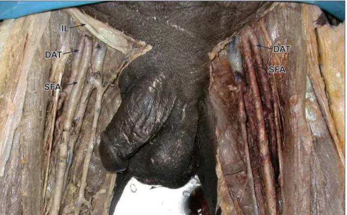
During routine dissection of the lower limb of a middle aged male cadaver, an unusual origin of the DAT was observed bilaterally. The artery was traced from origin to termination and relevant measurements were made with digital caliper. On the right side, the DAT arose from the posterolateral aspect of the FA, 4 mm distal to the inguinal ligament. It arose from the lateral aspect of the FA, 8 mm distal to the inguinal ligament on the left side (Figure 1). At their origins, the SFA and DAT measured 6.56 mm and 6.79 mm in diameter respectively on the
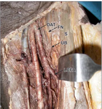
right side. The measurements on the left side were 5.73 mm and 6.51 mm respectively. On both sides, the DAT coursed inferomedially and exited the femoral triangle in the interval between the pectineus and adductor longus muscles. The relationship of the DAT to structures in the lower part of the femoral triangle, and its mode of termination, did not reveal any variations on either side. On both sides, the medial and lateral circumflex femoral arteries (MCFA and LCFA) arose from the DAT. On the right side the MCFA and LCFA arose 3.19 cm and 4.75 cm from the origin of the DAT respectively. The measurements on the left side were 1.96 cm and 5.45 cm respectively. An unnamed branch from the DAT on the left side was seen coursing superolaterally, passing deep to the sartorius muscle and running towards the anterior superior iliac spine (ASIS) (Figure 2). No other variations were observed in the branching pattern of the DAT on either side. The deep external pudendal artery arose from the MCFA bilaterally. On the right side the femoral nerve emerged in the femoral triangle by piercing the iliacus muscle.
Discussion
The origin of the DAT is commonly described as being from the lateral aspect of the FA around 3.5 cm distal to the inguinal ligament [1]. In the present case the DAT arose less than 1 cm distal to the inguinal ligament. A bilateral, high origin as reported here is a rare occurrence. In a study conducted on 430 thighs by Quain, it was found that the
Nachiket SHANKAR Roopa R
Department of Anatomy, St. John’s Medical College, Karnataka, INDIA.
Dr. Nachiket Shankar Assistant Professor Department of Anatomy St. John’s Medical College Sarjapur Road, Bangalore – 560034 Karnataka, INDIA.
+91 80 22065061/62 nachiket76@gmail.com
Received May 9th, 2009; accepted August 28th, 2009
ABSTRACT
During routine dissection of a middle aged male cadaver, an unusual origin of the deep artery of thigh was observed bilaterally. It arose from the femoral artery less than 1 cm distal to the inguinal ligament. On both sides, its diameter was greater than that of the femoral artery. An unusually distal origin of the lateral circumflex femoral artery was observed bilaterally. An unnamed branch from the deep artery of thigh on the left side was seen coursing superolaterally towards the anterior superior iliac spine. The deep external pudendal artery arose from the medial circumflex femoral artery on either side. On the right side, the femoral nerve emerged in the femoral triangle by piercing the iliacus muscle. As the deep artery of thigh is often used in vascular reconstructive procedures and is frequently visualized by various radiological imaging techniques, anatomical variations of itself as well as its branches have significant clinical implications. © IJAV. 2009; 2: 99–101.
Key words [deep artery of thigh] [high origin] [lateral circumflex femoral artery] [deep external pudendal artery] [femoral nerve]
eISSN 1308-4038
100 Shankar and Roopa
Figure 1. Bilateral high origin of the deep artery of thigh from the common femoral artery. (DAT: deep artery of thigh; IL: inguinal ligament; SFA: superficial femoral artery)
origin of the DAT was less than 1.3 cm distal to the inguinal ligament in 20 thighs (20 of 430; 4.7%). The majority of the DAT (68%) arose between 2.5 and 5.1 cm from the inguinal ligament [3]. Another study by Siddharth et al. on 100 dissected thighs revealed that the DAT originated at a median distance of 4.4 cm from the inguinal ligament. Only in one instance (1 of 100; 1%) did it arise at the level of the inguinal ligament [4]. In a study conducted in India, dissection of 48 femoral triangles revealed that none of the DAT arose less than 1.9 cm distal to the inguinal ligament. The average distance of the origin of the DAT from the midpoint of the inguinal ligament was 4.75 cm [5]. In 253 lower limb arteriograms from Czech Republic a high origin of the DAT at or above the inguinal ligament was found in 1.2% of the subjects [6].
In the present case the DAT arose from the posterolateral aspect of the FA on the right side and lateral aspect of the FA on the left side. In the study conducted by Siddharth et al., 52% of the DAT arose from the posterolateral or lateral aspect of the FA. Less common was an origin from the dorsal aspect (37%) and least common the posteromedial or medial aspect (11%) [4]. Similar results have been reported by other investigators [2,5].
The DAT develops in the rete femorale, a network of blood vessels that connect the precursors of the proximal part of FA and the distal part of the SFA. The distal part of the FA and the proximal part of the SFA also develop in rete femorale. Thus the DAT appears as a branch of the FA after regression of the rete femorale. Anatomical variations in
the origin of the DAT occur due to variability in the pattern of regression of the rete femorale [2].
The diameter of the DAT in this study was found to be 6.79 and 6.51 mm on the right and left sides, respectively. On both sides the diameter was greater than that of the SFA; this is an unusual occurrence. Berguer et al. have stated that the diameter of the SFA is usually around 30% more than the DAT [7]. Previous studies on cadavers have shown that the diameter of the DAT at its origin is usually between 4.5 mm to 6 mm [4,8]. If the DAT is to function adequately as a collateral, it has to have a large caliber. An explanation for this may be found in comparative anatomy. In the phylogenetically farther advanced vertebrate animals, all of which have very strongly developed extensors in the posterior extremities, the deep femoral artery supplying blood to this muscle group necessarily has to have a large caliber [9].
In the present case the origin of the LCFA was more distal than usual. Dixit et al. have stated that in only 4% of the cases was the origin of the LCFA more than 45 mm from the origin of the DAT [5]. Siddharth et al. found that the LCFA arose at a median distance of 1.5 cm distal to the origin of the DAT [4]. The distance of the origin of the MCFA from the origin of the DAT was not unusual in the present case. It is usually given off higher than the LCFA [4]. The unnamed branch that was observed coursing towards the ASIS probably took part in the arterial anastomosis around the ASIS, replacing the superficial circumflex iliac artery, which is usually a branch of the femoral artery and which was absent in the present case.
IL
DAT
SFA
DAT
101 Unusual origin of the deep artery of thigh
Figure 2. The left deep artery of thigh showing an unnamed branch coursing superolaterally and an unusually distal origin of the lateral circumflex femoral artery. (LCFA: lateral circumflex femoral artery; FN: femoral nerve; S: sartorius muscle; UB: unnamed branch)
The external pudendal arteries usually described as two in number, superficial and deep, are found infrequently. More commonly only a single artery is found [10]. In a large majority of cases, this artery arises from the femoral artery and only very rarely from the DAT [11]. The deep external pudendal artery arising from the MCFA, as seen in this case, is very uncommon. Passage of the femoral nerve through the iliacus muscle, as seen in the present case, may predispose to entrapment of the nerve in this course. A cadaveric study revealed that in 19 of 242 specimens (7.9%), the femoral nerve passed through slips of either the iliacus or the psoas major [12].
The relationship of the femoral nerve, artery and vein in the upper part of the femoral triangle is important in procedures like femoral arterial and venous puncture and femoral nerve blocks. A high origin of the DAT could thus pose difficulties in performing these procedures. Surgical exposure of the DAT is often necessary in vascular reconstructive procedures. Furthermore, plastic surgeons have shown great interest in the muscular branches of the DAT when designing procedures that incorporate myocutaneous flaps [13]. The DAT is often visualized using arteriography, ultrasound and Doppler imaging, digital subtraction angiography and magnetic resonance imaging [5]. Thus anatomical variations of the deep artery of thigh and its branches have significant clinical implications.
References
[1] Standring S, ed. Gray’s Anatomy. 39th Ed., Edinburgh, Elsevier Churchill Livingstone Publishers. 2006; 1451–1452.
[2] Massoud TF, Fletcher EW. Anatomical variants of the profunda femoris artery: an angiographic study.
Surg Radiol Anat. 1997; 19: 99–103.
[3] Bergman RA, Thompson SA, Afifi AK, Saadeh FA. Compendium of human anatomic variation: catalog, atlas and world literature. Baltimore, Urban and Schwarzenberg. 1988; 86.
[4] Siddharth P, Smith NL, Mason RA, Giron F. Variational anatomy of the deep femoral artery. Anat Rec. 1985; 212: 206–209.
[5] Dixit DP, Mehta LA, Kothari ML. Variations in the origin and course of profunda femoris. J Anat Soc India. 2001; 50: 6–7.
[6] Voboril R. Note on the variability of the arteries of the lower extremeties in man. Folia Morphol (Praha). 1990; 38: 265–272.
[7] Berguer R, Higgins RF, Cotton LT. Geometry, blood flow, and reconstruction of the deep femoral artery.
Am J Surg. 1975; 130: 68–73.
[8] Bloda E, Sierocinski W, Kling A. Variation of the arteria profundus femoris in man. Folia Morphol (Warsz). 1982; 41: 123–131.
[9] Vaas F. Some considerations concerning the deep femoral artery. Arch Chir Neerl. 1975; 27: 25–34.
[10] Henriet JP. Sapheno-femoral venous confluence and the external pudendal network: anatomical data and new statistics. Phlebologie. 1987; 40: 711–735. (French)
[11] La Falce OL, Ambrosio JD, Souza RR. The anatomy of the superficial external pudendal artery: a quantitative study. Clinics (Sao Paulo). 2006; 61: 441–444.
[12] Vazquez MT, Murillo J, Maranillo E, Parkin IG, Sanudo J. Femoral nerve entrapment: a new insight. Clin Anat. 2007; 20: 175–179.
[13] Colborn GL, Mattar SG, Taylor B, Skandalakis JE, Lumsden AB. The surgical anatomy of the deep femoral artery. Am Surg. 1995; 61: 336–346.
UB FN
LCFA DAT