Nanomed. J., 3(4):217-222, Autumn 2016
The effect of electrospun poly(lactic acid) and nanohydroxyapatite
nanofibers’ diameter on proliferation and differentiation of mesenchymal
stem cells
Amir Doustgani*Department of chemical engineering, University of Zanjan, Zanjan, Iran ABSTRACT
Objective(s): Electrospun nanofibrous mats of poly(lactic acid) (PLA) and nanohydroxyapatite (nano-HA) were prepared and proliferation and differentiation of mesenchymal stem cells on the prepared nanofibers were investigated in this study.
Materials and Methods:PLA/nano-HA nanofibers were prepared by electrospinning. The effects of process parameters, such as nano-HA concentration, distance, applied voltage, and flow rate on the mean diameter of electrospun nanofibers were investigated. Scanning electron microscopy (SEM) was used to determine the mean fiber diameter of produced nanofibers. Mechanical propertes of nanofibrous mats were evaluated using a universal testing machine. Response surface methodology was used to model the fiber diameter of electrospun PLA/nano-HA nanofibers.
Results: The average fiber diameter for optimized nanofibers was 125 ± 11 nm. MTT and ALP results showed that optimization of fiber diameter increased the osteogenic differentiation of stem cells.
Conclusion: It could be concluded that optimization of fiber diameter has beneficial effect on cell proliferation and differentiation. Optimized nanofibers of PLA/nano-HA could be good candidates for bone tissue engineering.
Keywords:Mesenchymal stem cells, Nanofiber, Optimization
*Corresponding Author Email:[email protected] Tel: (+98)024-33054275
Note. This manuscript was submitted on April 18, 2016; approved on July 23, 2016
INTRODUCTION
The rapidly growing research in the bone tissue engineering area thus provides a new and promising approach for bone repair and regeneration. Scaffolds for hard tissue engineering applications could be fabricated from a wide variety of classes of materials. Many bone tissue engineering scaffolds have been made as composites, either by the introduction of nanohydroxyapatite (nano-HA) within polymeric matrices or by the mineralization of nano-HA on the surface of polymeric substrates [1,2]. The biologically beneficial characteristics of nano-HA, such as that it is the major inorganic component of the bone matrix, its specific affinity toward many adhesive proteins,
and its direct involvement in the bone cell differentiation and mineralization processes, make nano-HA especially attractive for applications in the bone regeneration field [3]. Mesenchymal stem cells (MSCs) have been widely used in regenerative medicine and tissue engineering studies, and proven to have significant potential in clinical application because of their convenient isolation, lack of significant immunogenicity, high capacity of expansion, and potential to differentiate into tissue-specific cell types [4-10]. Electrospinning is a well-known and ubiquitous technique to produce nanofibers and has been used by many researchers to make nanofibrous matrix for tissue engineering applications [11-15]. Scaffolds for hard tissue engineering applications could be fabricated from a wide variety of classes of materials [16]. Although
How to cite this article
Doustgani A. The effect of electrospun poly(lactic acid) and nanohydroxyapatite nanofibers’ diameter on proliferation and differentiation of mesenchymal stem cells. Nanomed J., 2016; 3(4): 217-222.DOI:10.22038/nmj.2016.7575
Nanomed. J., 3(4): 217-222, Autumn 2016
the polymers used are similar to the polymers applied in soft tissue engineering, reinforcement of scaffolds is frequently necessary for hard tissue engineering due to their low mechanical properties. By combining two or more classes of materials into composites, such as a crystalline ceramic (e.g. hydroxyapatite) and a synthetic polymer, scaffolds with improved mechanical properties can be expected [17]. It was previously shown that both the strength and the conductivity of the film/mat of fibers produced by electrospinning are sensitive to fiber diameter [18]. Moreover, size of the fibers along with morphology influences the hydrophobic behavior of polymers [19]. Therefore, controlling the mean fiber diameter which is a function of process parameters is crucial. Response surface methodology (RSM) is a collection of statistical and mathematical techniques useful for the statistical modeling and analysis of problems in which a response of interest affected by several variables, with the objective being to optimize this response. In this study, poly(lactic acid) (PLA) polymer solutions with different amounts of nanohydr-oxyapatite were used to produce nanofiber mats by electrospinning.
The experiment parameters were material related variables (i.e., nano-HA concentration in solution) and process-related variables (i.e., applied voltage, distance and flow rate).
The result of the experiment was average fiber diameter. The effect of fiber diameter on proliferation and differentiation of MSCs on the produced nanofibers was investigated.
MATERIALS AND METHODS Materials
PLA with molecular weight of 75 kD and nanohydroxyapatite (nano-HA) (d 200 nm) were obtained from Sigma Aldrich and used without further puriûcation. Chloroform was purchased from Merck.
Solution preparation
Nano-HA powder was dispersed in PLA solution with a two-step method. For this purpose, nano-HA was added in chloroform and sonicated at room temperature for 40 min. In the second step, 1.0 g of PLA pellets was added to the nHA/DMF/Chloroform dispersion.
This was followed by magnetic stirring until polymer dissolved completely with subsequent
sonication for 40 min to obtain a uniform dispersion of the nano-HA in the PLA solution.
Fabrication of nanofibrous scaffold
An electrospinning machine was used to prepare nano- HA/PLA nanofibrous scaffolds. Prepared solutions with different amounts of nano-HA, were placed on a syringe pump used to dispense the solution at a controlled rate.
The collector was a rotating cylindrical drum which was placed at different distances from the needle. A high voltage electric field (Nano spinner TM, Iran) was applied to draw the ultra-fine fibers from the spinneret. The humidity during the experiments was kept the same (65%). To remove any residual solvent present in the fibers, the electrospun nanofibers were subsequently vacuum dried.
Characterization of nanofibers morphology
Scanning electron microscopy (SEM) (SEM; Vega II XMU instrument Tescan, Czech Republic) was used to characterize the morphology of electrospun nanofibers.
The samples were sputtered with a thin layer of gold prior to SEM analysis. An accelerating voltage of 20 kV was applied. Based on SEM images, the average diameter of yarns and electrospun fibers were determined by using image analysis software. The results are reported as average values of 100 measurements.
Nanomed. J., 3(4):217-222, Autumn 2016
Table 1. Selected factors and their levels
High axial (+2) High factorial (+1) Center (0) Low factorial (-1) Low axial (-2) Variable 30.0 22.5 15.0 7.5 0.0 X1(A)
35.0 27.5
20.0 12.50
5.0 X2(B)
21.0 18.0
15.0 12.0
9.0 X3(C)
1.5 1.2
0.9 0.6
0.3 X4(D)
X1(A)=Concentration (w/w) %; X2(B) = Distance (cm) X3(C) = Voltage (kV); X4(D) = Flow rate (ml/h)
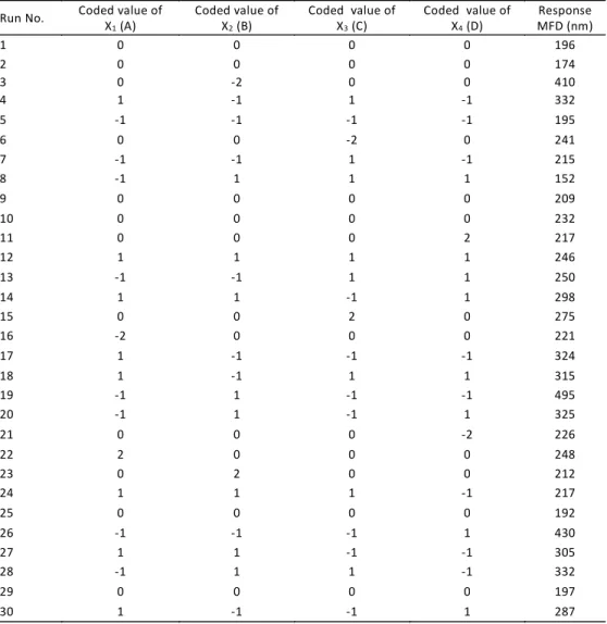
Table 2. Experimental conditions and their responses
Response MFD (nm) Coded value of
X4(D) Coded value of
X3(C) Coded value of
X2(B) Coded value of
X1(A) Run No. 196 0 0 0 0 1 174 0 0 0 0 2 410 0 0 -2 0 3 332 -1 1 -1 1 4 195 -1 -1 -1 -1 5 241 0 -2 0 0 6 215 -1 1 -1 -1 7 152 1 1 1 -1 8 209 0 0 0 0 9 232 0 0 0 0 10 217 2 0 0 0 11 246 1 1 1 1 12 250 1 1 -1 -1 13 298 1 -1 1 1 14 275 0 2 0 0 15 221 0 0 0 -2 16 324 -1 -1 -1 1 17 315 1 1 -1 1 18 495 -1 -1 1 -1 19 325 1 -1 1 -1 20 226 -2 0 0 0 21 248 0 0 0 2 22 212 0 0 2 0 23 217 -1 1 1 1 24 192 0 0 0 0 25 430 1 -1 -1 -1 26 305 -1 -1 1 1 27 332 -1 1 1 -1 28 197 0 0 0 0 29 287 1 -1 -1 1 30
The regression correlation coefficient (R2) is used in the context of statistical models and provides a measure of how well the observed response values fit the model. This coefficient ranges from 0 to 1. When R2 approaches 1, the actual data fit the polynomial model properly. Low values of R2 imply that the dependent variables in the applied model do not correlate well.
Mechanical characterization of nanofibrous scaffolds The tensile measurements were performed on the nanofibrous webs using a universal testing machine (STM-20, SANTAM Design & Manufacturing Co., Iran). Prepared scaffolds were cut into 10 × 60 mm specimens, and a tensile test was conducted at 50 mm/min crosshead speed at room temperature. At least six samples were tested for each type of electrospun nanofibrous scaffold.
Cell culture
MSCs were seeded onto nanofiber mats at a density of 105 cells/well in 24-well culture plates containing osteogenic differentiation medium (50μ g/ l L-ascorbic acid, 10-8 mol/l dexamethasone, 10 mmol/l-glycerophosphate, 10 mmol/l vitamin D3, 100μ g/ml penicillin, 100 μ g/ml streptomycin, 0.3 ìg/ml amphotericin, 2.2 g/L sodium bicarbonate and 15% fetal bovine serum) and incubated in a humidified CO2 incubator at 37 °C.
Cell proliferation
Proliferation of MSCs on nano-HA/PLA scaffolds in comparison to TCPs (tissue culture plates) was evaluated via MTT assay. Sterilized nanofibrous membranes were placed in a 24-well culture plate, seeded with a cell density of 5 × 10³ cells per cm2, and incubated at 37 °C, 5% CO2under basal medium. After 1, 7, and 21 days of cell seeding, 50μ L of MTT solution (5 mg/mL in DMEM) was added to each well followed by incubation at 37 °C for 3 h. For dissolution of the dark-blue intracellular formazan, the supernatant was removed and 1 mL of DMSO (Merck) was added. The optical density was read spectrophotometrically at a wavelength of 570 nm. The same procedure was performed for cultured cells in TCPS as control.
Alkaline phosphatase (ALP) activity
ALP is used to measure ALP activity by a kinetic rate method using 2-amino-2-methyl-1-propanol buffer. In this reaction, ALP catalyzes the hydrolysis
of colorless organic phosphate ester substrate, p -nitrophenyl phosphate, to a yellow product,
p-nitrophenol, and phosphate. This reaction occurs at an alkaline pH of 10.3. Nanofibrous scaffolds with MSCs were washed twice with PBS and added with 1 mL of 10 mM Tris buffer to isolate the sample. The sample (1 mL) was again sonicated for 2 min at 4°C. The sonicated sample was centrifuged at 3000 rpm for 5 min at 4°C, then the supernatant was used for the assay of ALP activity.
Statistical Analysis
All experiments were conducted at least for n= 3. Data are expressed as mean ± SD. One-way analysis of variance (ANOVA) was used to compare results. A p-value of less than 0.05 was considered statistically significant.
RESULTS AND DISCUSSION Experimental model
The experimental conditions and their responses are shown in Table 2. A polynomial model for the average variations of DOX/PLA nanoûbers was chosen and ûtted to the results. Adequate model for prediction of the response variables is given by the following equation:
MFD = 147.24 + 54.36A 45.37B+ 72.59AD
+ 84.95BC 28.22BD 48.17ABC
All the variables are in coded values. Values of R-square and adjusted R-R-square for the model are 0.92 and 0.8, respectively. The high value of R2 indicates that the cubic equation is capable of representing the system under the given experimental domain.
MFD optimization
An optimization technique was used in this research for optimization of PLA/nano-HA nanofibers diameter using design expert software.
Nanomed. J., 3(4):217-222, Autumn 2016
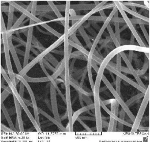
in Fig 1. As can be seen, the MFD was 125 ± 11 nm. Comparing the experimental results with the response provided by the model shows that the difference is less than 5% and they are close to each other.
Fig. 1. SEM image of optimized nanofibers
Mechanical properties of prepared nanofibers The mechanical properties of biocomposite nanofibrous scaffolds were evaluated by tensile testing. The tensile strength of PLA/nano-HA nanofibers (Exp. No. 19) was 2.53 ± 0.12 MPa and the elastic modulus was 7.18 ± 0.87 MPa. By optimizing of nanofibers diameter, the tensile strength increased to 6.12 ± 0.21 MPa with an elastic modulus of 15.3 ± 0.95 MPa. According to Baji et al. [20], the mechanical properties of the nanofibers were affected by mean fiber diameter. Upon increasing the fiber size, both tensile strength and modulus decreased and the larger diameter fibers tend to display bulk-like properties. In fibers with finer diameter, the lamellae and fibrillar structures align themselves along the fiber axis, which play a critical role in enhancing the mechanical properties of the fibers.
Cell proliferation results
The effect of PLA/nano-HA fiber diameter on cell attachment and proliferation has shown in Fig 2. As can be seen, a significant increase in cell number was observed on both optimized and non-optimized scaffolds at 7 and 21 days. There was no significant difference, between the scaffolds at 1 day. The
proliferation of MSCs on optimized PLA/nano-HA scaffold was found to be 15 and 40 % higher after day 7 and 21, respectively compared to non-optimized scaffold. One possible reason for the increased cell proliferation is that the mechanical stretching of the optimzed nanofibers leads to better cell proliferation.
This phenomenon is similar to the mechanical stimulation of cells by matrix stretching studies reported previously [21].
Fig. 3. ALP results for scaffolds (non-optimized and optimized) and TCPs
Fig. 2: Proliferation results for scaffolds (non-optimized and optimized) and TCPs
A
LP
a
ct
iv
it
y
(
IU
/l
)
/
to
ta
l
p
ro
te
in
(
m
g
/d
l)
O
p
ti
ca
l d
e
n
si
ty
Culture time (day)
Culture time (day)
TCPs non-optimized Optimized TCPs non-optimized Optimized
ALP results
fiber than on the non-optimized PLA/nano-HA fibers (P
d 0.05), confirming the crucial role of fiber size in the stimulation of bone cell response and thus its significance in the bone regeneration.
CONCLUSION
PLA/nano-HA nanofiber mats were successfully fabricated by electrospinning. The overall optimal electrospinning conditions were 12.5 (w/w)% concentration, 14.5 kV voltage, 25 cm distance and 0.8 ml/h flow rate. The favorable attachment and proliferation and differentiation of MSCs on the optimized nanofibers indicated good cytocompatibility compare to non-optimized nanofibers. The PLA/nano-HA fibrous composites prepared by electrospinning by using the optimized parameters could lead to the construction of suitable porous scaffolds for bone tissue engineering. It could be concluded that optimization of fiber diameter has beneficial effect on cell proliferation and differentiation.
ACKNOWLEDGEMENT
The authors gratefully acknowledge the financial support from the Iran Nanotechnology Initiative Council (INIC).
CONFLICT OF INTEREST
The authors declare that there are no conflicts of interest regarding the publication of this manuscript.
REFERENCES
1. Liao S, Chan CK, Ramakrishna S. Stem cells and biomimetic materials strategies for tissue engineering. Mater Sci Eng C. 2008; 28: 1189-1202.
2. Araujo JV, Martins A, Leonor IB, Pinho ED, Reis RL, Neves NM. Surface controlled biomimetic coating of polycaprolactone nanofiber meshes to be used as bone extracellular matrix analogues. J Biomater Sci Polym Ed. 2008; 19: 1261-1278. 3. Liu Y, Wang G, Cai Y, Ji H, Zhou G, Zhao X, Tang R, Zhang M.
In Vitro effect of nanophase hydroxyapatite particles on proliferation and osteogenic differentiation of bone marrow derived mesenchymal stem cells. J Biomed Mater Res. 2009; 90A: 1083-1091.
4. Horwitz EM, Le Blanc K, Dominici M, Mueller I, Slaper-Cortenbach I, Marini FC, Deans RJ, Krause DS, Keating A. Clarification of the nomenclature for MSC: the International Society for cellular therapy position statement. Cytotherapy. 2005; 7(5): 393-395.
5. Dominici M, Le Blanc K, Mueller I, Slaper-Cortenbach I, Marini F, Krause D, Deans R, Keating A, Prockop Dj, Horwitz E. Minimal criteria for defining multipotent mesenchymal stromal cells. The International Society for Cellular Therapy position statement. Cytotherapy. 2006; 8(4): 315-317.
6. Pittenger MF. Multilineage potential of adult human mesenchymal stem cells. Science. 1999; 284 (5411): 143-147. 7. Prockop DJ. Marrow stromal cells as stem cells for nonhematopoietic tissues. Science. 1997; 276 (5309): 71-74. 8. Vinatier C, Mrugala D, Jorgensen C, Guicheux J, Noël D. Cartilage engineering: a crucial combination of cells, biomaterials and biofactors. Trends Biotechnol. 2009; 27(5): 307-314.
9. Li W-J, Tuli R, Huang X, Laquerriere P, Tuan RS. Multilineage differentiation of human mesenchymal stem cells in a three-dimensional nanofibrous scaffold. Biomaterials. 2005; 26(25): 5158-5166.
10. Karp JM, Leng Teo GS. Mesenchymal stem cell homing: the devil is in the details. Cell Stem Cell. 2009; 4(3): 206-216. 11. Sundaray B, Subramanian V, Natarajan TS, Xiang RZ, Chang
CC, Fann WS. Electrospinning of continuous aligned polymer fibers. Appl Phys Lett. 2004; 84: 1222-1224. 12. Huang ZM, Zhang YZ, Kotaki M, Ramakrishna S. A review
on polymer nanofibers by electrospinning and their applications in nanocomposites. Compos Sci Technol. 2003; 63: 2223-2253.
13. Matthews JA, Boland ED, Wnek GE, Simpson DG, Bowlin GL. Electrospinning of collagen type II: A feasibility study. J Bioact Compat Polym. 2003; 18: 125-134.
14. Kidoaki S, Kwon IK, Matsuda T. Mesoscopic spatial designs of nano- and microfiber meshes for tissue-engineering matrix and scaffold based on newly devised multilayering and mixing electrospinning techniques. Biomaterials. 2005; 26: 37-46.
15. Min BM, Lee G, Kim SH, Nam YS, Lee TS, Park WH. Electrospinning of silk fibroin nanofibers and its effect on the adhesion and spreading of normal human keratinocytes and fibroblasts in vitro. Biomaterials. 2004; 25(7/8): 1289-1297.
16. Stamatialis DF, Papenburg BJ, Giron´es M, Saiful S. Medical applications of membranes: Drug delivery, artificial organs and tissue engineering. J Membr Sci. 308 (2008) 1-34.
17. Fabbri P, Bondioli F, Messori M, Bartoli C, Dinucci D, Chiellini F. Porous scaffolds of polycaprolactone reinforced with in situ generated hydroxyapatite for bone tissue engineering. J Mater Sci Mater Med. 2010; 21: 343-351.
18. Kwon IK, Kidoaki S, Matsuda T. Electrospun nano- to microfiber fabrics made of biodegradable copolyesters: structural characteristics, mechanical properties and cell adhesion potential. Biomaterial. 2004; 26: 3929-3939. 19. Acatay K, Simsek E, Yang CO, Mencelo YZ. Tunable,