Abstract
Submitted: October 19, 2016
Accepted: March 9, 2017
Effects of
Syzygium ar om at icum
,
Cinnam om um zeylanicum
, and
Salvia
t r iloba
extracts on proliferation and
differentiation of dental pulp stem
cells
Hypersensitivity, local irritative and cytotoxic effects are known for the chemical components of Syzygium arom at icum and Cinnam om um zeylanicum contained in dental materials. However, there is no intimate data in dentistry using the whole extracts of these plants and introducing new ones. Salvia t riloba
used in several dental traumas. Objectives: We aimed to show and compare the effect of S. ar om at icum, C. zeylanicum, and S. t r iloba extracts on dental pulp stem cells (DPSCs) proliferation, differentiation, and immune responses. Material and Methods: Using xCELLigence, a real time monitoring system, we obtained a growth curve of DPSCs with different concentrations of the
μ
by using an ELISA Kit to detect early and late markers of differentiation. Results: The level of osteonectin (ON, early osteogenic marker) decreased, which indicated that the osteogenic differentiation may be accelerated with addition of extracts. However, the level of osteocalcin (OCN, late osteogenic marker and sign of calcium granulation) differed among the extracts, in which S. ar om at icum presented the highest value, followed by S. t r iloba and C. zeylanicum. Surprisingly, the determined calcium granules were reduced in S. ar om at icum and S. t r iloba. In response to tumor necrosis factor alpha (TNF-D), S. t r iloba-treated DPSCs showed the most reduced level of IL-6 cytokine level. We suggest C. zeylanicum as a promising osteogenic inducer and S. t riloba
in biocomposite or scaffold fabrications for dentistry. Conclusions: Because calcium granule formation and cell viability play a critical role in hard tissue formation, S. ar om at icum in dentistry should be strictly controlled, and the
Ke y w or ds: Dental Pulp. S. ar om at icum. C. zeylanicum. Salvia t r iloba. Osteoblast.
1 2 3
Nurdan SARAÇ4 3 1
Duygu UÇKAN2
http://dx.doi.org/10.1590/1678-7757-2016-0522
1Gazi University, Faculty of Dentistry, Department of Medical Microbiology, Ankara, Turkey.
2Hacettepe University, PEDI-STEM Center for Stem Cell Research and Development, Ankara, Turkey. 3Gazi University, Faculty of Dentistry, Department of Oral and Maxillofacial Surgery, Ankara, Turkey. 4Mugla Sitki Kocman University, Faculty of Sciences, Department of Biology, Mugla, Turkey.
Corresponding address:
Gazi University, Faculty of Dentistry, Department of Medical Microbiology. Emek - Ankara - Turkey . Phone: +90 312 203 4365 - Fax: +90 312 221 3202
Introduction
Medicinal plants have been extensively utilized
for clinical purposes in various diseases, including
dental disorders13. Of these, Syzygium ar om at icum,
Cin n am om u m zey lan icu m, and Salv ia t r ilob a are
activities. Eugenol is the main component of S.
ar om at icum, and it plays a prominent role in dental
and oral preparations13. C. zeylanicum is also used
in toothpastes and mouthwashes. They are used as
in dentistry. However, allergies to materials and/or
extracts used in dentistry are an increasing issue and
have been subject of research11,12,16,17.
Medications administered to teeth and oral mucosa,
such as zinc oxide-eugenol (ZOE) and cinnamon,
including mouthwashes, can reach the pulp tissue
or periodontium after penetrating the enamel and
dentin or passing through apical foramens2. If the
medications are cytotoxic, they might disturb the
function of mesenchymal stem cells (MSCs), which
exist in dental pulp and in the periodontium. Therefore,
it is important to study the cytotoxicity of agents used
in oral treatment.
In this study, we hypothesize that a natural agent,
which maintains dental pulp stem cells (DPSCs)
viability, promotes osteogenic differentiation while
modulating the immunological response, and that it
could achieve success in regeneration during healing
and may also prevent bone resorption and improve
regeneration.
Although various physiological activities of the
extracts have been shown, their effects on osteogenic
differentiation of mesenchymal stem cells have never
been assessed.
Material and Methods
Extraction of plant samples
S. a r o m a t i cu m , C. ze y l a n i cu m bark, and S.
t r iloba
were purchased from a local market in Mugla,
Turkey. The air-dried plant samples were extracted
with ethanol (Merck, Dermstadt, Germany) using a
Soxhlet apparatus. The extracts were evaporated
and stored in sterile opaque glass bottles under
refrigerated conditions until use. The dried extract
low glucose (DMEM-LG) with 10% fetal bovine serum
(FBS) (Invitrogen, Carlsbad, California, USA), 1%
L-Glutamine (Sigma St. Louis, Missouri, ABD), and
1% penicillin-streptomycin (Invitrogen, Carlsbad,
California, USA).
Isolation and culture of dental pulp stem cells
Human dental pulp tissue was obtained from
patients (15-20 years of age) who were undergoing
extraction of third molars for orthodontic reasons at
the Department of Oral and Maxillofacial Surgery, Gazi
University, Ankara. All patients signed an informed
consent form. After the tooth surfaces were disinfected
(75% ethanol), the teeth were drilled and the dental
pulp was gently extracted with forceps. The extracted
streptomycin, and 10% FBS (Invitrogen, Carlsbad,
California, USA) [hereafter referred to as the stem cell
(SC) culture medium], after which it was minced into
fragments of 1 to 2 mm3. The tissue fragments were
cultured in T75 plates (Nunc, Waltham, Massachusetts,
atmosphere containing 5% CO2. The cell cultures
were monitored regularly with an inverted microscope
(Olympos CKX41, Tokyo, Japan) and the SC culture
media were changed every 3 days. After reaching
70-Trypsin/EDTA (Sigma St. Louis, Missouri, USA) and
sub-cultured for further experiments. The experiments
were performed with passage 2-3 cells.
Immunophenotypic analysis
The culture-expanded adherent cells were analyzed
USA). The antibody panel included CD29-FITC
(e-bioscience, USA), CD73-PE (BD, USA), CD90-PE
(BD, USA), and CD44-PE (e-bioscience, USA) as
mesenchymal stromal markers, as well as their isotype
controls. CD45-FITC (BD, USA), CD14-PE (BD, USA),
and CD34-FITC (BD USA) were used as hematopoietic
markers to exclude cells of hematopoietic origin. The
relative frequencies of the cells that expressed the
respective surface markers were analyzed using FACS
Diva software 6.0.0 (BD) by acquiring 10,000 events
for each sample.
S. t r iloba
on DPSCs proliferation, using the
xCELLigence system
The xCELLigence system was used according to the
manufacturer’s instructions. An impedance-based real
time cell analyzer (RTCA), an RTCA single plate (E-plate
96), an RTCA computer, and a tissue-culture incubator
constitute the xCELLigence system (Roche Applied
Science, Mannheim, Germany)15. There are sensor
electrodes on the surface of each well of the E-Plate
96. Physiologic changes in the cells are determined and
measured by the electronic impedance of the sensor
electrodes. Electrode geometry, ion concentration on
the well, and whether the cells are attached to the
electrodes affect the impedance measured between
electrodes in each well. In the absence of cells, the
electrode impedance is mainly determined by the ion
environment at the electrode-solution interface and
in the bulk solution. In the presence of cells, cells
attached to the electrode sensor surfaces change
the local ion environment at the electrode-solution
interface, leading to increased impedance. Thus, if
more cells are present in the environment growing on
the electrodes, the value of the electrode impedance
will be increased. This mechanism gives real time
monitoring adherent cells.
To measure the background impedance, we
connected the E-plate 96 to the xCELLigence system
and ensured that the proper electrical contacts were
containing different concentrations of the Extracts
(5, 10, and 25 μg/mL) and of standard culture media
(control) were added to each well of E-plate 96. The
cells were resuspended (5000 cells/cm2) in SC culture
media. Cell growth and proliferation were monitored
every 30 min for up to 169 h.
Effect of
S. ar om at icum , C. zeylanicum ,
and
S. t r iloba
on DPSCs differentiation
The concentration that decreased the doubling
time and increased the proliferation was selected
based on the results from the xCELLigence system
analysis. This concentration was added into the
osteogenic and adipogenic differentiation media14.
Images were obtained with a CKX41 digital imaging
microscope (Olympus CKX41, Tokyo, Japan). The
media were changed every 3 or 4 days for 21 days.
The removed culture supernatant was stored in
osteocalcin (OCN) and osteonectin (ON) levels in
kit, according to the manufacturer’s instructions
(R&D Systems, Inc. Minneapolis, USA). The limits of
detection for the ELISA ranged from 1.2 to 75 ng/mL
for OCN and 1.56 to 50 ng/mL for ON. The calcium
ion concentration in the differentiation medium was
measured using a QuantiChrom calcium assay kit
according to the manufacturer’s instructions (DICA
500, BioAssay Systems, USA).
Determining immunomodulatory activities
DPSCs were plated at a density of 5000 cell/
cm2 in 96-well culture plates and allowed to attach
of the
was added. After 24 h, the cell culture supernatants
and IL-10 ELISAs, according to the manufacturer’s
instructions. The ELISA limits were 0.052-0.118 pg/
mL for IL-6 and 0.39-25 pg/mL for IL-10. The media
as controls.
Statistical analysis
All calculations were performed using the RTCA
integrated software of the xCELLigence system. The
dose response equations to the experimental data
standard deviation (SD) (n=4). For the proliferation
experiments, the statistical analysis was performed
using one-way analysis of variance (ANOVA) (p<0.05).
Results
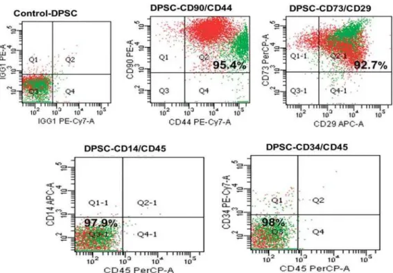
Common mesenchymal SC markers (CD29, CD73,
CD44, and CD90) were consistently positive (>95%)
and hematopoietic markers (CD14, CD34, and CD45)
were negative (>95%) in all samples tested, indicating
a mesenchymal origin of the cells (Figure 1).
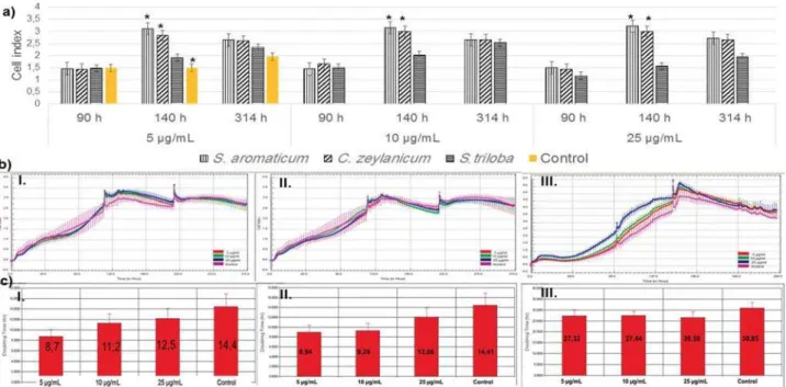
xCELLigence assays
Using Trypan Blue, we found that only the S.
ar om at icum extract induced cell proliferation (Figure
2). 5, 10, and 25 μg/mL concentrations were selected
for xCELLigence analysis according to the highest cell
cell proliferation with S. arom at icum and C. zeylanicum
was in a positive correlation with the Control Group
at 140 h (Figure 3a). Growth curves were obtained in
the real time monitoring system (Figure 3b.I-III). The
doubling time (DT) of the Extracts suggested that S.
ar om at icum (Figure 3c-I) and C. zeylanicum (Figure
3c-II) reduced the DT in a dose-dependent manner.
S. t r iloba (Figure 3c-III) showed a reduction in DT
independent from the concentrations. We calculated
the IC50 values, which were 8001 μg/mL, 102 μg/mL,
and 1475 μg/mL for S. ar om at icum, C. zeylanicum,
and S t riloba, respectively. The 10 μg/mL concentration
was selected for further studies.
Differentiation assays of DPSCs
Adipogenic differentiation was not seen in the
Control (Figure 4a.I) and in the Extract Group (Figure
4b-d.I). Morphologically, approximately 20% of the
cells adopted rounder shape in all Groups. However, no
lipid droplets were observed. On the other hand, the
DPSCs in the Control Group were well differentiated
in osteogenic differentiation media (Figure 4a.II).
Calcium granules were seen as dark nodules, and the
extracellular matrix was dyed red. S. ar om at icum
-treated cells showed reduced calcium granules
and extracellular matrix staining (Figure 4b.II).
C. zey lan icu m -treated cells presented remarkable
calcium granules and extracellular matrix dying (Figure
4c.II). S. t r iloba displayed an average osteogenic
Figure 1- Surface markers of dental pulp stem cells (DPSCs). The cells were positive for CD90, CD44, CD29, and CD73 (mesenchymal stem cell markers) and negative for CD14, CD45, and CD34 (hematopoietic stem cell markers)
Figure 2- Viability of dental pulp stem cells (DPSCs) treated with different Extracts. Trypan bBue assay showed S. aromaticum had no toxic effect on the cells. C. zeylanicum and S. triloba reduced cell viability after 10 μg/mL. Increased concentration reduced the cell number in S. aromaticum and C. zeylanicum in a negative strong correlation
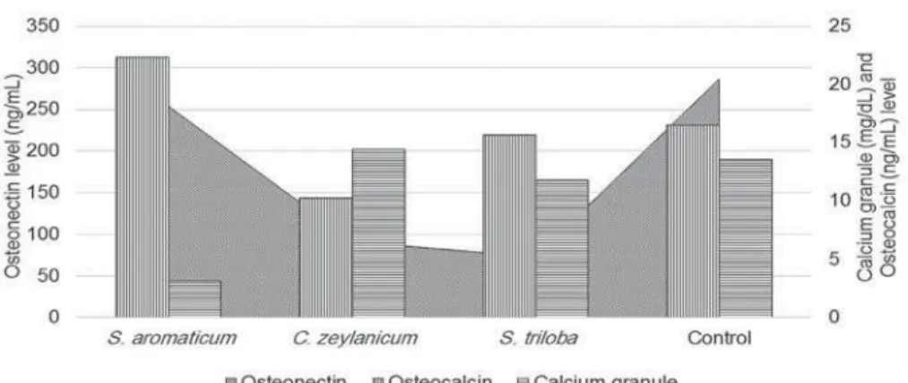
Alizarin Red S. The quantity of calcium granules and
extracellular matrix protein levels is shown in Figure 5.
Osteonectin levels showed a decrease in cells treated
measure of osteogenic differentiation was supported
by microscopic analysis. S. ar om at icum showed the
lowest calcium concentration, while C. zey lanicum
Figure 3- xCELLigence analysis of dental pulp stem cells (DPSCs) treated with 5, 10, and 25 μg/mL of the Extracts. a: Cell index of DPSCs
μ S. aromaticum and C. zeylanicum growth curve of the cells with S. aromaticum C. zeylanicum
(b.II) and S. triloba S. aromaticum (c.I), C.
zeylanicum (c.II), and S. triloba (c.III)
Figure
presented the highest. Interestingly, regarding
osteocalcin levels, S. ar om at icu m and S. t r ilob a
presented high sign of granulation, but low calcium
concentration. However, we found no statistical
Determining the preventive effect of plant
extracts on the inflammatory response of
DPSCs following TNF-
D
stimulation
Both IL-6 and IL-10 were present in DPSCs cell
culture supernatants (Figure 6). When the Extracts
were used alone, IL-6 levels were found decreased
compared to the Control Group. We observed increased
IL-6 levels after TNF-D induction. The Extracts were
able to prevent the increase of IL-6 levels in response
observed in the presence of S. t r iloba, C. zeylanicum,
and S. ar om at icum , respectively, but there was no
Discussion
In this study, we presented the effects of S.
ar om at icum, C. zeylanicum, and S. t r iloba extracts on
DPSCs. Using real time monitoring and quantitative
analyses, we showed that C. zeylanicum and S. t riloba
extracts have positive effects on human DPSCs.
Initially, we aimed to define the effective
concentration of the plant extracts. Oriental medicine
practices are primarily based on personal experience,
but often rely on unknown mechanisms, resulting
system is a sophisticated cell-based assay. It
continuously monitor cell proliferation, viability, and
label-free cytotoxicity in real time, by showing the
physiologic state of the cells and eliminating expensive
reagents, which are used in conventional cell analyses9.
We found a reduced cell index in a ratio of 71% for C.
zeylanicum and S. t r iloba in the Trypan Blue assay at
5 μg/mL, while an increase with a proportion of 47%
and 21% was seen in the xCELLigence assay. Growth
curves provide information on three parameters: the
lag phase before cell proliferation is initiated after Figure 5- Calcium granule, osteocalcin, and osteonectin levels of dental pulp stem cells (DPSCs) treated with the Extracts
Figure 6- IL-6 and IL-10 levels determined in the culture supernatant of dental pulp stem cells (DPSCs). IL-10 level reached 67.9 pg/mL with S. triloba in response to TNF-D induction
growth phase, and the terminal density. A shortened
DT at 5 and 10 μg/mL was observed with C. zeylanicum
and S. arom at icum , suggesting cell proliferation would
increase more rapidly and the healing of the fracture or
implant region could be shortened. It is also important
to determine IC50 values, since the IC50 represents
the concentration of a drug that is required for 50%
inhibition in vit r o. The IC50 for S. ar om at icum (8001 μg/mL), C. zeylanicum (102 μg/mL), and S. t r iloba (102 μg/mL) was calculated and compared with the
literature. Surprisingly, the clinically used eugenol/S.
ar o m at i cu m concentration was 6.5 M1, which is μ
study. The wide difference between the data lead us to
suggest that S. ar om at icum/eugenol could penetrate
pulp tissue and trigger adverse effects, such as cell
cytotoxicity. Eugenol containing endodontic cements
(zinc oxide-eugenol) is applied to a variety of dental
tissues. When placed onto a wet soft tissue, a much
greater amount of eugenol would release from ZOE
onto dentin, and the released eugenol is considered
one of the ingredients responsible for cytotoxicity and
allergic reactions10. Thus, the release of eugenol/S.
ar om at icum should be controlled, and an alternative
similar properties to S. ar om at icum .
To show whether the extracts induce differentiation
of DPSCs, we constructed an in vit r o model in which
lipid-laden adipocytes, calcium granules, and early
and late markers of osteogenesis were examined
after 21 days. At the end of the culture period,
adipogenic differentiation was not seen in DPSCs.
al.7 (2000), who expanded DPSCs from single-cell
clones and demonstrated that these cells presented
osteogenic differentiation, but do not form
lipid-laden adipocytes. The osteogenic differentiation
potential of DPSCs in vit r o and in vivo has been
well-documented in several studies3,7. During osteogenic
cell differentiation, the markers of undifferentiated
cells are gradually turned off, and differentiation
markers are sequentially expressed. We observed
sequential secretion of proteins at the end of the
assay, in which the ON levels decreased in the
Extract-treated group, compared to the control group. ON is
an early marker of osteogenesis that is synthesized
of >24 hours under conditions of transcription arrest4.
DPSCs treated with the Extracts showed reduced ON
and increased OCN levels, while the Control group was
vice ver sa, suggesting that osteogenic differentiation
was more rapid in the Extract-treated cells. However,
conspicuously, calcium concentrations were low in S.
ar om at icum and S. t r iloba-treated DPSCs, despite
the increased OCN levels. S. ar om at icu m and S.
t r iloba may disturb the calcium deposit development
of the cells. Similarly, Anpo, et al.1 (2011) provided
evidence that eugenol/S. arom at icum reduces collagen
synthesis, which play a critical role in osteogenesis.
Because S. arom at icum/eugenol has been extensively
used in dental practice as an endodontic medication
and for prevention of dry socket, further molecular
studies should be done to clarify its effect on
osteogenic differentiation.
Oral surgery procedures are frequently associated
after oral and maxillofacial surgery. These compounds
have no deleterious effect on bone cells. However,
long-term use of NSAIDs has repeatedly been reported
to interfere with bone remodeling and to delay bone
healing6. Indications, choice of compound dosage,
duration of treatment, precautions for use, drug
interactions, contraindications, adverse events, and
the risk of infection related to the use of an
anti-8. Therefore,
in diabetic or immunocompromised patients, agents
healing without masking infection, are desired5. In our
study, both IL-6 and IL-10 were present in the DPSCs
cell culture supernatants. When the Extracts were
used alone, the IL-6 level was decreased in DPSCs. A
reduced IL-6 level in response to TNF-D showed that
the plant extracts could serve as immunomodulatory
S. t r iloba showed
TNF-D stimulation. We therefore believe that it could
be a natural alternative to NSAIDs and glucocorticoids.
In this study, we determined that C. zeylanicum ,
as a promising osteogenic inducer, and S. t r iloba, as
individually or in combination with biocomposites
or scaffold fabrications in dentistry. We showed
the osteogenic differentiation of DPSCs. Because
eugenol has been extensively used in dental practice
as an endodontic medication, we suggest that the
concentration of eugenol should be controlled and
that alternative agents should be used. Further
studies to determine the mechanism of the adverse
effects of eugenol are necessary to reduce the toxic
and antiosteogenic effect and to prevent the failure
of endodontic treatments. This study showed the
biological effect of whole plant extracts. Obviously, the
chemical composition and the fractions of the extract
should be determined.
Acknowledgement
Technological Research Council of Turkey (TUBITAK,
Project no: SBAG 113S448). We specially thank
We thank Fatima Aerts Kaya for kindly editing the
manuscript.
References
1- Anpo M, Shirayama K, Tsutsui T. Cytotoxic effect of eugenol
on the expression of molecular markers related to the osteogenic
diffferentiation of human dental pulp cells. Odontology.
2011;99:188-92.
2- Bartelstone HJ. Radioiodine penetration through intact enamel with
uptake by bloodstream and thyroid gland. J Dent Res. 1951;30:728-33.
et al. Human postnatal dental pulp cells co-differentiate into osteoblasts
and endotheliocytes: a pivotal synergy leading to adult bone tissue
formation. Cell Death Differ. 2007;14:1162-71.
4- Dole NS, Kapinas K, Kessler CB, Yee S, Adams JD, Pereira RC, et al.
A single nucleotide polymorphism in osteonectin 3' untranslated region
regulates bone volume and is targeted by miR-433. J Bone Miner Res.
2015;30(4):723-32.
5- Egermann M, Goldhahn J, Schneider E. Animal models for fracture
treatment in osteoporosis. Osteoporos Int. 2005;16(Suppl 2):129-38.
6- Gerstenfeld LC, Thiede M, Seibert K, Mielke C, Phippard D, Svagr
B, et al. Differential inhibition of fracture healing by non-selective and
Orthop Res. 2003;21:670-5.
7- Gronthos S, Mankani M, Brahim J, Robey PG, Shi S. Postnatal human
dental pulp stem cells (DPSCs) in vit r o and in vivo. Proceed Nat Acad Sci. 2000;97:13625-30.
8- Harder AT, An YH. The mechanisms of the inhibitory effects of
review. J Clin Pharmacol. 2003;43(8):807-15.
9- Hensten-Pettersen A. Comparison of the methods available for
assessing cytotoxicity. Int Endod J. 1988;21:89-99.
10- Hume WR. In v it r o studies on the local pharmacodynamics, pharmacology and toxicology of eugenol and zinc oxide-eugenol. Int
Endod J. 1988;21(2):130-4.
11- McGivern B, Pemberton M, Theaker ED, Buchanan J A, Thornhill
MH. Delayed and immediate hypersensitivity reactions associated with
the use of amalgam. Br Dent J. 2000;188:73-6.
12- Moore MM, Burke FJ, Felix DH. Allergy to a common component of
resin-bonding systems: a case report. Dent Update. 2000;27:432-4.
13- Pavithra B. Eugenol: a review. J Pharm Sci Res. 2014;6:153-4.
14- Pittenger MF, Mackay AM, Beck S, Jaiswal RK, Douglas R, Mosca
JD, et al. Multilineage potential of adult human mesenchymal stem
cells. Science. 1999;284:143-7.
15- Roche Diagnostics. GmbH. Introduction of the RTCA SP instrument.
In: RTCA SP instrument operator's manual A. San Diego: Acea
Biosciences, Inc.; 2008. p. 14-6.
16- Syed M, Chopra R, Sachdev V. Allergic reactions to dental materials:
a systematic review. J Clin Diagn Res. 2015;9:ZE04-9
17- Tremblay S, Avon SL. Contact allergy to cinnamon: case report. J
Can Dent Assoc. 2008;74:445-61.