Amiodarone Induced Morphological Changes
in Rabbit Pneumocytes
Fereshteh Mehraein, Ph.D.1 *, Alireza Shams, Ph.D.2
1. Histology Department, Medical School, Iran University of Medical Sciences, Tehran, Iran 2. Anatomy Department, Medical School, Ghazvin University of Medical Sciences, Ghazvin, Iran
* Corresponding Address: P.O.Box: 1449614525, Histology Department, Medical School, Iran University of Medical Sciences, Tehran, Iran
Email: [email protected]
Abstract
Received: 2/Jun/2008, Accepted: 10/Sep/2008
Objective: Amiodarone as an iodinated benzofuran derivative is a potent antiarrhyth-mic agent currently used for the treatment of ventricular arrhythmias. Pulmonary tox-icity is one of the complications of Amiodarone therapy. The aim of this study was to determine the toxicity of Amiodarone for pneumocytes.
Materials and Methods: 14 male white New Zealand rabbits were divided in a con-trol group and an experimental group. The experimental group was subjected to intra peritoneal injection with a single daily dose of 80 mg/kg Amiodarone for two weeks. The control group received only normal saline. At the end of the injection period, the two groups were anesthetized and perfused with Karnovsky fixative. The lung tissue was removed and fixed, then prepared for light and electron microscope studies. Morphometric studies were made on sections to find nucleus profile dimensions.
Results: Light microscope observation showed acute changes in the alveolus includ-ing congestion of alveolar capillaries and infiltration of red blood cells (RBCs) into the lumen of the alveoli. Electron microscope study of lung tissue revealed abnor-mal inclusion bodies within type ΙΙ & Ι pneumocytes. The micrographs also showed the presence of vacuoles in 5% of the type ΙΙ pneumocytes. Morphometric studies showed that the nucleus of the cells in the experimental group were smaller than in the control group (p<0.01).
Conclusion: These results indicate that Amiodarone administration can cause dam-age to pnuemocytes and the alveolus of rabbit lung, so the effectiveness of Amiodar-one in long term treatment of heart failure patients is limited because of the develop-ment of lung toxicity.
Keywords: Amiodarone, Toxicity, Ultrastructure, Morphology
Introduction
Amiodarone, an iodinated benzofuran derivative origi-nally discovered in 1961, is a powerful antiarrhythmic drug used for the treatment of a wide variety of cardiac arrhythmias (1). The mechanism of its action is blocking sodium and calcium channels and β adrenergic receptors in the myocardial cells and prolonging repolarization (2). Numerous adverse effects of Amiodarone, reported by Mason (3), have been confirmed by other investigators. During the treatment of patients with severe arrhythmia Martin and Rosenow (4) and Pitcher (5) observed the development of pulmonary fibrosis of the lung, the most serious clinical side effect of Amiodarone. The pathol-ogy of Amiodarone induced pulmonary toxicity, which includes alveolitis, phospholipidosis and, at advanced stages, irreversible fibrosis, was described by Wilson (6) and Reasor (7). Juliah (8) reported side effects in
response to low dose administration of Amiodarone to postmyocardial infarction patients at risk of ventricular arrhythmias. Rossi (9), Taylor (10), Card (11) and Uhal (12) studied the occurrence of fibrosis in heart failure patients treated with Amiodarone. Jarand (13) reported a case of Amiodarone induced lung injury presenting as a pulmonary mass. Several pathologic mechanisms for Amiodarone induced lung toxicity have been suggested including alteration in membrane properties, increased intracellular free calcium (5), generation of radicals (14) and lung phospholipidosis direct cytotoxicity (7). Since type I and II pneumocytes accomplish many lung functions known to be affected in Amiodarone induced pulmonary toxicity, in the present study, the effects of Amiodarone administration on morphological changes in pneumocytes in the rabbit lung were investigated. Yakhteh Medical Journal, Vol 11, No 1, Spring 2009, Pages: 13-16
Original Article
Materials and Methods
Fourteen 6-month old male white New Zealand rabbits, 1.5 Kg in weight were purchased from the Iran Pasteur Institute and housed in a temperature controlled room at 23±2ºC. They were fed with pellets purchased from Pars Dam, Tehran, Iran and they were cared for in accordance with principals and guidelines of the cellular and molecu-lar research center of Iran University of Medical Sciences. The rabbits were equally divided into control and experi-mental groups. The experiexperi-mental group were given a dai-ly intraperitoneal injection of a single dose of 80 mg/kg Amiodarone (EBEWE Pharma,Austria) for two weeks. The control group received only normal saline. At the end of the injection period the two groups were anesthetized and perfused with Karnovsky solution. The whole lungs were excised, cut into small cubes and transferred to Kar-novsky solution for overnight fixation. The tissue sam-ples were divided into two parts, one for Transmission Electron Microscope (EM) and the other one for Light Microscope (LM) observations. For EM study fixed tis-sues were rinsed in 0.1M phosphate buffer and post-fixed with osmium tetroxide for two hours. They were then rinsed in phosphate buffer and dehydrated via a graded acetone series and infiltrated with Epon resin-acetone and Epon100%, then embedded in Epon resin and polymer-ized for 48hr at 60ºC. The specimens embedded in Epon resin were thick sectioned (5μ) with an ultramicrotome, stained with 0.5% toluidine blue and observed with light LM. Next ultrathin sections of around 60 nm thick were prepared, picked up onto 300 mesh copper grid, stained with uranyl acetate (20 min) and lead citrate (20 min) and examined using a Zeiss EM 900 electron microscope at the EM centre of the Iran medical school. A second part of the tissues was processed for LM study. The specimens were dehydrated in an alcohol series, cleared in xylene, then infiltrated with paraffin and embedded in paraffin. Paraffin embedded tissue blocks were cut using a mi-crotome and 5μ thick serial sections were obtained and stained with Hematoxylin and Eosin, dehydrated, cleared and mounted.
Morphometric studies: for finding the size of cell nuclei Nuclear profile estimation measurements were made on sections with the aid of an image analyzer, Leica DMLB, and Leica Qwin software. Forty nuclei were randomly se-lected from tissue at a magnification of ×1000. The major (a) and minor (b) axes, mean profile diameter (√a.b) and axial ratio (major axis/minor axis) of 40 longitudinally sectioned nuclear profiles were obtained (15). Statistical analysis was performed using the statistical software SPSS and student's t test was used to compare the means.
Results
LM study of lung tissue from the control group showed alveolar pneumocytes of normal appearance and the lu-men of the alveoli were clear (Fig 1). In the experilu-mental group, the lumen of the alveoli had exfoliated cells (Fig 2). On the other hand in the experimental group some acute changes were observed including congestion of the
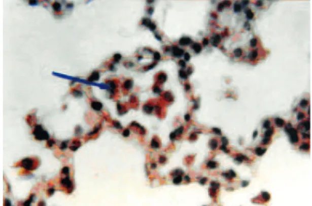
alveolar capillaries and infiltration of RBCs in to the lu-men of the alveolus (Fig 3).
Fig 1: Photomicrograph of the alveoli in the control group. The cells had a normal appearance and the lumen of the alveoli was clear (×400, H&E staining).
Fig 2: Photomicrograph of an alveolus in the experimen-tal group, the exfoliated cells have been shown with arrow (×400 , H & E staining).
Fig 3: Photomicrograph of an alveolus in the experimen-tal group with congestion of capillaries (thin arrow). RBCs were infiltrated in the lumen of alveolus (big arrow) (×1000, H&E staining).
EM observations showed that the type І pneumocyte of rabbits in the control group is a squamous cell with an elongated nucleus which occupied most of the volume of the cell (Fig 4A) and the type ІІ pneumocyte was dome-shaped with a round to oval nucleus and lamellar body (Fig 4B).
EM studies also revealed that 10% of the type І & ІІ Amiodarone Morphological Changes in Rabbit Pneumocytes
Mehraein and Shams
pneumocytes in the experimental group contained in-clusion bodies with a dense structure (Fig 5), while 5% of type ІІ pneumocytes had no inclusions but contained vacuoles (Fig 6).
Fig 5: Electron micrograph of type I pneumocyte (big ar-row) and type II pneumocyte (arrow head) with electron dense inclusions (arrow) (×3000).
Fig 6: Electron micrograph of a type ІІ pneumocyte in the experimental group. The vacuoles are seen inside the cell (arrows) (×6000).
Morphometric studies showed that nuclear dimensions in the control and experimental groups were different (table1) and the differences were statistically significant (p<0.01) .
Table 1: Mean diameter and axial ratio of nucleus in the experimental and control groups expressed as means and standard deviations.
Experimental group n = 40
Control group n = 40 Variables
3.44 ± 0.84* 7.25 ± 2.75
Mean diameter of nucleus
1.21 ± 0.30* 1.20 ± 0.28
Axial ratio of nucleus
n=number of nuclei * Significant p<0.01
Discussion
Pulmonary drug toxicity is increasingly a cause of acute and chronic lung disease. Numerous agents including cytotoxic and noncytotoxic drugs have the potential to cause pulmonary toxicity. The clinical and radiological manifestations of these drugs generally reflect the un-derlying pathological processes including bronchiolitis, pulmonary hemorrhage and diffuse alveolar damage (16, 17). Among these drugs, Amiodarone is a potent drug recommended for the management of cardiac arrhyth-mias. It is known that pulmonary toxicity is the most significant life threatening side effect associated with Amiodarone use (5, 18). It has been shown that Amio-darone is directly cytotoxic to bovine endothelial cells and interstitial fibroblasts (19). The susceptibility of pneumocytes to Amiodarone induced cytotoxixity has been reported by some investigators (6). A variety of re-cent studies suggest a critical role for alveolar cell apop-tosis and lung fibrosis (12). In this study acute pathologi-cal changes were observed, including alveolar capillary congestion and infiltration of RBCs in to the lumen of alveoli, which is a sign of inflammation. Card in 2003 showed that Amiodarone induces acute pulmonary in-flammation following intratracheal administration after 24 hours in a hamster model (11). In other research, light microscopy of the lung tissue in Amiodarone treated rats showed pathological changes after three weeks (14). The pathological changes present clinically as coughing and
Fig 4: A. Electron micrograph of typeII pneumocyte,B. Electron micrograph of type I pneumocyte with lamellar body (arrow) in control group (×6000).
low fever. Another finding in this research was appear-ance of inclusion bodies inside the pneumocytes. The ac-cumulation of inclusions in the cytoplasm is thought to be due decreased degradation of phospholipids because Amiodarone is a powerful inhibitor of lysosomal phos-pholipases which appears to increase with increased du-ration of exposure of the cells to Amiodarone (21). These inclusions have been detected in other tissues exposed to Amiodarone, for example Pitsiavas in 1997 found that Amiodarone induced specific ultrastructural changes of thyroid cytotoxicity in rats. The specific changes includ-ed evidence of inclusions and mentioninclud-ed that Amiodar-one is directly cytotoxic to the thyroid (22). There has also been debate in the past as to whether the inclusion bodies seen here in Amiodarone treated animals only re-flect the ongoing cytotoxic process or whether these bod-ies are directly toxic to the cell in their own right. Amio-darone is an amphiphilic drug and the administration of amphiphilic drugs can cause the formation of inclusion bodies in many cell types. These changes are the result of an interaction between the drugs and phospholipids (23). We observed only vacuoles in 5% of type ІІ pneumocytes and this is the induction role of Amiodarone to these cells (I don’t really understand this clause). The highest preva-lence of reactive type ІІ pneumocytes (RPІІ) was noted in patients with systemic inflammatory response and alveo-lar haemorrhage, in addition, RPІІ tended to occur more frequently in ventilator associated drug induced pulmo-nary disorders and they were associated mainly with con-ditions invloving acute lung injury (24). The changes in nuclear dimensions show cell injury due to the direct cel-lular damage caused by Amiodarone (7).
Conclusion
In conclusion, these results suggest that the effective-ness of Amiodarone in long term treatment of the heart failure patients is limited because of the development of pulmonary toxicity. Further studies are necessary to show the precise molecular basis of Amiodarone cytotoxicity which can help in the choice of substitute new drugs with less side effects.
Acknowledgments
This research was supported by the research center of Iran University of Medical Sciences. There is no conflict of interest in this article.
References
1. Plomp TA. Analytical profiles of Amiodarone in analyti-cal profiles of drug substances. Florey K (ed). San Diego. Academic Press Inc; 1991.
2. Underwood RD, Sra J, Akhtar. Evaluation and treat-ment strategies in patients at high risk of sudden death post myocardial infarction. Clin Cardiol. 1994; 20:743-758. 3. Mason JW. Amiodarone. N Engl J Med. 1987; 316: 455-488. 4. Martin WJ, Rosenow EC. Amiodarone pulmonary tox-icity: recognition and pathogenesis part1. Chest. 1988: 1067-1075.
5. Pitcher WD. Amiodarone induced pulmonary toxicity. Am J Med Sci.1992; 303: 206-212.
6. Wilson BD, Lipmann ML. Pulmonary accumulation of Amiodarone and N-desethylamiodarone: relationship to the development of pulmonary toxicity. Rev Respir Dis. 1990; 141: 1553-1558.
7. Reasor MJ, Kocew S. An evaluation of possible mecha-nisms underlying Amiodarone induced pulmonary toxicity. Proc Soc Exp Biol Med. 1996; 212: 297-304.
8. Juliah DG, Camm AJ, Frangin G, Janse MJ, Schwartz PJ, Simson P. Randomised trial of effect of Amiodarone on mortality in patients with left ventricular dysfunction after recent myocardial infarction: EMIAT. Lancet; 1997; 349: 667-674.
9. Rossi SE, Erasmus J, Page Mcadams H, Sporn T, Good-man PH. Pulmonary drug toxicity: Radiologic and pathologic manifestations. Radiographics. 2000; 20: 1245-1259. 10. Taylor MD, Roberts JR, Hubbs AF, Reasor MJ, An-tonini JM. Quantitative image analysis of drug induced fibrosis using laser scanning confocal microscopy. Toxicol Sci. 2002; 67: 295-302.
11. Card JW, Racz WJ, Brien JF, Margolin SB, Massey TE. Differential effects of Pirfenidone on acute pulmonary injury and ensuing fibrosis in the hamster model of Amio-darone induced pulmonary toxicity. Toxicol Sci. 2003; 75: 169-180.
12. Uhal BD, Wang r, Laukka J, Zhuang J, Soledad-Con-rad V, Filippatos G. Inhibition of Amiodarone induced lung fibrosis but not alveolitis by angiotensin system antago-nists. Basic Clin Pharmacol Toxicol. 2003; 92(2): 81-87. 13. Jarand J, Minion J, Lee A. Amiodaronoma;One of mul-tiple form of Amiodarone pulmonary toxicity. Chest. 2005; 128(4): 446s.
14. Vereckei A, Blazovics A, Gyorgy I, Feher E, Toth M, Szenasi G, Zsinka A, Foldiak G, Feher J. The role of free radicals in the pathogenesis of Amiodarone toxicity. J Car-dovasc Electrophysiol. 1993; 4: 161-177.
15. Inuwa IM, Willams MA. A morphometric study on the endometrium of rat uterus in hypothyroid and thyrox-ine treated hypothyroid rats. UPSALA J Med Sci. 2006; 111(2): 215-226.
16. Wilson BD, Clarkson CE, Lipmann ML. Amiodarone-induced pulmonary inflammation. Am Rev Respir Dis. 1991; 143: 1110-1114.
17. Cooper JA. Drug induced lung disease. Adv Intern Med 1997; 42: 231-268.
18. Marchlinski JL, Gansler TS, Waxymann HL, Joseph-son ME. Amiodarone pulmonary toxicity. Ann Inter Med.1982; 97: 939-945.
19. Martin WJ ІІ, Howard DM. Amiodarone induced lung toxicity. In vitro evidence for the direct toxicity of the lung. Am J Pathol. 1983; 120: 344-350.
20. Kennedy JI. Clinical aspect of Amiodarone pulmonary toxicity. Clin Chest Med 1990; 11: 119-129.
21. Bartussio A, Marzini S, Agostini M, Alberti A, Cimenti C, Bruttomesso D, et al. Amiodarone inhibits lung degra-dation of SPA and perturbs the distribution of lysosomal enzymes. AM J Physiol Lung Cell Mol Physiol. 2001; 281: l1189-l1199.
22. Pitsiavas V, Smerdely P, Li M, Boyages S. Amiodar-one induces a different pattern of ultrastructural changes in the thyroid to iodine excess alone in both the BB/W rat and the Wistar rat. Eur J Endocrinol. 1997; 137: 89-98. 23. Hruban Z. Pulmonary and generalized lysosomal stor-age induced by amphiphilic drugs. Environ Health Per-spect. 1984; 33: 53-76.
24. Linsson K, Jacob J , Poletti V, Van Mook W, Conelis-sen E, Dent M. Reactive type ІІ pneumocytes in broncho-alveolar lavage fluid. Acta Cytol. 2004; 48: 497-504.
Amiodarone Morphological Changes in Rabbit Pneumocytes