NICHOLAS JACOB WALKER
CYTOGENETIC AND MOLECULAR CHARACTERIZATION OF TWO POPULATIONS OF MINNOWS ASTYANAX GITON EIGENMANN 1908 AND ASTYANAX AFF.FASCIATUS (CHARACIFORMES: CHARACIDAE)
IN THE SÃO BARTOLOMEU CREEK, MINAS GERAIS STATE, BRAZIL
Dissertação apresentada à Universidade Federal de Viçosa, como parte das exigências do Programa de Pós-Graduação em Biologia Animal, para obtenção do título de Magister Scientiae.
VIÇOSA
Ficha catalográfica preparada pela Seção de Catalogação e Classificação da Biblioteca Central da UFV
T
Walker, Nicholas Jacob, 1982-
W182c Cytogenetic and molecular characterization of two
2013 populations of minnows Astyanax giton Eigenmann 1908 and Astyanax aff. fasciatus (Characiformes: Characidae) in the São Bartolomeu Creek, Minas Gerais State, Brazil / Nicholas Jacob Walker. – Viçosa, MG, 2013.
viii, 57f. : il. (algumas color.) ; 29cm.
Orientador: Jorge Abdala Dergam dos Santos.
Dissertação (mestrado) - Universidade Federal de Viçosa. Referências bibliográficas: f. 51-57.
1. Characidae. 2. Polimorfismo (Genética). 3. Evolução (Biologia). 4. Vertebrado. 5. Peixe. 6. Citogenética. 7. Biologia molecular. 8. Biodiversidade. 9. Morfologia. I. Universidade Federal de Viçosa. Departamento de Biologia Animal.
Programa de Pós-Graduação em Biologia Animal. II. Título.
NICHOLAS JACOB WALKER
CYTOGENETIC AND MOLECULAR CHARACTERIZATION OF TWO POPULATIONS OF MINNOWS ASTYANAX GITON EIGENMANN 1908 AND ASTYANAX AFF.FASCIATUS (CHARACIFORMES: CHARACIDAE)
IN THE SÃO BARTOLOMEU CREEK, MINAS GERAIS STATE, BRAZIL
Dissertação apresentada à Universidade Federal de Viçosa, como parte das exigências do Programa de Pós-Graduação em Biologia Animal, para obtenção do título de Magister Scientiae.
APROVADA: 7 de março de 2013.
______________________________ Prof. Rubens Pazza
_____________________________ Prof. José Cola Zanuncio
________________________________ Prof. Jorge Abdala Dergam dos Santos
ACKNOWLEDGMENTS
Jorge Dergam for invaluable help with cytogenetics, collection efforts, morphology, analyzing the results and writing this manuscript.
All of the students at the Beagle for help in catching and processing fish and cytogenetic techniques.
José Cola Zanuncio for reviewing earlier drafts of this manuscript.
Natalia Travenzoli, Priscilla Caroline and Udson Santos for help with extracting and purifying DNA and mtDNA analysis.
Mariana Campanha Félix for arranging karyotypes.
Professor L.A.C. Bertaco for identifying Astyanax giton by morphological characters.
LIST OF CONTENTS
LIST OF CONTENTS...iv
LIST OF FIGURES...v
LIST OF TABLES...vii
RESUMO... viii
ABSTRACT...ix
LITERATURE REVIEW... 1
1. INTRODUCTION... 16
2. MATERIALS AND METHODS... 23
2.1 COLLECTION AREA... 23
2.2 MORPHOLOGICAL PROTOCOLS... 24
2.3 CYTOGENETIC PROTOCOL... 25
2.4 MOLECULAR DNA... 26
3. RESULTS... 28
3.1 MORPHOLOGY... 28
3.1.1 PRINCIPAL COMPONENT ANALYSIS... 30
3.2 CYTOGENETICS... 30
3.3 B CHROMOSOMES... 36
3.4 MOLECULAR DNA... 38
4. DISCUSSION... 39
4.1 MORPHOLOGY... 41
4.2 CYTOGENETICS... 43
4.3 MOLECULAR DNA... 49
5. CONCLUSION... 50
LIST OF FIGURES
Figure 1. Collection areas within the Federal University of Viçosa (UFV) in Viçosa, Minas Gerais State, Brazil. a) Reservoir 1; b) Reservoir 2. Map adapted from Google Earth imagery... 24 Figure 2. Specimens of Astyanax giton (left) and Astyanax aff. fasciatus (right) from the São Bartolomeu Creek, a tributary of the Doce River Basin... 29 Figure 3. Principal component analysis (PCA) for Astyanax giton (blue) and Astyanax aff. fasciatus (red), combining data from both reservoirs. Most important coefficients were length (0.4595), height (-0.56) and aspect ratio (0.5554) for component 1 (y-axis) and length (-0.4028), scales below lateral line (0.4133), scales on lateral line (0.4919) and anal fin rays (0.456)... 30 Figure 4. Karyotype of Astyanax giton from the São Bertolomeu Creek, a tributary of the Doce River Basin. Karyotypic formula: 4m+18sm+22st+6t... 31
Figure 5. NOR sites in Astyanax giton from the São Bartolomeu Creek, a tributary of
the Doce River Basin.The top photo shows one NOR in the short arm of one subtelocentric chromosome in the 14th pair; the bottom photo shows NOR sites in the short arms of submetacentric pair 10 and subtelocentric pair 14. A B chromosome is shown in the box... 32 Figure 6. C-banding in Astyanax giton from the São Bertolomeu Creek, a tributary of the Doce River Basin. Heterochromatin is seen in the centromeres of all chromosomes, in the telomeric regions of the long arms of pairs 1, 4, 8, 18, 21 and 24, and interstitially in the long arms of the 7th pair... 32 Figure 7. Karyotype of Astyanax aff. fasciatus from the São Bartolomeu Creek, a tributary of the Doce River Basin. Karyotypic formula: 10m+20sm+12st+6t... 33 Figure 8. NOR sites in Astyanax aff. fasciatus from the São Bartolomeu Creek, a tributary of the Doce River Basin. In the top photo, NORs are visible in the short arms of three submetacentric chromosomes in pairs 6, 11 and 13, in the bottom photo they are visible in the long arms of one subtelocentric chromosome (pair 17) and one telocentric chromosome (pair 24)... 34 Figure 9. a) C-Banding in Astyanax aff. fasciatus from the São Bartolomeu Creek, a tributary of the Doce River Basin. Heterochromatin visible in nearly all centromeres, in the distal portions of the long arms of the first metacentric pair, four submetacentric pairs (plus one in pair 14), two subtelocentric pairs and all three telocentric pairs. Interstitial markings visible in the long arms of one subtelocentric chromosome of the 20th pair. b) Flourescence in situ hybridization (FISH): 28S probes are shown in red and visible in the short arms of two subtelocentric/telocentric pairs and in the centromeric region of one metacentric pair. 5S probes are shown in green, visible in the short arms of one telocentric pair and the centromeres of one metacentric pair... 35
Figure 10. B chromosomes in Astyanax giton.Top two karyotypes show micro B
LIST OF TABLES
RESUMO
WALKER, Nicholas Jacob., M.Sc., Universidade Federal de Viçosa, Março de 2013. Citogenética e caracterização molecular de duas populações de peixes Astyanax giton Eigenmann 1908 e Astyanax aff. fasciatus (Characiformes: Characidae) na ribeira São Bartolomeu, Minas Gerais, Brasil.Orientador: Jorge Abdala Dergam Dos Santos.
Os peixes neotropicais de água doce são os mais abundantes em termos de espécies, e os peixes da família Characidae têm a maior diversidade entre eles. O gênero Astyanax contém 130 espécies, que se encontram em condição de incertae sedis em termos sistemáticos. Porém, as espécies podem ser diferenciadas com estudos de padrões morfológicos, citogenéticos e moleculares. As populações do gênero Astyanax do ribeirão São Bartolomeu de Minas Gerais, Brasil, nunca foram identificadas. Neste
ABSTRACT
WALKER, Nicholas Jacob., M.Sc., Universidade Federal de Viçosa, March 2013. Cytogenetic and molecular characterization of two populations of minnows Astyanax gitonEigenmann 1908 andAstyanax aff. fasciatus(Characiformes: Characidae) in the São BartolomeuCreek, Minas Gerais State, Brazil. Adviser: Jorge Abdala Dergam Dos Santos.
Neotropical freshwater fish are the most abundant vertebrate species, with Characidae fish representing the greatest diversity among them. The genus Astyanax contains approximately 130 species, all of which are currently considered incertae sedis in systematic terms. Because of vague original descriptions, many local populations are difficult to identify. In these cases, the combined analysis of morphological, cytogenetic and molecular data may provide useful information for determining their taxonomic status. We applied this approach on two populations of Astyanax occurring in the São Bartolomeu Creek of Minas Gerais State, Brazil, a tributary of the Doce River Basin. Morphological characters included the analysis of lateral line scales, teeth, anal fin rays. Cytogenetic techniques included conventional coloration (Giemsa), heterochromatin patterns (C-Banding), nucleolar organizer regions
LITERATURE REVIEW
Few published works exist regarding Astyanax giton. According to Fishbase,
A. giton is found in the Paraíba do Sul River Basin and coastal streams of Espírito Santo and Rio de Janeiro States, Brazil [1]. Recently, A. giton has been found in the Sossego Creek in Viçosa, Minas Gerais State, Brazil [2].
Eigenmann [3] defined A. giton by the following characteristics: Snout more than half the length of the eye; two or three teeth in the front row of the premaxillary; a caudal band, depth 2.5 to 2.6; eye 2.5; anal fin with 23-24 rays; 34 scales on the lateral line with 5 above and four below as well as a diffuse (sometimes absent) vertically elongate humeral spot crossing the third scale of the lateral line. Eigenmann based these characters on two specimens from the Rio Paraíba in Eastern Brazil.
Astyanax giton from the Paraíba do Sul River Basin, Serra da Bocaina, São Paulo State, Brazil presented a diploid number of 2n=50 and a karyotype of 6m+8sm+8st+28t, FN=72 [4]. Both A. giton and A. intermedius from this river showed five chromosome pairs bearing 5SrDNA sites, all telocentrics except for one subtelocentric in A. giton. The occurrence of the same number of 5S rDNA sites in similar locations supports the proximity of these species [4,5].
Populations of A. giton from the Paraitinga River and Jacuí Stream presented the same diploid number and karyotype as those from the Paraíba do Sul River Basin [4,6]. In the Paraitinga River and Jacuí Stream, C-banding revealed heterochromatin in the interstitial regions of chromosomes 4, 5, 6 and 9 as well as in the pericentromeric regions of many chromosomes. Silver nitrate staining in the nucleolar organizer regions (Ag-NOR) from two sites on the Paraitinga River showed multiple NORs, up to six markings, and fluorescent in situ hybridization with the 18S rDNA probe revealed up to ten markings, many of them very reduced [5].
formula of 6m+8sm+4st+32t and heterochromatin in many pericentromeric regions. No GC or AT positive heterochromatin was found in either species and the heterochromatin did not hybridize with the As51 satellite DNA probes, a rarity in fish. These data indicate that A. giton and A. intermedius belong to a different clade than A. scabripinnis and A. parahybae, which is supported by a 5S metacentric marker pair observed in the latter two species but not in the former [4,6,7].
Astyanax fasciatus was described by Cuvier in 1819 with Brazil as its type locality. This species is widely distributed, ranging from the Rio Negro in Patagonia to Mexico and southern Texas [3,8,9]. Different localities have more or less distinct varieties of A. fasciatus. These varieties are often only differentiated statistically; i.e., individuals are impossible to distinguish but a large number of specimens may be distinguished from those of another locality.
Astyanax fasciatus is absent from some rivers where it has probably taken on distinct forms, such as in the Paraíba do Sul River. In this river it is replaced by the similar A. parahybae, which Eigenmann considered a distinct form of A. fasciatus formed by river isolation. This is the result of species isolation in rivers independently emptying into the Atlantic in eastern and southeastern Brazil, Central America and Mexico.
Astyanax fasciatus is characterized morphologically by a depth of caudal peduncle more than half the length of the head. It usually has a single tooth in the maxillary. The depth is 2.3 to 3. Scales below the lateral line are in parallel series. The caudal band is simple and median [3]. In the Três Marias River, A. fasciatus present an anal fin with 22 to 32 rays, a lateral line with 37 to 41 scales, with seven or eight above and 5.5 to 6.5 below, a length of 2.6 to 3.2 times its height, and one maxillary tooth [10].
three variants in sympatry, providing further evidence for considering A. fasciatus as a species complex [13]. Chromosomal inversions favor speciation, especially over sites of geographical co-occurrence [9].
Diverse cytotypes, including 2n=45, 2n=46, 2n=47, 2n=48 and 2n=50, have been found in sympatry [15]. In a study of A. fasciatus in the Água da Madalena stream, Botucatu, São Paulo State, Brazil, three karyomorphs (2n=46, 2n=48 and 2n=50) were found in sympatry. Based on the absence of intermediate chromosome numbers, the restriction of B chromosomes to the 2n=46 karyomorph and the absence of heteromorphism for the 18S rDNA sites, these karyomorphs are unlikely to hybridize and are considered three separate species [15]. Diploid numbers for A. fasciatus are highly variable across streams and basins.
Astyanax fasciatus has diploid numbers of 2n=45, 46, 47 or 48, sometimes in sympatry, in the Rio Grande do Sul Basin [13,16]. However, populations of this fish in the Upper Tibagi River may present karyotypes of 6m+18sm+14s+10t (2n=48) or 8+18sm+14st+10t (2n=50), as well as heterochromative variations within the same karyotype [14]. Cytotypes without gene flow can be considered distinct taxonomic units, even in sympatry.
The 2n=46 cytotype is present in other river systems as well, including the Araguari River in Uberlândia, Minas Gerais State, Brazil. Here, specimens of
A. fasciatus presented a karyotype of 14m+16sm+10st+6t (FN=86) with Ag-NORs in the terminal region of the short arm in the first submetacentric chromosome pair and in the short arms of the third metacentric pair [18].
Populations of A. fasciatus of the Tibagi River showed variable Ag-NOR sites, with one cytotype having 2-3 and the other 1-3. The Ag-NOR sites show evidence of transposition, fission, deletion, translocation and inactivation, and the presence of multiple NORs could indicate a plesiomorphy within this genus. Differences in the karyotypes, constitutional heterogeneity, and 18S/5S sites do not seem to represent fitness or selective adaptation in Astyanax [19].
histone genes located in the pericentromeric regions of two chromosomes in A. fasciatus were identical to those in A. altiparanae and A. bockmanni, indicating these are a conserved feature of this genus. Astyanax fasciatus also presented clusters of 5S rDNA on one telocentric pair, a trait also found in A. bockmanni
but not A. altiparanae. The 5S and H1 genes co-localized on chromosome 3 and 5S hybridization revealed clusters in the pericentromeric regions of chromosome 23 [11].
Populations of Astyanax fasciatus in the Upper Paraná, Araguaia-Tocantins, Contas and São Francisco basins present multiple karyotypes in sympatry, reviewed in [15]. In the Água da Madalana Stream, Botucatu, São Paulo Brazil, a population of A. fasciatus included a 2n=46 cytotype (FN=84, 5m+8sm+6st+4a), a 2n=48 cytotype (FN=86, 5m+8sm+6st+4a) and a 2n=50 cytotype (FN=84, 5m+8sm+4st+8a). All contain similar constitutive heterochromatin and occur in sympatry, with no differences between males and females.
The cytotypes may be differentiated by morphology using canonical variate analysis, indicating the variants may be the result of gene flow or recent isolation. Karyotype morphology of the 2n=48 cytotype was different at Furnas 2 of the Vila Velha State Park in Paraná State, which is separated from other bodies of water and prohibits gene flow. The 2n=48 and 2n=50 cytotypes contain heterochromatin mainly distributed as very conspicuous blocks in the telomeric regions of the long arms of the telocentric chromosomes. One 2n=49 individual was found, with seven metacentric chromosomes and heterochromatin equal to the 48 and 50 cytotypes -- possibly a rare hybrid. A distinct 2n=50 cytotype in this region presented only a few chromosomes with heterochromatin, almost always located interstitially [14].
two submetacentric chromosomes, colocalized with 5S in the two metacentric and telocentric chromosomes [7].
In the Mogi-Guaçu river, the headwaters (at Ouro-Fino) contain A. fasciatus with diploid numbers of 2n=48 and 2n=47; in the middle region (Cachoeira de Emas, Pirassununga) they have diploid numbers of 2n=48, 2n=46, 2n=45, 2n=47 and 2n=46 but with different chromosome morphology; and near the confluence of the Mogi-Guaçu with the Pardo River (Barrisha), 2n=46 and 2n=48 [13]. NOR, 18S and 5S markers were conserved within all cytotypes. The 2n=45 cytotype is likely the result of centric fusion among telocentric chromosomes as no 2n=44 cytotype has been found. At Emas Falls, in the middle of the Mogi-Guaçu River (where fish cannot go upstream), most A. fasciatus are 2n=46 with few 2n=48 and chromosomal variants [9].
This scenario is reversed at localities upstream (Ouro Fino, all 2n=48) and downstream (Barrinha, most 2n=48). Some specimens from the Tietê River may have invaded the Mogi-Guaçu at a certain point between Emas falls and Barrinha, possibly through floods that temporarily connected tributaries from each main river, or by human activities. An occasional hybrid of 2n=48 and 2n=50 is found in the Tibagi River, a tributary of Paranapanema [9].
NORs are variable between cytotypes in the Mogi-Guaçu, ranging from two (most common) to eight with FISH 18S markers located in the NORs. Other fluorescent markings were smaller, always telomeric and located in subtelocentric and telocentric chromosomes, although more difficult to see and not detectable in all specimens. All cytotypes have a ribosomal site on the subterminal region of the short arm of one submetacentric pair and 5S markings in the pericentromeric region of the short arms of one telocentric pair [13]. These markings indicate the proximity of the cytotypes and a recent divergence[13]. The 2n=46 cytotype has six variations, likely the result of random interbreeding between cytotypes, i.e. karyotype plasticity. Although differences in karyotype may cause reproductive isolation, this is not the case in this river [21].
possible that Cachoeira de Emas contains populations that converged during migrations. 2n=46 could have come from the Paraná River, through either the Tietê or Grande rivers, or small floods in areas where the Tietê and Mogi-Guaçu rivers are close[13].
If 2n=46 is an invading karyotype, the ability to interbreed may be helping it succeed [13]. Shared features of the karyotypes suggest recent divergence, suggesting two populations or two species [19]. Chromosomal speciation may cause reproductive isolation because hybrids may be infertile, although that does not appear to be the case in this river[13]. The 2n=46 invader is likely able to interbreed with 2n=48, as indicated by the frequency and location of 2n=46 cytotypes in nearby Paraná Basin collection sites and constitutive heterochromatin in terminal regions on long arms of variouschromosomes and in the terminal regions of the short arms of submetacentric chromosomes in both cytotypes, with these regions being GC-rich and containing NORs [16].
Banding patterns differ among Astyanax. C-banding is highly variable, with polymorphisms occurring even within populations. GC-rich heterochromatic regions are often associated with NORs [9]. Fifty nine percent AT satellite DNA was identified in a portion of the A. scabripinnis genome [22] containing monomeric units of 51 bp, known as As51 [23]. This sequence has also been identified in A. fasciatus, A. janeiroensis and A. paranae. Although satellites are likely targets for breaks and fusions, As51 has not been associated with modifications to the diploid number in A. fasciatus or A. scabripinnis [23].
Several species of Astyanax including A. altiparanae, A. lacustris, A. fasciatus, A. schubarti and A. scabripinnis show conserved localization of 5S/28S markers between species, indicating a close phylogenetic relationship [7], with 5S visible in more than one pair [24]. NORs are almost always fewer than regions detected by FISH. These 5S locations are absent in A. giton and A. intermedius, suggesting polyphyly or divergence within this genus [13]. R-banding patterns have been observed in four species (A. altiparanae, A. fasciatus, A. schubarti and A. scabripinnis) in one large submetacentric chromosome and one small pair of metacentric chromosomes, colocalized with C-band negative late replicating regions [7].
In Astyanax, heterochromatin may be balanced between AT and GC content [6] or 59% AT [22]. Astyanax fasciatus and A. parahybae have As51 DNA, while A. giton and A. intermedius do not. Both the 2n=46 and 2n=58 forms of A. fasciatus have 8 pairs of As51, which is similar to that of A. scabripinnis
from the Piracicaba River [12,16]. It is possible that As51 was the ancestral condition and that the pairs have increased with time as a result of Robertsonian and non-Robertsonian rearrangements that could duplicate or disperse satellite DNA. Most As51 sites are homologous, but slight variations exist. Variations between karyotypes could be the result of intrachromosomal rearrangements involving telocentric chromosomes with terminal As51 sites on the long arms, which were absent in the 2n=45 forms [16].
B-chromosomes are dispensable supernumerary chromosomes characterized by their lack of homology with regular ones [20] and do not obey Mendelian laws of inheritance [25]. B chromosomes are mostly composed of repetitive DNA sequences, especially ribosomal DNA, satellite DNA and in rare cases, protein coding genes [11]. It is estimated B chromosomes are present in 10-15% of eukaryotes [25] but this is impossible to determine with certainty as by definition they are not found in all individuals and are likely to be missed if the karyotype is based on only one individual [26].
B chromsomes can vary substantially in their size, frequency and other characteristics. They may be macro, intermediate or micro and most are heterochromatic. Many Bs contain rRNA genes detectable by both NOR and FISH [25]. B chromosomes may originate in one of two ways. They can be intraspecific, from the host genome (most common) or interspecific, from another species, .e.g. the result of hybridization [27]. As B chromosomes do not synapse or recombine, they evolve independently of the A set. Therefore, regardless of origin, the evolutionary pathways of B chromosomes are the same in either case and B chromosome morphology is not necessarily reflective of origin.
There are several ways to test B chromosome origins. Banding patterns and repetitive sequences may be compared between the B and A sets. If no matches are found, the B chromosome can be compared to the a set of related species. Chromosomal fragments may also be visible in interspecific crosses, and phylogenetic analyses may be done on the transposable elements of both the B and A sets [27].
B chromosomes in Astyanax can vary within species (by size or morphology). They may also vary by sex or altitude, meaning may be an adaptive trait [9]. There is no evidence on the precise role of B chromosomes on individual fitness in Astyanax[9]. Species of Astyanax, such as A. fasciatus, A. scabripinnis and A. schubarti present B-chromosomes [28,29]. The origin of supernumerary chromosomes is unknown, but it is accepted that they result from amplification of other parts of the genome [30] and A-chromosomes follow their own evolutionary pathway (Mestriner, 2000). The metacentric macro B chromosome is the most common in Astyanax[11].
In A. fasciatus, B chromosomes are reported for the 2n=46 karyomorph in the São Francisco Basin [15,31,32] and a 2n=49 karyomorph has been observed in the Tibagi River, in the Upper Paraná River Basin, Ponta Grossa, Paraná, Brazil [14].
There are two groups of B chromosomes in Astyanax. The first is composed of species with large metacentric Bs, including A. fasciatus, A. scabripinnis[32-36], A. eigenmanniorum, A. schubarti, and A. bockmanni[32], suggesting a common origin. This hypothesis could be tested by comparing DNA sequences of B chromosomes. The second is in the A. scabripinnis group, where the presence of distinct types of supernumerary chromosomes has been commonly reported in several populations ranging from large metacentric/submetacentric elements to small metacentric or even microchromosomes [9].
scabripinnis by finding that B chromosomes resulted from tandem duplications of heterochromatin segments in acrocentric pair 24, as well as sharing some repetitive DNA sequences [36,37]. This is based on the presence of an interstitial heterochromatic band in the long arm of this pair, which is believed to cause interstitial As51 sites at symmetric distances from the centromere in both arms of the B chromosome. B chromosomes may also vary by gender, with individual females or some populations containing an additional B [22]. A large metacentric B chromosome was found in A. scabripinnis, A. eigenmanniorum, A. schubarti and A. fasciatus[32,37]. This suggests that this B originated prior to species differentiation, but molecular analysis is needed to confirm. B chromosomes have not been documented in A. giton. No phenotypic effects resulting from B chromosomes have been observed in Astyanax [37].
As in the second hypothesis, rapid heterochromatization may occur by genetic imprinting to deactivate B chromosomes, which explains the presence of heterochromatin in many Bs. Sometimes the B forms anew in each generation due to non-disjunction [27]. Rapid heterochromatization of B chromosomes often occurs (as seen by C-Banding) and may be necessary for stability and positive selection. B chromosomes may also be a precursor to sex chromosomes (especially the y chromosomes, which also move to opposite poles during spermatogenesis) which are also high in repeats [25,27]. B chromosomes may originate from either autosomes or sex chromosomes, although the latter are not found in any populations of A. fasciatus or A. giton.
B chromosomes often contain rDNA to encode ribosomal RNAs, typically visualized as NORs (secondary constrictions). NORs may also be suspect to chromosome breakage, which could create Bs that could then lose rDNA segments, affecting their size. Bs may also accumulate fragments from various sources [27].
more DNA to replicate, meaning a longer cell cycle and larger cells. This slows development in some species. Bs may be more common in species with smaller genomes and fewer chromosomes [25].
B tolerance is dependent on the effects of supernumerary chromosomes. If fitness decreases with Bs, complete intolerance results. If fitness is unaffected with some B chromosomes, but is reduced when more are present, there will be incomplete tolerance (the most common case). If fitness is independent of Bs, the A set will be tolerant. Overtolerance occurs when the B increases host fitness. This last case is most likely when the B arises from hybridization as it includes new DNA. As the effects of B chromosomes are usually negative, they may be more common in favorable environments with less selective pressure [25].
Methylation has been proposed for deactivating sex chromosomes and may also function with late-replicating B chromosomes to make them genetically inert [27]. B chromosomes that are transcriptionally active with no apparent phenotypic effects could have their transcripts nullified by transporon insertion.
Meiosis is always asymmetric, especially in females with polar bodies and oocytes [25]. Since Bs are not paired during meiosis, they can go to their preferred meiotic pole, possibly increasing their transmission rate. Bs may also result from nondisjunction during meiosis anaphase. Bs are often unstable during meiosis, with a transmission rate of less than 0.5. A rate of greater than 0.5 indicates accumulation, or drive. B drive is a disregard for Mendelian transmission (also known as an unfair transmission pattern) and is necessary for their survival. B chromosomes must have accumulation methods, or they would only make sense as being beneficial; they likely begin by being selfish even if they become inactive later [25]. Due to meiosis, it is possible for the B to always originate from one sex. They can occur during female meiosis by migrating from one oocyte to another, as well as in males by avoiding heterochromatin [27].
may evolve another form, continuing the cycle. For example, in species of mealy bugs the B tries to decondense and segregate with the sperm, but drive suppressor genes in the A set prevent this from occurring. The selection to eliminate B chromosomes, however, is usually weak. Genes for their removal are only helpful to individuals with them, and in most cases the B has very small effects. On the other hand, the B is selecting for its very survival [25].
Bs are selected at the level of the chromosome and impact higher levels of organization [25]. They can affect gene expression of the A set, NOR expression and the level of recombination in the A set (chiasma effect). The latter is hypothesized to occur for one of four reasons:
1) The adaptation hypothesis: B chromosomes enable higher levels of genetic variation, leading to faster population evolution. This is criticized because it suggests parasites are selected for positive host effects.
2) Passive effects: Chiasma effects are the result of extra DNA, but this cannot explain contradictory effects.
3) The increased chiasma frequency is the result of parasitic Bs, because some recombinant progeny will be resistant to B chromosomes and individuals with Bs will have higher rates of recombination. (This is a variation of Matt Ridley’s Red Queen hypothesis [38], that recombination increases when conditions are poor.)
4) Changes in chiasma frequency serve the selfish purposes of the B chromosome, i.e. Bs pair with Bs to reduce meiotic loss with the side effect of more B chiasma, or the formation of bivalent Bs is blocked, or Chiasma results from restricted A-B pairing.
numbers of Bs are present in different ratios, one may have more drive than the other [25].
Many B chromosomes are stable, in which the drive mechanism and selection are balanced. In these cases, the B becomes more dependent on drift and population size, unless it can acquire an even number of Bs and regular meiotic behavior to incorporate itself into the A set [25]. B equilibrium (stability) results from two opposing forces: first, the accumulation of Bs by either the selfish chromosome theory or a high transmission ratio; and a change in individual fitness. As the survival of B chromosomes is ultimately linked to their hosts, they may become less harmful over time (which would reduce the probability of phenotypic effects) [25]. Newly formed Bs would therefore be expected to be the most harmful.
B chromosomes have three options -- they can remain as a parasite, they can acquire a function and integrate themselves into the host (the selfish gene, working horizontally; although this is rare and only possible when already common) or go extinct. They may change from parasitic to neutral and back many times. The A set may completely suppress drive, or do nothing. But the B has a secret weapon -- because it is genetically indispensible, it can support a high mutation rate without serious consequences, which explains the large number of B variants in many species. Even a neutralized B can still last more than 10,000 generations, plenty of time to regain drive/parasitism [25]. According to Muller's ratchet, deleterious mutations build up in asexual species, so sexual reproduction is needed to eliminate them. As B chromosomes are not subject to recombination, they may also be subject to these mutations [27].
In populations of A. scabripinnis, populations at the highest altitudes were the most likely to have large metacentric B chromosomes [24], but they had fewer NORs than low altitude populations [37].
C-Banding has shown that B chromosomes can be totally or partially heterochromatic [28]. Some Astyanax populations show faint paracentromeric C-bands in all chromosomes, being more conspicuous distally on the long arms of telocentric A chromosomes [22]. Additionally, C-band locations may coincide with those of NOR, and interstitial C-bands are sometimes present on the arms of B chromosomes.
Astyanax fasciatus from the Campo Novo River, Paraná Basin, São Paulo State, Brazil (22º23’8.35”S, 49º0’55.63”W) presented a diploid number of 2n=46, karyotype of 10m+14st+20st+2t and FN=90 [11]. These authors suggest that the 2n=46 A. fasciatus are probably the result of fusions from the ancestral state of 2n=50. Two out of 25 individuals had medium-sized telocentric B chromosomes of similar size and morphology to pair 23, but it is unlikely that they originated from the A set because they do not have similar rDNA or hisDNA.
Astyanaxbimaculatus, A. fasciatus, A. schubarti, and A. scabripinnis
subjected to R-Banding showed one large submetacentric and two small submetacentric markers, indicating a common origin among them [7,8]. R- and G-banding of a population of A. scabripinnis paranae (now known as A. paranae, separate from the A. scabripinnis complex [41]) from Cascatinha stream showed a pattern of longitudinal differentiation in B chromosomes [29].
Silver nitrate (Ag-NOR banding) can identify the 45S rDNA located in the NORs, however the 5S rDNA sites require more specific techniques, e.g. fluorescent in situ hybridization (FISH) [24]. Intrapopulational NOR site variation may represent genomic variations in populations with stable karyotypes [24].
Astyanax spp. are found in two reservoirs of the São Bartolomeu Creek at the Federal University of Viçosa in Minas Gerais State, Brazil, and have never been formally identified. We hypothesize that they are A. giton and A. fasciatus,
show cytogenetic variance across basins and each may constitute a separate species complex. This hypothesis may be tested by cytogenetic characters and morphology.
Dam construction, flow regulation, mining, deforestation and industrial and agricultural pollution increasingly affect major river basins by modifying fish habitats and altering fish life-cycles. However, compared to mammals, birds and amphibians, relatively few South American freshwater fish have been scientifically assessed as endangered species by IUCN or other criteria, even [42].
1. INTRODUCTION
Neotropical freshwater fish comprise approximately 6,000 of the 13,000 species worldwide, in 25 orders and 71 families [9]. These fishes represent the most diversified vertebrate group and are adequate for studies of genetic diversity and evolution [43].
Neotropical fishes present high karyotypic variability, with changes in the diploid number, sex chromosome systems, supernumerary chromosomes, natural triploidy, and diversity of constitutive heterochromatin and nucleolar organizer regions (NORs) [44]. Some families of Neotropical fish are characterized by stable diploid numbers, such as Anostomidae (including the genera Abramites, Anostomus, Laemolyta, Leporellus, Leporinus, Pseudanos,
Rhytiodus and Schizodon), Prochilodontidae, (including Prochilodus and
Semaprochilodus), Parodontidae (including Apareiodon and Parodon), Curimatidae (including Curimata, Curimatella, Curimatopsis and Cyphocharax), Cynodontidae (including Cynodon, Hydrolycus and Rhaphydon), Hemiodontidae
(including Anodus, Argonectes, Bivibranchia and Hemiodus) and Pseudopimelodidae (including Lophiosilurus, Microglanis and
Pseudopimelodus), all of which present 2n=54 (excluding rare instances of B chromosomes) [45].
In other cases, the diploid numbers may be more variable. In the family Erythrinidae, Hoplias lacerdae presents a conservation of the diploid number 2n=50 in the Usina Limoeira (São Jose do Rio Pardo) and CA UNESP (both in São Paulo State), in the Rio São Francisco and Uberlândia (Minas Gerais State) and the Rio Pitinga (Amazonas). This diploid number is also found in H. aff.
Grosso); Goiás Velho (Goiás), Corumba and Miranda (Mato Grosso do Sul), Paramaribo (Suriname) and the Rio Aguapey in Argentina. This last location also includes 2n=42 cytotypes, as do the Doce River, Lake Caríoca (of the Doce River), Lake Patos, Rio Grande, Rio Piranga and Rio Sapucal (all located in Minas Gerais); Rio Juquiá, Rio Jurumirím and Lake Guaporé (São Paulo); Rio Vorá, São Mateus do Sul, Curitiba, Rio Jurumirím, Rio Iguaçu and Rio Tibagi (Paraná) and Araguaiana (Mato Grosso) [45]. In total, seven cytotypes have been found for H. malabaricus [46].
The Characiformes are one of the main orders of Ostariophysan fishes, including approximately 1,900 valid species belonging to over 270 genera, currently arranged in 18 families in Africa, Central America and South America [47,48].It is one of the most numerous freshwater fish fauna in the Neotropical region, second only to catfish [49]. Phylogenetic relationships within Characiformes are not clear [50].
Characidae is the most diverse family and comprises 58% of this order, with approximately 250 South American genera and 1,100 species, [8,37,48,51,52]. It comprises the majority of freshwater fish in Brazil, with 20 subfamilies and approximately 400 species [10,37]. Phylogenetic relationships of Characidae, however, are not known (but see [21]). Genera of this family have variable diploid numbers, with 2n=52 in Bryconamericus, Cheirodon,
Exodon, Inpaichthys, Markiana, Piabina, Triportheus, Charax, Cynopotamus,
Galeocharax, Roeboides, Odontostilbe, Serrapinus and Mimagoniates; 48 in
Bryconops; 50 in Ctenobrycon, Hasemania, Hollandichthys, Moenkhausia,
Nematobrycon, Pselogrammus, Salminus, Aphyocharacinae, Brycon,
Deuterodon and Oligosarcus, the latter two closely related to Astyanax. Diploid numbers of 60 or greater are found in Pristobrycon, Pygocentrus, Serrasalmus
and Metynnis, while lower numbers are found in Phenacogaster (46) or
Paracheirodon, which presents 32, 36, 50 or 52 chromosomes [45].
number of 2n=50 is considered basal for the genus [53] as found for A. bimaculatus, with most Astyanax containing between 2n=46 and 2n=50 chromosomes [7,15] except for A. schubarti which has 2n=36 [7,54].
The genus Astyanax was described in 1854[55] based on samples collected from Baird and Girard in Texas and Mexico. The original description lists the presence of an adipose fin, large scales, dorsal fin above the ventrals a non-serrated abdominal line and a double row of teeth on both the upper and lower jaw, flattened with several conical spines or processes upon their edge, lacking canine and palative teeth. The double row of mandibular teeth is not reported in later descriptions of this genus.
Astyanax was described again by Carl Eigenmann in 1917, with samples from South America[3]. Eigenmann characterized Astyanax as small compressed fishes, more or less elongate, reaching a length of 150 mm and usually much smaller (with the exception being the larger Astyanax maximus). Fishes of this genus have a complete lateral line, pre-maxillary teeth in two series (internal series with four or usually five teeth), a mandible with strong frontal teeth and minute conical teeth on the sides. The crowns of the premaxillary and mandibular are usually ridged and denticulate, with few or no teeth in the maxillary. Maxillary bone is short, with its anterior edge forming a more or less continuous curve from end to end. The lateral line rarely has more than 39 scales although it can be as high as 41 in some specimens of A. fasciatus, A. taeniatus and A. scabripinnis. The lateral line curves slightly in the front, and runs along the fourth longitudinal series of scales, above the ventral fin or higher. The scales are of normal size and cover the base of the rays of the tail, although the rest of the tail is naked. Regular and complete series of scales are found between the occipital process and the dorsal. The gill rakers are setiform and no predorsal spine is present. Body length is about three times body height [3,10].
the reason Astyanax is one of the most speciose genera in the Neotropics, including several species complexes [9]. Astyanax is regarded as incertae sedis
because of its likely polyphyletic origin [9] and considered a recent group with chromosome polymorphism in several species [21]. Astyanax has extensive morphological diversification and highly complex taxonomy with a number of species complexes [15,37,52].
Although doubts remain on the monophyly of Astyanax[3,4,7,8,56], this genus is an excellent model for all kinds of studies of evolutionary mechanisms [41]. It is one of the best documented Neotropical fish genera from a cytogenetic standpoint, with the species complexes Astyanax scabripinnis, A. fasciatus and
A. altiparanae (the latter is sometimes referred to as A. bimaculatus) being the most studied [41]. Species complexes are characterized by wide cytogenetic variation, mainly regarding distinct diploid numbers. Karyotypes, however, could also differ because of non-Robertsonian rearrangements. Whether or not individual cytotypes are considered distinct species depends on whether or not they can hybridize (karyotype plasticity) [41]. Some examples of karyotypes are as follows:
Astyanax scabripinnis from Rio Agua do Rancho, Paraná present 2n=50,
6m+28sm+16t, 4 NORs, C-Bands and 0-2 micro B chromosomes; A.
bimaculatus from the Rio Paraguai, Mato Grosso do Sul present 2n=50,
6m+26sm+12st+6t, 1 NOR, C-, Q- and Mytramicin Bands and no Bs; A.
altiparanae from Fundo Altenas, Minas Gerais are 2n=50, 8m+28sm+6st+8t, 2
NORs, C-Banding, Chromomycin A3 and no Bs; A. aff. abramis from the Rio
Bento Gomes, Mato Grosso are 2n=50, 22m+10sm+6st+12t, 1 NOR,
C-Banding, Chromomycin A3 and no Bs; A. eigenmanniorum from the Marrecas,
Paraná are 2n=50, 6m+20sm+8st+16t, 5 NORs, C-Bands, Chromomycin A3 and
no Bs; A. schubarti from the Rio Mogi-Guaçu, São Paulo are 2n=36,
12m+16sm+4st+4t, 1 NOR, C-Bands and no Bs; and A. fasciatus from the Rio
São Francisco, Minas Gerais are 2n=46, 6m+24sm+8st+8a, 4 NORs, C-Bands,
Astyanax fasciatus has highly variable karyotype morphology [41], with two standard diploid numbers of 2n=46 and 2n=48 and some 2n=50 specimens with affinities to the A. fasciatus group in the Tibagi River Basin [9]. Several cytotypes may occur in sympatry, as in the Mogi-Guaçu River.The karyotype of
Astyanax giton is also geographically variable although a diploid number of 2n=50 is conserved. A comparison of Astyanax spp. cytotypes across basins is shown (Table 1).
Table 1. Comparison of diploid numbers and cytotypes of Astyanax fasciatus, A. parahybae and A. giton across several basins in Brazil
Species Location
Dipl
oid Bs m
s
m st t
A. fasciatus
Meia Ponte River, Goiânia, GO, Paranáiba
River 46
2 8
1 8 Mogi-Guaçu River, Pirassununga, SP,
Upper Paraná River 46
1 4
2 0
1 0 2 Juquiá River, Registro, SP, Upper Paraná
River 48
1 0
2 4
1 2 2 Mogi-Guaçu River, Upper Paraná River 46 3
2
1 4 Paiol Grande Stream, São Bento do
Sapacuaí, SP, Grande River 48 1 2
2 0
1 2 4
45 1
2 2 0
1 0 3 1
2 2 0
1 0 4 46 1 2 2 0 1 0 4 1
2 1 9
1 0 6 1
2 2 1
1 0 4 47 1 2 2 0 1 0 5 Mogi-Guaçu River, Pirassununga, SP,
Upper Paraná River
48 8 2
2 1 2 6
47 1
2 2 0
1 0 5 Mogi-Guaçu River, Ouro Fino, MG, Upper
Paraná River
48 8 2
2 1 2 6
46 1
2 2 0
1 0 4 Mogi-Guaçu River, Barrinha, SP, Upper
Paraná River
48 8 2
River 2 0 0 Araguari River, Uberlândia, MG,
Paranaíba River 46
1 4
1 6
1 0 6 São Francisco River, MG 46 0-1 1
2 1
6 4 4
Astyanax aff. fasciatus
São Bartolomeu Creek, Viçosa, MG,
Doce River 48 0-2
1 0
2 0
1
2 6
A. parahybae Paraíbuna River, Paraíba do Sul River 48 8 1 8 1 2 1 0 Paraitinga River, Paraíba do Sul River 48 8 1
8 1 2 1 0 A. giton
Paraitinga River, Cunha, SP, Paraíba do
Sul River 50 6 8 8
2 8 Jacuí Stream, Cunha, SP, Paraíba do Sul
River 50 6 8 8
2 8 Sossego Dam, Viçosa, MG, Doce River 50 6 8 2
4 1 2 São Bartolomeu Creek, Viçosa, MG,
Doce River 50 0-1 4
1 8
2
2 6
Brazilian states abbreviated as follows: GO = Goías, MG = Minas Gerais, SP = São Paulo.Table adapted from data reviewed by Pazza & Kavalco [9]. Data for Astyanax giton from Sossego from Aguiar [2], A. fasciatus from São Francisco from (UNESP) [45] and Astyanax aff.fasciatus and A. giton from São Bartolomeu (bold) from this work.
In terms of morphology, Eigenmann [3] points out that "the true A. scabripinnis shades through [A. intermedius] into A. taeniatus.... I think it would be possible to arrange specimens of A. scabripinnis, A. intermedius and A. taeniatus from the Paraíba into series in respect to any one character and get complete integrations." In Astyanax taeniatus, the teeth on the sides of the lower jaw tend to become graduate, a condition leading to the distinguishing character of Deuterodon[3]. Astyanax taeniatus usually has three teeth in the front row of the premaxillary, 19 to 24 rays in the anal fin, two maxillary teeth, 32 to 39 scales on the lateral line with 5 to 7 above and 4 or 5 below. The teeth of A. obscurus, A. taeniatus, A. giton and A. intermedius all share the common trait of gradually decreasing, unlike most other Astyanax (including A. fasciatus) which exhibit an abrupt reduction in size between the anterior and posterior dentary teeth [57].
scabripinnis). karyotypic variation may be caused by structural rearrangements, e.g. pericentric inversions. This is observed in A. giton and A. intermedius, which are mainly differentiated cytogenetically by a paracentric inversion in the chromosomal pair beaing 5S rDNA [9].
Astyanax giton is found in the Paraíba do Sul River Basin and coastal streams of Espírito Santo and Rio de Janeiro States [1] and in the Doce River Basin in Minas Gerais State, Brazil [2]. Astyanax giton is characterized morphologically by two or three teeth in the front row of the premaxillary, an anal fin with 23-24 rays, 34 scales on the lateral line with 5 above and four below as well as a diffuse (sometimes absent) vertically elongate humeral spot crossing the third scale of the lateral line [3].
Populations morphologically similar to Astyanax fasciatus are distributed from the Rio Negro in Patagonia to Mexico and southern Texas [3,8]. Astyanax fasciatus is characterized morphologically by a single maxillary tooth, an anal fin with 22 to 32 rays, a lateral line with 37 to 41 scales, with seven or eight above and 5.5 to 6.5 below, a length of 2.6 to 3.2 times its height, and one maxillary tooth [10]. Astyanax fasciatus is considered a species complex, based on morphological differentiation among basins and karyotypical diversity [11-16]. Diploid numbers for A. fasciatus are highly variable across streams and basins.
Astyanax fasciatus is absent from the Paraíba do Sul River in which it is replaced by A. parahybae, which may be a distinct form of A. fasciatus resulting from rivers independently emptying into the Atlantic in eastern and southeastern Brazil, Central America and Mexico[3,58].
2. MATERIALS AND METHODS
2.1 COLLECTION AREA
Ninety-one individuals (32 A. giton and 59 A. fasciatus) were captured from two reservoirs of the São Bartolomeu Creek at the Federal University of Viçosa, in Viçosa, Minas Gerais State, Brazil from October 2011 to July 2012. The São Bartolomeu creek is part of the Doce River Basin, in southeast Brazil, (18º 45’ and 21º 15’ S and 39º55’and 43º 45’W). Fish were captured with rods from two reservoirs, one near the Quatro Pilastras (20°45'32.81"S 45°52'22.54"W) hereafter referred to as reservoir 1, and a second upstream dam (20°46'06.31"S 42°52'17.27"W), hereafter referred to as reservoir 2 (Figure 1). These reservoirs were selected because they are somewhat isolated and have never been studied cytogenetically. They are located within the Atlantic Rainforest, an area considered a biodiversity hotspot, with only 8% of original forest remaining [60].
Although fish can swim from Reservoir 2 to Reservoir 1 via a waterfall, they cannot go in the opposite direction, suggesting the possibility of differences in species composition between these reservoirs. Fifty fish (23 A. giton and 27
A. fasciatus) were captured from Reservoir 1 and 41 fish (9 A. giton and 32 A. fasciatus) from Reservoir 2. One male A. giton was captured from Reservoir 1; the other 31 were female. Thirteen male A. fasciatus were captured from Reservoir 1 along with 14 females, and six males and 26 female A. fasciatus
Figure 1. Collection areas within the Federal University of Viçosa (UFV) in Viçosa, Minas Gerais State, Brazil. a) Reservoir 1; b) Reservoir 2. Map adapted from Google Earth imagery.
Fish were kept in aquariums for cytogenetic analysis, fixed in 10% formalin for one week, transferred to 70% ethanol, deposited in the ichthyological collection at the João Moojen Zoological Museum for morphological studies or sent to Dr. Vinicius A. Bertaco at the University of Rio Grande Do Sul for taxonomic confirmation.
2.2 MORPHOLOGICAL PROTOCOLS
confirmed by type of gonad. Morphometric data were analyzed [3,10,56] including the length and height of the fish, the aspect ratio (length/height), the number of rays in the anal fin, the number of scales on, above and below the lateral line, and the number of teeth in the dentary, maxillary and pre-maxillary. Data were analyzed using the software program PAST v. 2.17b [61]. Data were transformed using the option to remove size from distances in PAST with isometric Burnaby then plotted with principal component analysis (PCA) with a correlation matrix and disregarding groups.
For PCA, fish were separated by species and the following thirteen morphological characters were used: 1) Total length, 2) Body height, 3) Aspect ratio (length/height), 4) Scales above lateral line, 5) Scales below lateral line, 6) Scales on lateral line, 7) Number of rays on anal fin, 8) Dentary teeth (center), 9) Dentary teeth (side), 10) Maxillary teeth, 11) Number of teeth in the first premaxillary row, 12) Number of teeth in the second premaxillary row, and 13) Number of teeth in the third premaxillary row. This last attribute was observed only in a single female Astyanax aff.fasciatus from reservoir 1 containing a single tooth in the third row; the rest of the fish had only two rows as expected for this genus.
2.3 CYTOGENETIC PROTOCOL
Cytogenetic analyses were carried out on 60 fish with specimens from both reservoirs. Mitotic chromosomes were obtained from cell suspensions of the anterior kidney, using the conventional air-drying method[62] after fish were anaesthetized with clove oil [63]. In addition to the standard Giemsa method, chromosomes were analyzed using silver nitrate staining [64] to visualize the nucleolar organizing regions (Ag-NOR), banding was used to detect C-positive heterochromatin [65] and fluorescent in situ hybridization (FISH) was performed [66] using 28S and 5S DNA sequence probes.
subtelocentric (st) or telocentric (t) according to centromeric index values [67] using the program ImageJ 1.46.
2.4 MOLECULAR DNA
DNA extraction followed [68] on six specimens. Fragments of cytochrome oxidase subunit I (COI) were amplified using primers L8524 (5'-AAY CCT GAR ACT GAC CAT G-3') and H9236 (5'-GTT AGT GGT CAK GGG CTT GGR TC-3') [69]. Double-stranded DNA was synthesized in 50 μl reactions containing 10 μl dNTPs (1 mM), 5 μl reaction buffer (200 mM Tris–HCl pH 8・4, 500 mM KCl), 2
μl MgCl2 (50 mM), 2 μl of each primer (10 mM), 0・5 μl (2・5 U) Taq DNA
polymerase (Phoneutria), 2 μl template DNA (100 ng/μl) and 26・5 μl H2O.
PCR conditions were as follows: 94◦ C (2 min), five cycles of 94◦ C (45 s), 54◦ C
(45 s) and 72◦ C (1・5 min) and 29 cycles of 94◦ C (45 s), 58 ◦ C (45 s) and 72 ◦
C (1・5 min). PCR products were purified using Qiaquick (Qiagen;
Table 2. Samples used for mtDNA phylogenetic analysis of Astyanax
aff.fasciatus and A. giton compared with A. intermedius, A. fasciatus, A. scabripinnis and A. parahybae from various basins
Genbank Species Basin Location
(GPS) Reference
GU702127.1 A. intermedius GU702128.1 A. intermedius GU702129.1 A. intermedius GU702130.1 A. intermedius
Paraíba do Sul
23.374 S
46.053 W [74]
A. intermedius A. intermedius A. intermedius Paraíba do Sul 19°51'10.97'', 42°48'0.73'' Unpublished research (Beagle) A. intermedius A. intermedius A. intermedius A. intermedius
Doce 20º29'02.31" S 42º49'58.16" S [2]
GU702120.1 A. scabripinnis GU702225.1 A. scabripinnis GU702190.1 A. scabripinnis
Paraíba do Sul
22.60 S
45.161 W [74] A. giton
A. giton Doce
20º29'02.31" S 42º49'58.16" S [2] A. giton
A. giton A. giton
Doce 20º45'32.81" S 45º52'22.54" W Astyanax aff.
fasciatus
Astyanax aff. fasciatus
Astyanax aff. fasciatus
Doce 20º46'06.31" S 42º52'17.27" W
Present work
GU702088.1 A. parahybae HM064982.1 A. parahybae HM064976.1 A. parahybae HM064977.1 A. parahybae
Paraíba do Sul
23.369 S
46.024 W [74]
JQ353530.1 A. fasciatus 23.5238 S 45.8896 W JQ353557.1 A. fasciatus 25.3779 S
49.8054 W JQ353550.1 A. fasciatus
Upper Paraná
21.5748 S 46.3236 W JQ353545.1 A. fasciatus
JQ353546.1 A. fasciatus
São Francisco
18.4003 S 45.471 W JQ353589.1 A. fasciatus Upper Paraná 15.6673 S
47.9524 W
[75]
A. bimaculatus
A. bimaculatus Doce
20°12’26.49”S 42°52’37.80”W
3. RESULTS
3.1 MORPHOLOGY
Astyanax giton was characterized by a total length 67.96 to 92.45 mm, height 20.05 to 30 mm, 5 to 6.5 (one with 7) scales above the lateral line, 4 to 6 (one with 6.5) scales below the lateral line, 31 to 39 (most 33 to 38) scales on the lateral line, 16 to 21 (most 18 to 21) rays on the anal fin, 4 (one with 3) to 5 central dentary teeth, 0 to 3 lateral dentary teeth, 0 to 2 teeth in the maxillary, 2 to 4 (one with 5) teeth in the first row of the premaxillary and 3 to 5 teeth in the second row of the premaxillary and a black vertically elongated humeral spot.
3.1.1 PRINCIPAL COMPONENT ANALYSIS
There were no significant differences in morphology between fish of the same species from each reservoir. Fish of the same species from different reservoirs could not be differentiated visually. PCA placed each species slightly differently, although overlapped (Figure 3).
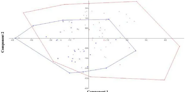
Figure 3. Principal component analysis (PCA) for Astyanax giton (blue) and
Astyanax aff. fasciatus (red), combining data from both reservoirs. Most important coefficients were length (0.4595), height (-0.56) and aspect ratio (0.5554) for component 1 (y-axis) and length (-0.4028), scales below lateral line (0.4133), scales on lateral line (0.4919) and anal fin rays (0.456).
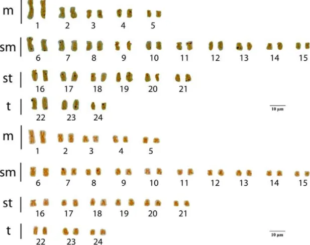
3.2 CYTOGENETICS
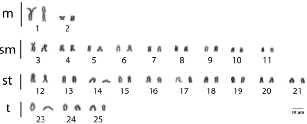
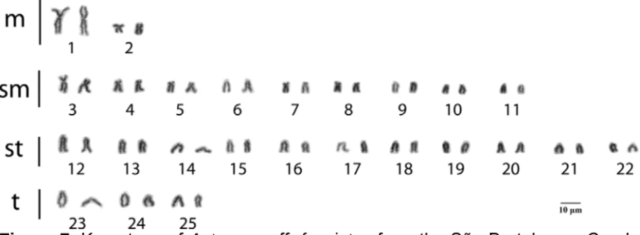
Figure 4. Karyotype of Astyanax giton from the São Bertolomeu Creek, a tributary of the Doce River Basin. Karyotypic formula: 4m+18sm+22st+6t.
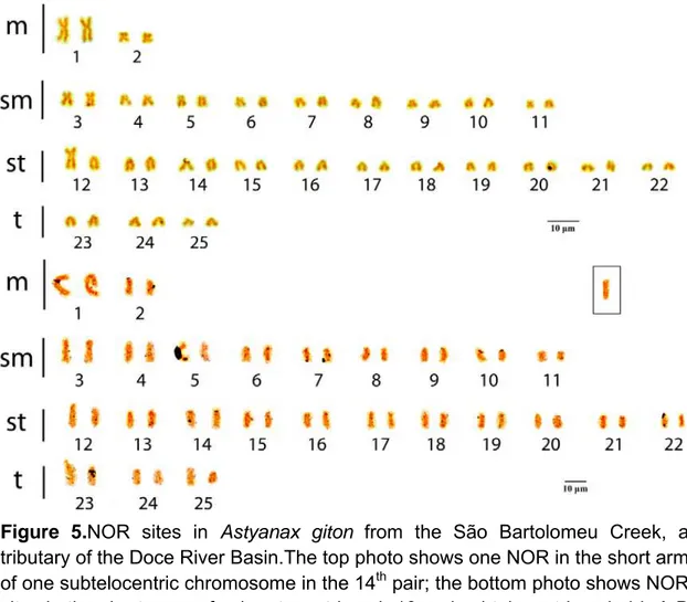
Figure 5.NOR sites in Astyanax giton from the São Bartolomeu Creek, a tributary of the Doce River Basin.The top photo shows one NOR in the short arm of one subtelocentric chromosome in the 14th pair; the bottom photo shows NOR sites in the short arms of submetacentric pair 10 and subtelocentric pair 14. A B chromosome is shown in the box.
Astyanax aff.fasciatus presented a karyotypic formula of 10m+20sm+12st+6t (FN=90 (Figure 7). There were no differences between karyotypes of males and females.
Figure 7. Karyotype of Astyanax aff. fasciatus from the São Bartolomeu Creek, a tributary of the Doce River Basin. Karyotypic formula: 10m+20sm+12st+6t.
Figure 8. NOR sites in Astyanax aff. fasciatus from the São Bartolomeu Creek, a tributary of the Doce River Basin. In the top photo, NORs are visible in the short arms of three submetacentric chromosomes in pairs 6, 11 and 13, in the bottom photo they are visible in the long arms of one subtelocentric chromosome (pair 17) and one telocentric chromosome (pair 24).
visible in the short arms of one telocentric pair and the centromeres of one metacentric pair.
(a)
(b)
3.3 B CHROMOSOMES
Supernumerary (b) chromosomes were found in five fish, one Astyanax
aff. fasciatus and four A. giton, including the only male of this species, all from Reservoir 1 (Table 3). All B chromosomes were micro except in one A. giton that had a macro B chromosome resembling the first metacentric pair. The Astyanax
aff.fasciatus had one metaphase with a single B chromosome and a second with two B chromosomes and was the only specimen to present more than one supernumerary chromosome.
Table 3. Presence of B chromosomes Accession
number Species Sex
Metaphases containing B chromosomes
CT 2651 A. giton M 1/18 (5.6%)
CT 2664 Astyanax aff. fasciatus M 2/13 (15.4%)
CT 3003 A. giton F 2/2 (100%)
CT 3005 A. giton F 12/26 (46.2%)
CT 3130 A. giton F 3/41 (7.3%)
Four out of 30 A. giton (13.3%) and one out of 30 Astyanax aff. fasciatus
Figure 11. Karyotype of Astyanax aff. fasciatus CT 2664. B chromosomes from two separate metaphases are shown in boxes.
3.4 MOLECULAR DNA
Based on mitochondrial data, the species morphologically similar to
A.giton grouped with A. giton from the nearby Sossego Dam (Viçosa, Minas Gerais State, Brazil) while those resembling Astyanax aff.fasciatus grouped with
A. fasciatus and A. parahybae, a species similar to A. fasciatus but found only in the Rio Paraíba do Sul, where A. fasciatus is absent (Figure 12).
4. DISCUSSION
The Neotropical freshwater fish fauna has long been considered the richest in the world [59], the result of a long evolutionary history of isolation and specialization [46]. Freshwater fishes are highly suitable for the recovery of past biogeographic processes and indicators of past paleohydrology [59], due to their obligatory relationship with water [46], as related forms can only occur in isolated basins if the water bodies were connected in the past.
Coastal and continental drainages may shared such a past connection, based on cytogenetic evidence from H. malabaricus, which presents seven similar cytotypes between the Ivaí, Iguaçu, Tibagi and Ribeira do Iguape rivers. Stocks of Astyanax aff. fasciatus from the Furnas region might also represent evidence of prior connection between basins [9].
The geologic evolution of the chain of high mountains associated with the eastern limits of the Brazilian Shield allow vicariance and isolation of fish species [58]. The Atlantic drainages (Doce, Paraíba do Sul and Ribeira) have existed since the break-up of Gondwanaland [46]. They include (from North to South) the Paraguaçu, Contas, Pardo and Doce rivers. These rivers have a large branching system of headwaters adjacent to the eastern headwaters of the São Francisco. Among these drainages are numerous smaller drainages that have no contact with the São Francisco Basin [58].
During periods of low sea level, the coastal plane and associated river basins were more extensive, with coastal fish species able to disperse and occupy extensive areas. Populations occupying different coastal subbasins became isolated due to repeated episodes of sea level rise, resulting in speciation and population differentiation [58] Studies of fish fauna from the Doce river show phylogenetically related fauna in the coastal and continental basins, although the exact causes of these relationships are not always clear [46]
endemics of either coastal or inland basins, a few species occurring on both sides of the watershed divide. species occurring on both sides of the main watershed divide (e.g. Astyanax fasciatus) are said to have a "pattern C" type of geographic distribution [58], which includes stream piracy between coastal and crystalline Brazilian shield rivers [76]Astyanax taeniatus, a coastal freshwater fish, reaches the northern limit of its distribution range in the Mucuri and Jucuruçu rivers [59]
Most species inhabiting the high mountain stream Guaratuba do not occur elsewhere, but one exception is A. paranae, which is common in the upper headwaters of the Tietê. This could be due to either erosion or tectonic fault reactivation [58]. Astyanax altiparanae is present in the coastal upper Rio Itatinga as well as the upper Tietê, presumably the result of stream capture by erosion. This species is often cited as being endemic to the upper Paraná, but has a vicariant form A. lacustris that inhabits the São Francisco basin. Morphological characters of both species are variable and overlapping [58].
The area north of the mouth of the Doce River is composed of beach ridges, deltas, sea cliffs and sandstone reefs, geologic structures that could divide fish populations [59]. Bathymetric surveys of this area show that various channels converge on the Abrolhos depression. The Pleistocene lagoons are delimited to the south by the mouth of the Doce River and to the north by the Abrolhos Formation. Along with A. fasciatus, the trahira, Hoplias malabaricus
has one of the largest distributional ranges, occurring from Panamá to the Buenos Aires Province in Argentina [46].
data indicate no evidence of genetic exchange between the Doce and Paraıba do Sul river populations of H. malabaricus during glacial periods [59]
The lack of data on geographic distributions and phylogeny of Neotropical freshwater fishes make it difficult to formulate hypotheses of biogeographic histories [56]. One possibility is that species of widespread distribution are relatively ancient, with spatial continuity broken by recent events of marine transgression (explaining the slight differences in Astyanax across basins). Species of restricted distribution may be also be related to these events.
4.1 MORPHOLOGY
The systematics of Astyanax are poorly understood, with the Atlantic Rainforest region containing several undescribed species [77]. Some "species" of this genus are widespread but vary geographically. Phylogenetic and taxonomic relationships among these fishes, as well as those of limited distribution, are not well known. Previous studies of Astyanax relied on morphological characters such as dentition, lateral line scales, tail fin and osteology [56]. As the species of Astyanax are morphologically similar, however, it has been difficult to separate them based on this criteria.
Based on morphological characters, Professor Vinicius de Araújo Bertaco at the Universidade Federal de Rio Grande do Sul confirmed our identification of
A. giton, based on the key of Melo [56] that describes this species as follows: Dentary teeth gradually decrease in size from the symphysis, differentiating this species from A. scabripinnis. with the base of the teeth in the dentary and internal series of the premaxillar being thin. The central cuspid is approximately the same size as the side cuspids, with the maximum number of cuspids exceeding 8. The interopercular does not expand further and does not cover a portion of the sub-opercular. Body height is 28.9 to 37.4% of the pattern length, with 34 to 39 scales on the lateral line, with a rounded naked area between the second and third infra-orbital.
by the interpolation of supplementary series. Astyanax fasciatus parahybae also has a head of 4.25 to 4.44; depth of 2.5 to 2.66; 27 to 34 (usually 31 or 32) rays in the anal fin, a lateral line with 37 to 41 (usually 39 or 40) rays. an eye equal to the interorbital; one maxillary tooth; 4 to 5 teeth in the outer row of the premaxillary in a 2:1 ratio[3]. Melo also identified A. parahybae as having irregularly placed scales above the anal fin and the anal fin having 29 or more rays, both of which differentiate this species from A. fasciatus and definitively separate it from the A. fasciatus complex[56].
According to Eigenmann, when A. fasciatus, A. scabripinnis and A. taeniatus are found together in the same river it is possible to differentiate one from another, but across different rivers and basins each undergoes so many modifications it is impossible to clearly define them morphologically [3].
Astyanax taeniatus in one river could be confused for A. scabripinnis, A. fasciatus, A. giton or A. intermedius in another. Lütken described both A. taeniatus and A. scabripinnis as A. scabripinnis rivularis. Jenyns recognized A. taeniatus as an intermediate form of A. scabripinnis. Bertaco recognized A. fasciatus as A. intermedius. Eigenmann described A. intermedius as the center of a triangle formed by A. scabripinnis, A. fasciatus and A. taeniatus.
Species assigned to Astyanax are not a monophyletic entity [8,78]. Current definition relies on morphological characters such as two rows or teeth in the premaxillary, a complete lateral line and the presence of a caudal fin, which are widespread in Characidae [3]. Astyanax intermedius has been included in the Astyanax scabripinnis species complex [78]. Astyanax intermedius has a humeral spot similar in form to A. microschemos; however A. intermedius is distinguished from this species by having a larger interorbital width (29.7-34.1% vs. 32.5-47.8%)
Astyanax giton is most easily distinguished from A. fasciatus by the presence of a vertically elongate black humeral spot. Astyanax fasciatus